An interesting case of lump abdomen Dr Vipul

An interesting case of lump abdomen Dr. Vipul metta Surgery resident

Introduction An elderly female was referred from gynaecology opd to surgery with a huge lump in abdomen. They suspected a uterine growth but wanted a surgical opinion. Her outside sonography was suggestive of a uterine leiomyoma We felt that the growth was not uterine and rather a growth from the large bowel and investigated the patient further. Clinically it was a GIST from large bowel

CLINICAL HISTORY • A 51 year old post menopausal female patient presented with complaint of lump in the left lower abdomen since 2 months. • Complains of dull pain over left side of lower abdomen. • No history of altered bowel habits or blood in stools • No history of fever or vomiting. • No history of hematuria or burning micturition • No history of weight loss • No history of cough or other respiratory complaints • No history of vaginal discharge

General Examination • Conscious and well oriented. • Moderately Built and nourished • Afebrile • Vitals- stable • No pallor, icterus, cyanosis, clubbing, generalized lymphadenopathy, edema

PER ABDOMEN On inspection • Umbilicus was central and Inverted • No abdominal distention • A 15 X 10 cm lump is seen in the left lumbar and left iliac regions not moving with respiration On palpation • A 15 cm x 10 cm swelling in the left iliac fossa extending upto peri umbilical and left lumbar regions • It was non tender, hard swelling with irregular surface upper border was ill defined • Lump was non-ballotable and dull to percussion. • No organomegaly or free fluid.

• Hernial orifices normal • Per rectal examination was normal. • On per vaginal examination was normal. • other Systemic Examination within normal limits Provisional diagnosis : GIST arising from descending and sigmoid colon

Investigations • Hb: 12. 2 g/dl • WBC count: 12, 400/mm 3 • Platelet: 2. 80 lakh/mm 3 • B. Urea - 31 mg/dl • S. creatinine: 0. 89 mg/dl • LFTs: normal • Serology: negative • Chest X-ray: Normal • ECG : Normal

USG - Abdomen and pelvis • A 11 X 11 cm hypo echoic mass lesion in Left iliac Region upto left Supra umbilical Region likely to be arising from sigmoid colon • Liver is normal in size and echogenicity • Uterus was normal • Mild hydroureteronephrosis on the left side, right kidney and ureter are normal • Rest of sonographic findings are normal CA-125 – normal CEA - within normal limits COLONOSCOPYExtrinsic compression seen in the sigmoid no intraluminal mass or growth Rest of bowel appears normal
 x 11. 5 cm(transverse) x 12")
MRI Abdomen and pelvis • A 11 cm(AP) x 11. 5 cm(transverse) x 12 cm(vertical) lobulated heterogenous mass lesion in left lumbar and left Iliac fossa Region arising from recto sigmoid with heterogeneous enhancement post contrast. • There are non enhancing cystic areas within the mass • It is displacing the Adjacent Bowel loops, Causing Mass effect on the Left mid ureter with Resulting mild hydronephrosis and proximal Hydroureter on left side. • No hepatic metastases • Rest abdomen is normal • Suggestive of Mesenchymal Stromal tumor - ? ? GIST

MRI – Abdomen and Pelvis

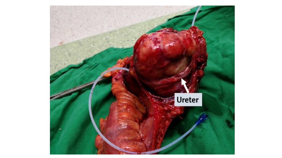
Intra operative finding • Patient Underwent explorative laparotomy under General anaesthesia with a stand by urologist • A large Irregular mass with Nodular surface arising from the wall of sigmoid colon occupying most of the left half of the abdominal cavity. The left ureter was seen to be encased by the mass causing dilated proximal ureter and compressing the left kidney • No peritoneal or liver nodules found • No enlarged lymph nodes found • Rest of the bowel was normal

• Mass encasing the mid ureter which was excised along with it from the colon.

Procedure • Sigmoidectomy with Colo rectal anastomoisis with ileal interposition. • Yang - monti’s procedure with left Dj stenting
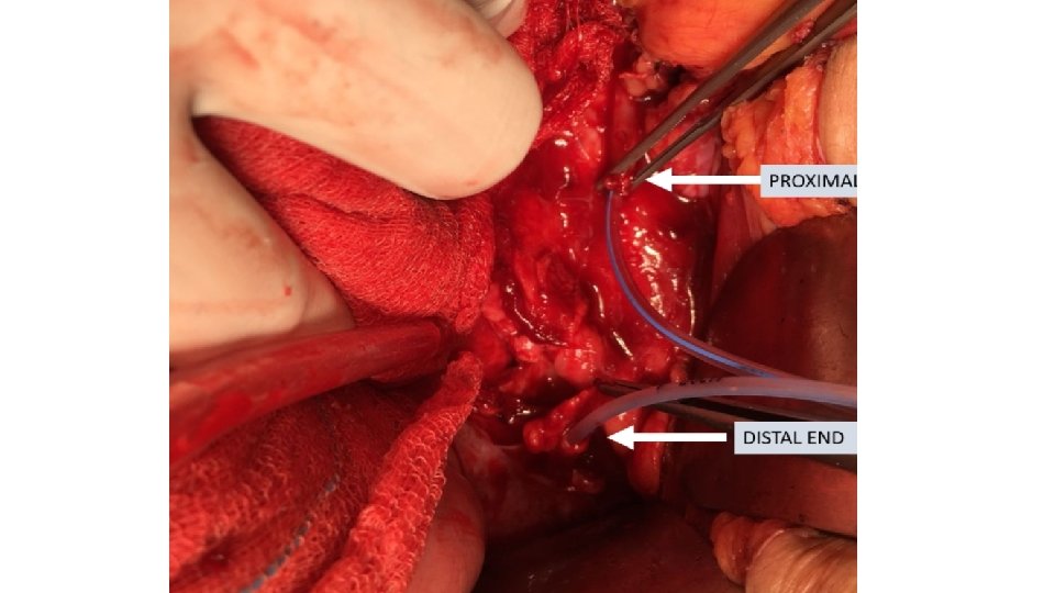

Distance between the proximal and distal cut ends of ureter Length of ileal graft needed

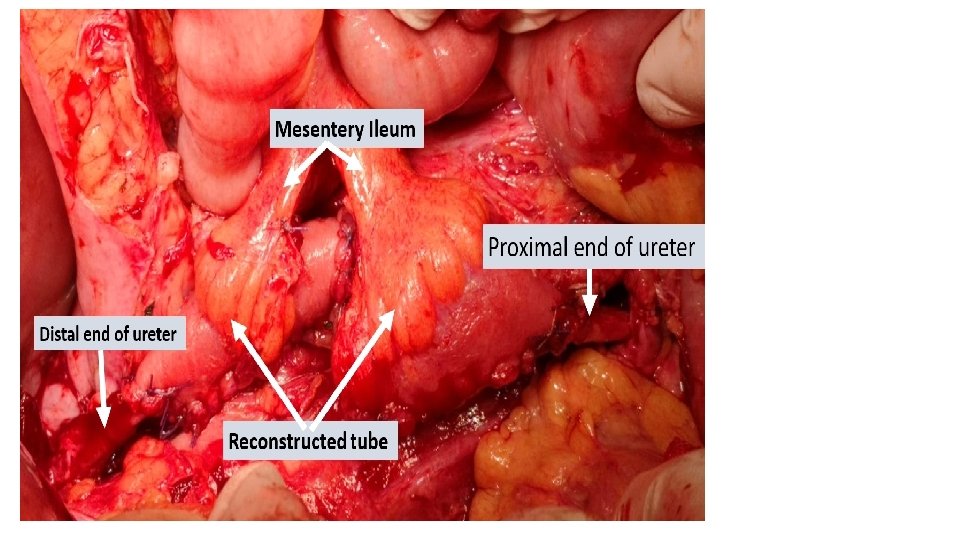
The ileal graft used for reconstruction of ureter. Two such grafts were used

The ileum was opened vertically and sutured horizontally and tubularized for uretero- ileal anastomosis

Ileo ileal anastomosis

Ileal graft Distal ureter Dj stent Ileum anastomosed to distal ureter with dj stent

-At the end of procedure patient had • 2 uretero ileal anastomoses • 2 ileoileal anastomoses -Drains were placed in the pelvis and hepatorenal pouch -Patient was shifted to sicu and monitored

Post operative period • Patient had good urinary output initially • From pod 3 there was an increase in the pelvic drain collection with a slight decrease in urine output suggesting an anastomotic leak • Iv pyelography was done which showed a block in the ureter due to mucous plug from the ileal secretions • Cystoscopy was done on pod 7 and the dj stent was replaced with a silicon stent • Left PCN was done to divert the urine • Over 3 weeks the patient recovered and the nephrostomy was closed • Drains were removed as the drain output reduced • Patient recovered well and was discharged.

Histopathology examination • HPE suggestive of a spindle cell tumor arising from sigmoid colon, possibilities of -low grade MALIGNANT PERIPHERAL NERVE SHEATH TUMOR -GASTROINTESTINAL STROMAL TUMOR -LEIOMYOSARCOMA. • IHC was done CD 117 negative DOG 1 negative Desmin positive Smooth muscle actin positive Pathological examination suggestive of LEIOMYOSARCOMA of sigmoid colon

DISCUSSION • Primary mesenchymal sarcomas of the gastrointestinal tract are rare and constitute only 0. 1– 3% of all gastrointestinal tumors. • Leiomyosarcoma (LMS) is the most common variant of these tumors and represents only 0. 12% of all colon malignancies. • LMS most commonly originates in the uterus, GI tract, and retroperitoneum. With in the GItract, the stomach is the most common site followed by the small intestine, colon, and rectum. • Surgery is the main treatment for primarycolonic LMS, asmost of the reported cases were diagnosed using surgically resected specimens. While adjuvant chemotherapy and/or radiation are often used in the management of primary colonic LMS, there are conflicting data on their efficacy and impact on overall survival. [4] • Due to increased incidence and histological similarity with GIST, in past leiomyosarcoma, were misdiagnosed as GIST. Immunohistochemistry is the crux of the diagnosis. GIST stains positive for CD 34 and C‑kit (CD 117) andstains negative for desmin in contrast to leiomyosarcomas which are positive for desmin and negative for CD 117, CD 34, and DOG 1. [5] • Immunohistochemistry is unavoidable tool in diagnosis of leiomyosarcoma and should be considered in GIST, particularly in unusual sites. In contrast to GIST which is curable with surgery followed by imatinib (in intermediate and high risk patients), leiomyosarcomasbehave aggressively requiring radical excision of tumor followed by intensive cytotoxic chemotherapy regimens. Radiotherapy has only a limited role. [6]
![REFERENCES • [1] A. Yaren, S. De˘girmencio˘glu, N. Callı Demirkan, A. G¨okçen Demiray, B.](http://slidetodoc.com/presentation_image_h/37afb0bb20a36d99e7091fbd9673bbe5/image-26.jpg "REFERENCES • [1] A. Yaren, S. De˘girmencio˘glu, N. Callı Demirkan, A. G¨okçen Demiray, B.")
REFERENCES • [1] A. Yaren, S. De˘girmencio˘glu, N. Callı Demirkan, A. G¨okçen Demiray, B. Tas¸k¨oyl¨u, and G. G. Do˘gu, “Primary mesenchymal tumors of the colon: a report of three cases, ” Turkish Journal of Gastroenterology, vol. 25, pp. 314– 318, 2014. • [2] K. Iwasa, K. Taniguchi, M. Noguchi, H. Yamashita, and M. Kitagawa, “Leiomyosarcoma of the colon presenting as acute suppurative peritonitis, ” Surgery Today , vol. 27, no. 4, pp. 337– 344, 1997. • [3] P. Singh, B. Bello, C. Weber, and K. Umanskiy, “Rectal leiomyosarcoma in association with ulcerative colitis: a rare condition with an unusual presentation, ” International Journal of Colorectal Disease, vol. 29, no. 7, pp. 887 -888, 2014. • [4] Babichev Y, Kabaroff L, Datti A, et al. PI 3 K/AKT/m. TOR inhibition in combination with doxorubicin is an effective therapy for leiomyosarcoma. J Transl Med 2016; 14: 67. • [5] Aggarwal G, Sharma S, Zheng M, Reid MD, Crosby JH, Chamberlain SM, et al. Primary leiomyosarcomas of the gastrointestinal tract in the post‑gastrointestinal stromal tumor era. Ann Diagn Pathol 2012; 16: 532‑ 40. • [6] Cite this article as: Kiran P, Shiny PM, Dhanya KS, Aravindan KP. Diagnosis of leiomyosarcoma of colon. J Can Res Ther 2015; 11: 1035.
- Slides: 26