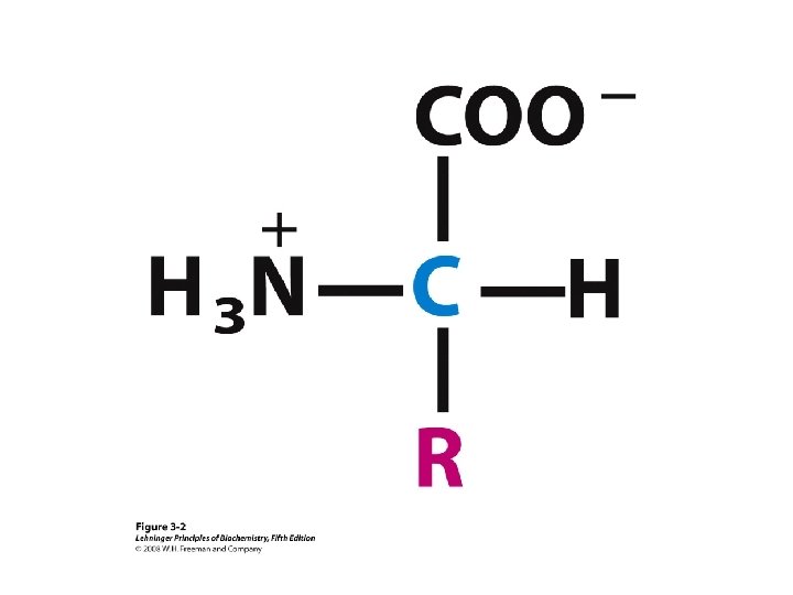
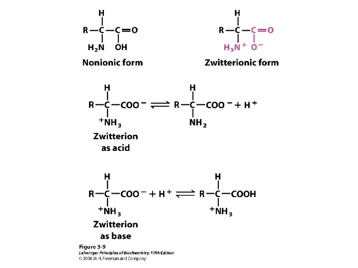
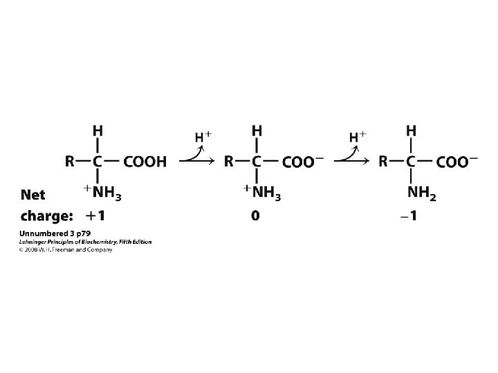
Amino Acids Peptides Proteins Amino Acids Zwitterion or


 of the Proteome in a Complex Organism")

 Alanine (A) Valine (V) Isoleucine (I) Leucine (L) Proline")


 • Chromatography – Ion")


 Genetic Method Proteomics")


 which is purified by")


- Slides: 91
Amino Acids, Peptides & Proteins Amino Acids Zwitterion or ampholyte Peptides Proteins Structure of Proteins Separation & purification Protein Sequencing Protein Synthesis
Our Life Is Maintained by Molecular Network Systems Molecular network system in a cell
Proteins Play Key Roles in a Living System • Three examples of protein functions Alcohol dehydrogenase oxidizes alcohols to aldehydes or ketones – Catalysis: Almost all chemical reactions in a living cell are catalyzed by protein enzymes. – Transport: Some proteins transports various substances, such as oxygen, ions, and so on. – Information transfer: For example, hormones. Haemoglobin carries oxygen Insulin controls the amount of sugar in the blood
Estimated Functional Roles (by % of Proteins) of the Proteome in a Complex Organism
Protein Functions • Transport • Regulatory • Motor • Fibrous Protein • Enzyme • Immunoglobulin
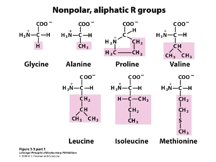
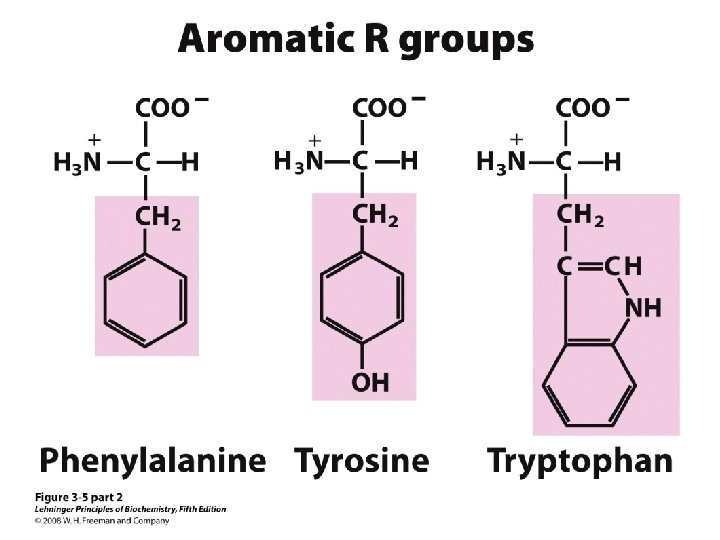
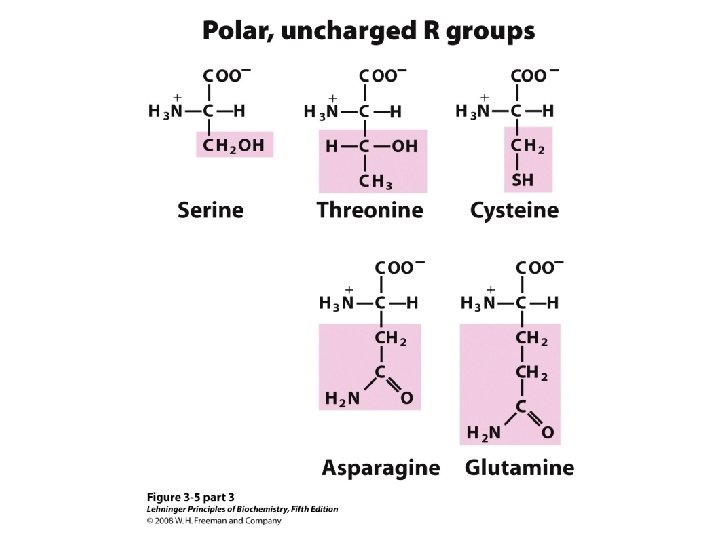
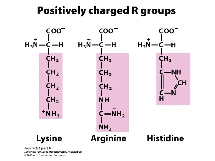
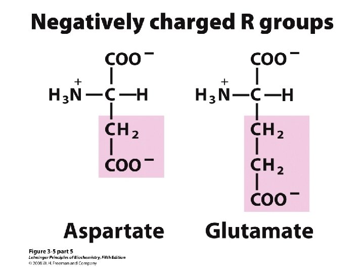
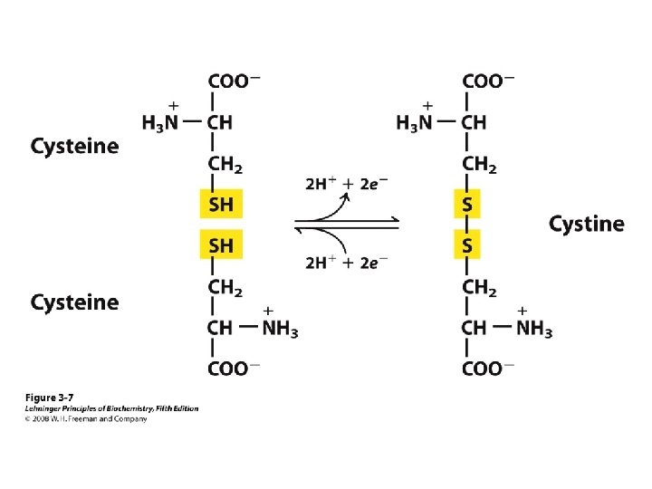
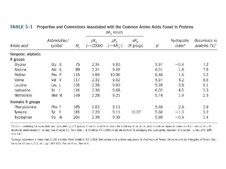
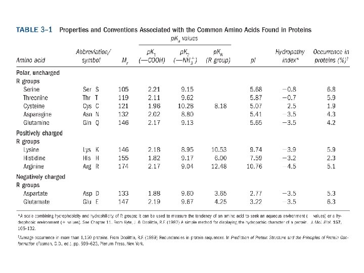
20 Amino acids Glycine (G) Alanine (A) Valine (V) Isoleucine (I) Leucine (L) Proline (P) Methionine (M) Phenylalanine (F) Tryptophan (W) Asparagine (N) Glutamine (Q) Serine (S) Threonine (T) Tyrosine (Y) Cysteine (C) Asparatic acid (D) Glutamic acid (E) Lysine (K) Arginine (R) Histidine (H) White: Hydrophobic, Green: Hydrophilic, Red: Acidic, Blue: Basic
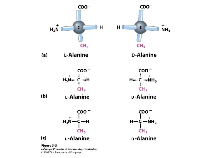
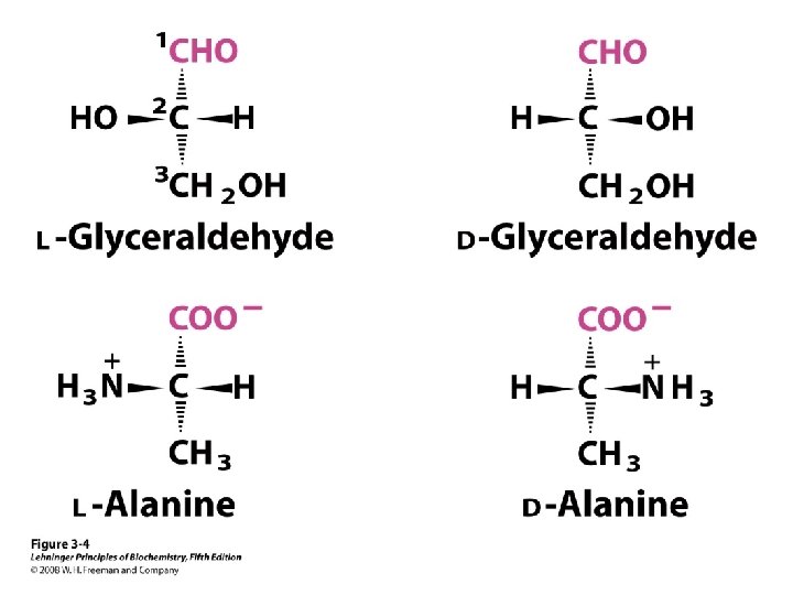
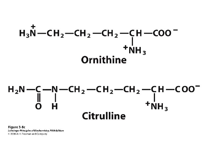
a-amino aicds All proteins are composed of amino acids Ø Twenty common amino acids Ø All amino acids are primary amino acids except for proline Ø A primary amino group is attached to the a-carbon of carboxyl group Ø Except for Glycine, all other amino acids have at least one chiral centers Ø All chiral amino acids are belong to L-amino acids Ø
Amino acids vary in Size Structure Electric charge Solubility in water
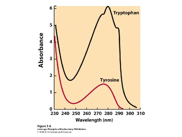
Classification of amino acids Classified by polarity of side chains hydrophobic: water fearing, non-polar side chains hydrophilic: water loving, polar neutral positively charged negatively charged aromatic
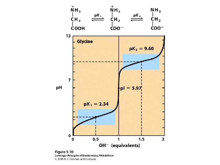
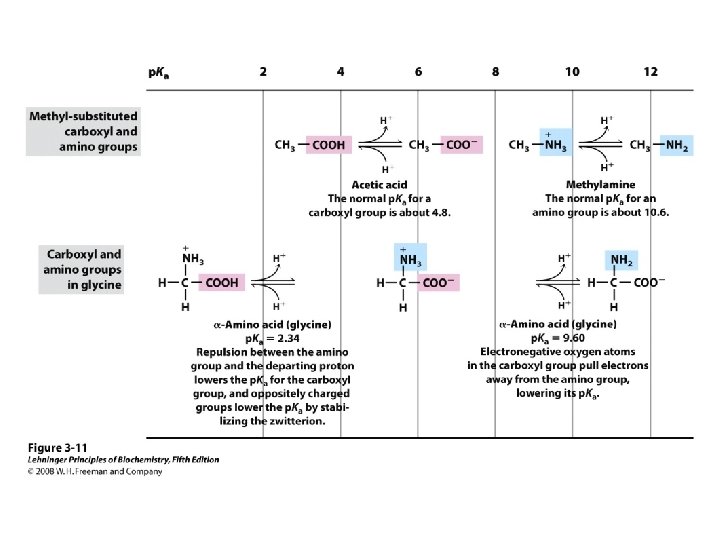
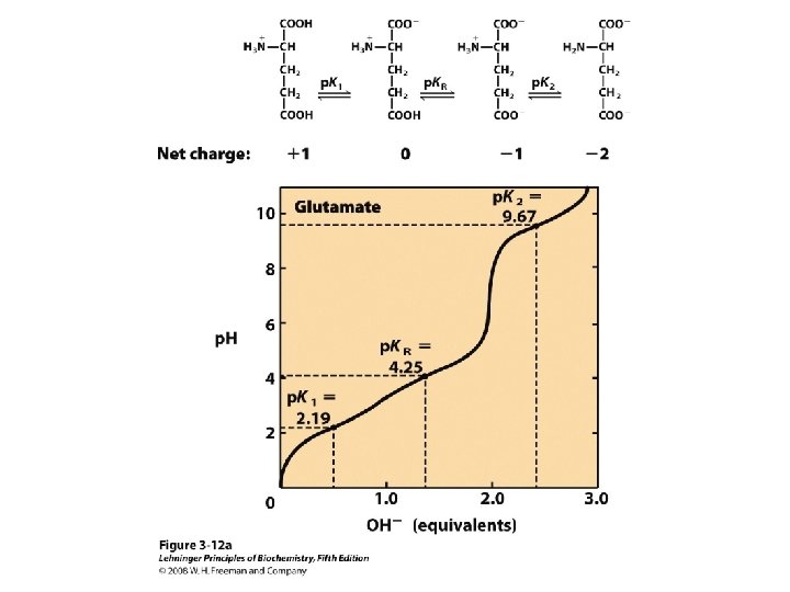
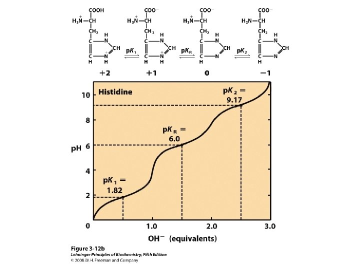
Titration curve of amino acids Neutral side chain Acidic side chain Basic side chain
Peptide Bond
Peptides Dipeptide Tripeptide Tetrapeptide Oligopeptide Polypeptide Protein
Peptide Bond
Replace -ine by -yl but keep the last -ine ! Ala-Gly-Arg Alanylglycylarginine
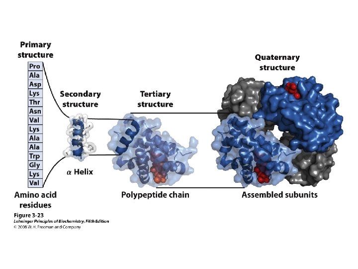
Structure of Proteins Primary structure Secondary structure Tertiary structure Quaternary structure
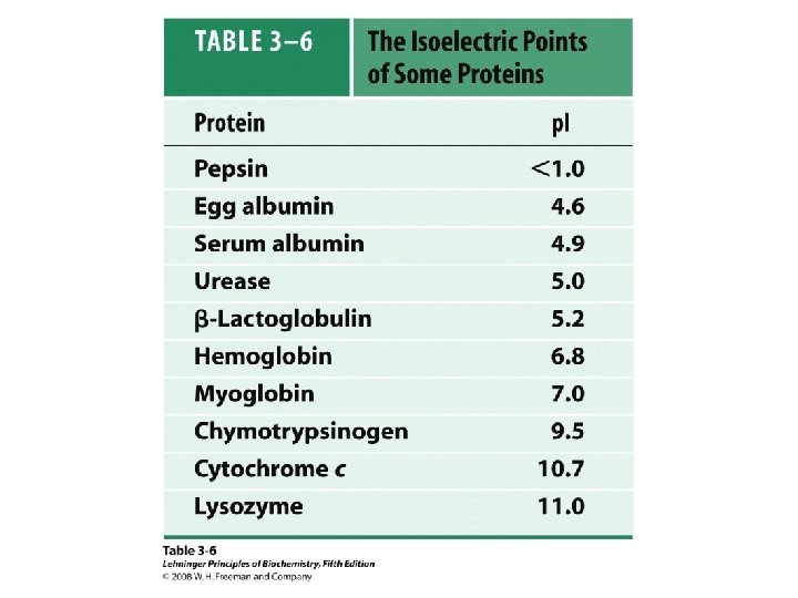
Peptide & Protein Charge p. I = ?
Group p. Ka p. H=1 p. H=3 p. H=7 -COOH 2. 2 0 - - -NH 2 8. 8 + + + 0 0 0 Glu 4. 3 0 0 - - Lys 10. 8 + + 0 0 Arg 12. 5 + + + 0 +3 +2 +1 0 -1 -2 Net Charge p. H=10 p. H=11 p. H=13 p. I = (8. 8 + 10. 8)/2 = 9. 8
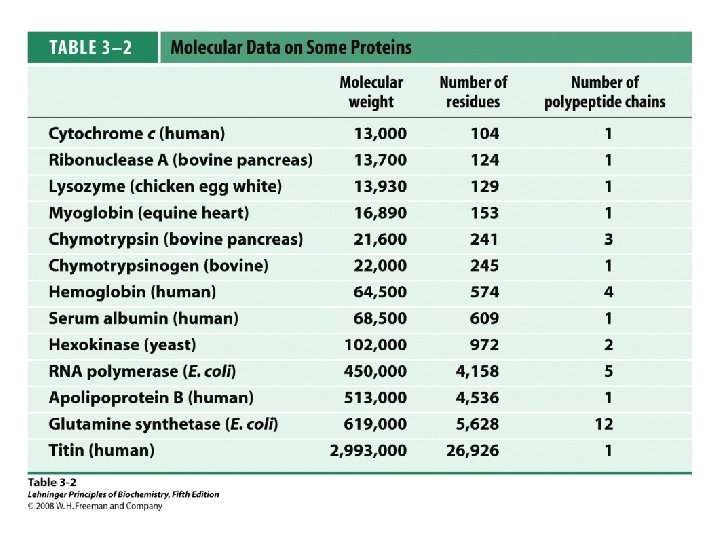
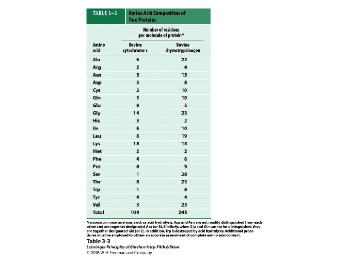
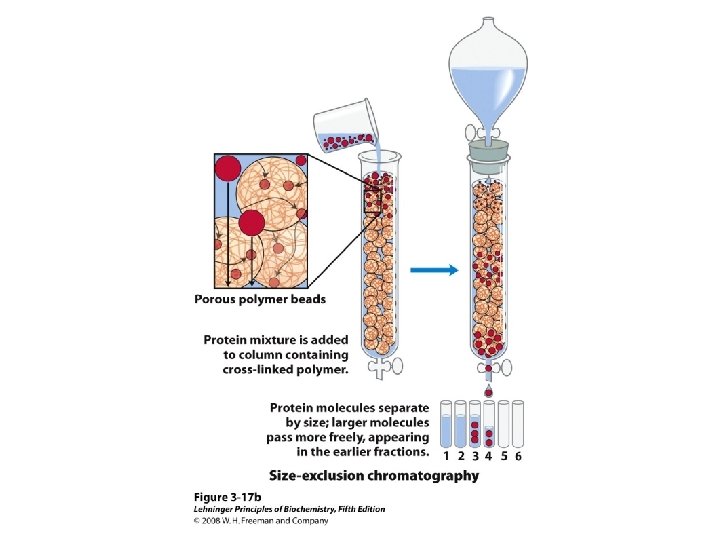
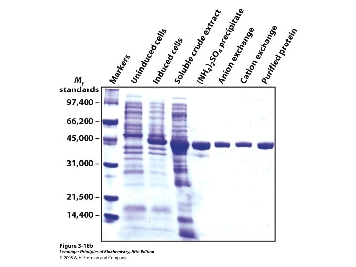
Protein Separation/Purification • In general, proteins contain > 40 residues – Minimum needed to fold into tertiary structure • Usually 100 -1000 residues; percent of each AA varies • Proteins separated based on differences in size and composition • Proteins must be pure to analyze, determine structure/function • Factors to control (to avoid denaturation or chemical degradation) – – – p. H Presence of enzymes Temperature Reactive thiol groups Exposure to air, water
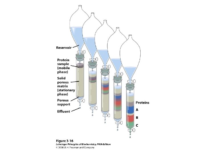
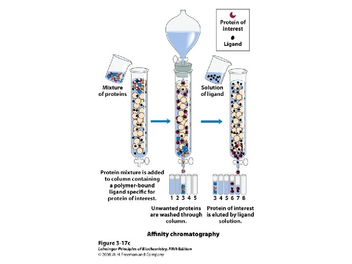
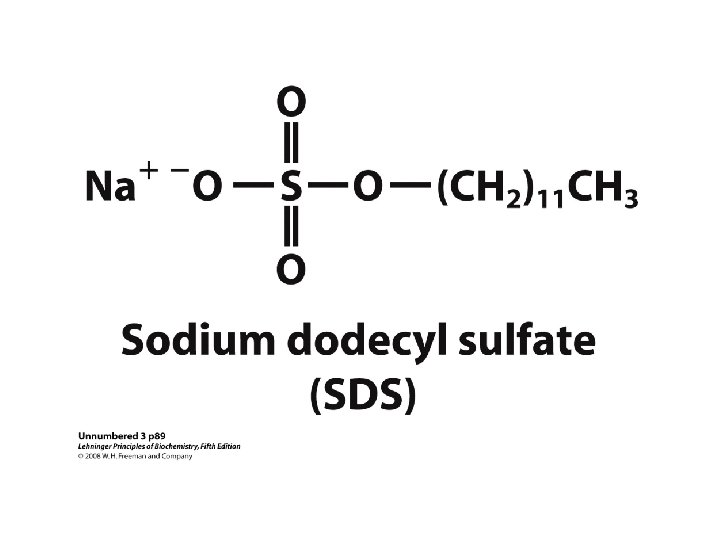
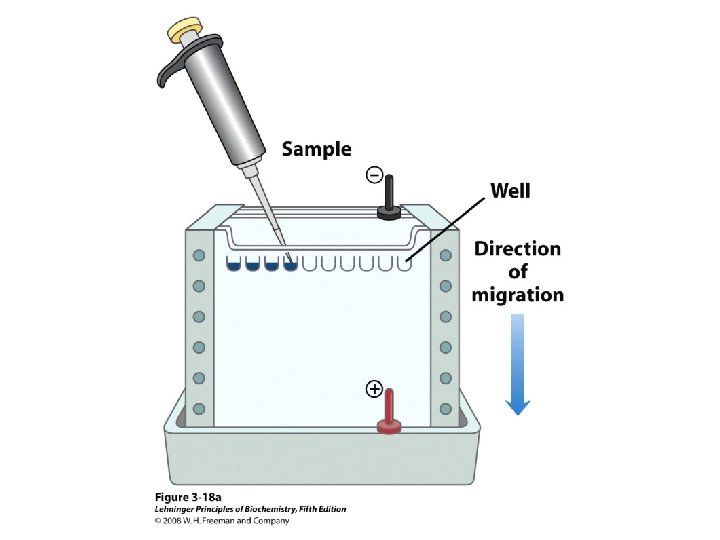
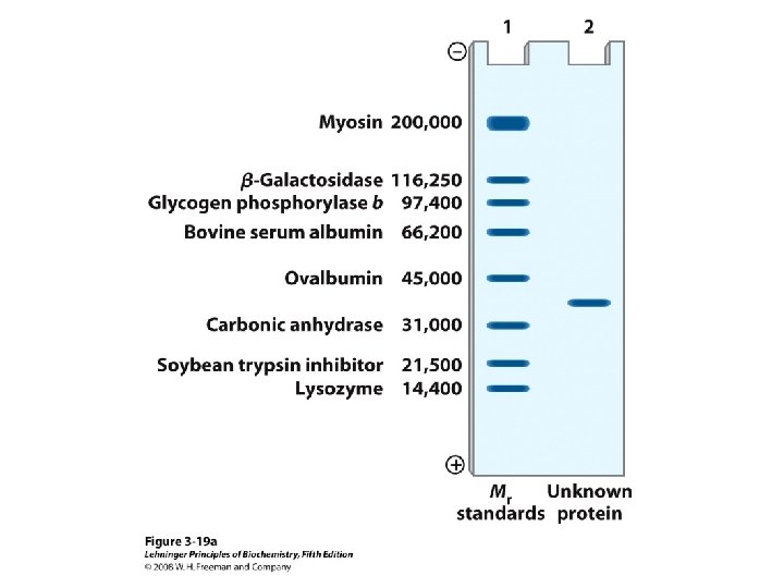
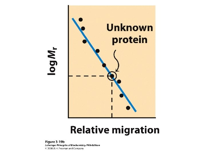
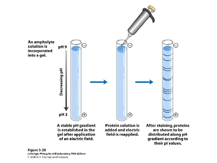
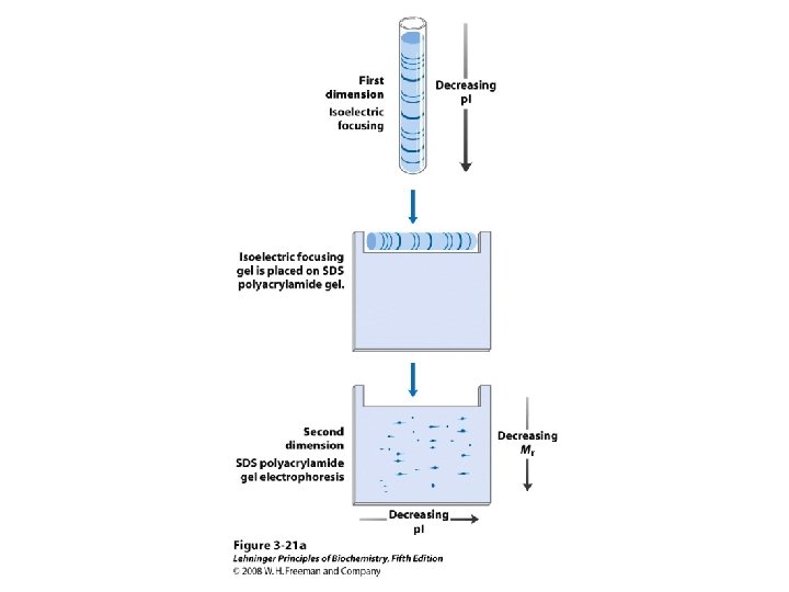
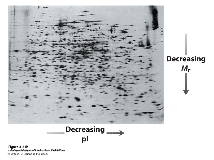
Methods of Separation/Purification • Solubility (salts, solvents, p. H, temperature) • Chromatography – Ion exchange – Gel filtration – Affinity • Electrophoresis
Protein Sequencing Traditional Chemical Method (Sanger Method) Genetic Method Proteomics
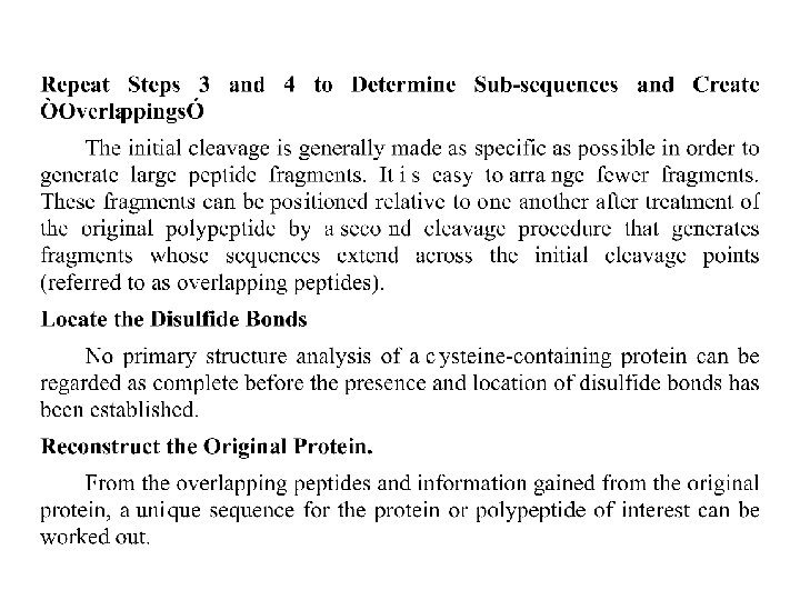
A Common Strategy for Protein Sequencing
Hydrolysis • A polypeptide can be hydrolyzed by refluxing with 6 M hydrochloric acid for 24 h • The individual amino acids can be separated from each other using a cation-exchange resin – An acidic solution of the amino acids is passed through the cationexchange column; the strength of adsorption varies with the basicity of each amino acid (the most basic are held most strongly) – Washing the column with a sequence of buffered solutions causes the amino acids to move through it at different rates
• In the original method, the column eluant is treated with ninhydrin, a dye used for detecting and quantifying each amino acid as it comes off the column • In modern practice, analysis of amino acid mixtures is routinely accomplished using high performance liquid 64 chromatography (HPLC)
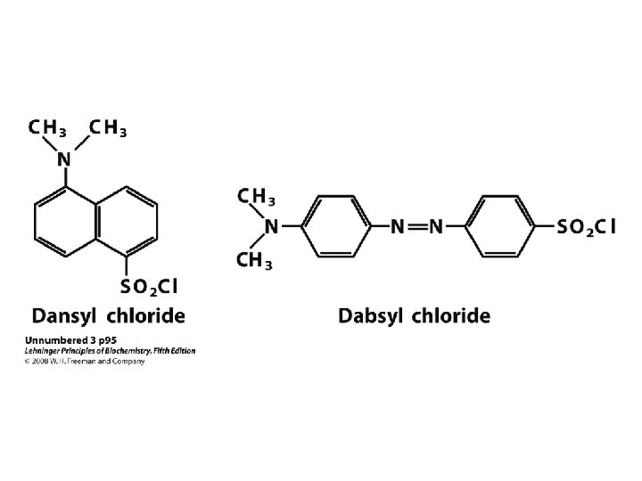
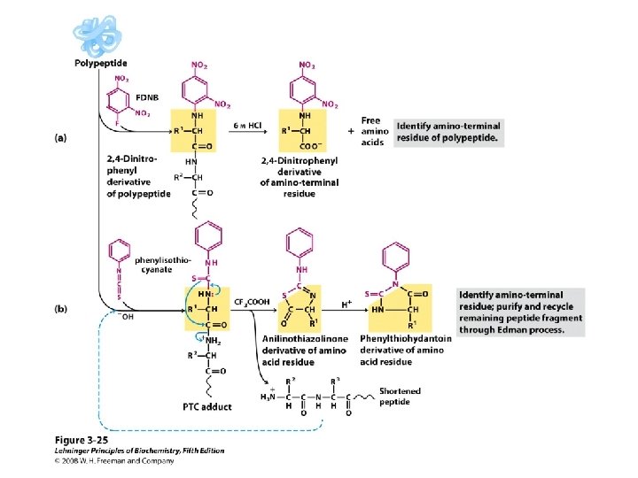
– Sanger N-Terminal Analysis • The N-terminal end of the polypeptide is labeled with 2, 4 -dinitrofluorobenzene and the polypeptide is hydrolyzed – The labeled N-terminal amino acid is separated from the mixture and identified • The Sanger method is not as widely used as the Edman method
– C-Terminal Analysis • Enzymes called carboxypeptidases hydrolyze C-terminal amino acids selectively – The enzyme continues to release each newly exposed C-terminal amino acid as the peptide is hydrolyzed; it is necessary to monitor the release of C-terminal amino acids as a function of time to identify them
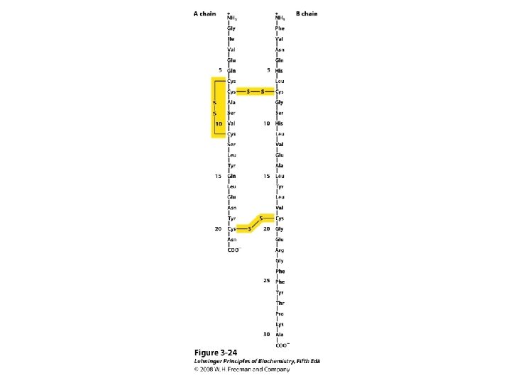
• Primary Structure of Polypeptides and Proteins • The sequence of amino acids in a polypeptide is called its primary structure – Several methods exist to elucidate the primary structure of peptides – Edman Degradation • Edman degradation involve sequential cleavage and identification of N-terminal amino acids • Edman degradation works well for polypeptide sequence analyses up to approximately 60 amino acid residues – The N-terminal residue of the polypeptide reacts with phenyl isothiocyanate – The resulting phenylthiocarbamyl derivative is cleaved from the peptide chain 68
– The unstable product rearranges to a stable phenylthiohydantoin (PTH) which is purified by HPLC and identified by comparison with PTH standards • Automated amino acid sequencing machines use the Edman degradation and high performance liquid chromatography (HPLC) – One Edman degradation cycle beginning with a picomolar amount of polypeptide can be completed in approximately 30 minutes – Each cycle results in identification of the next amino acid residue in the peptide
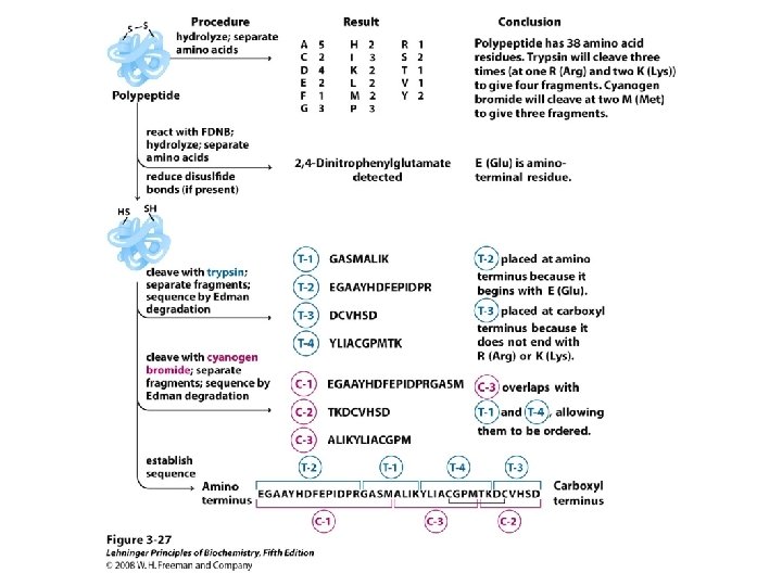
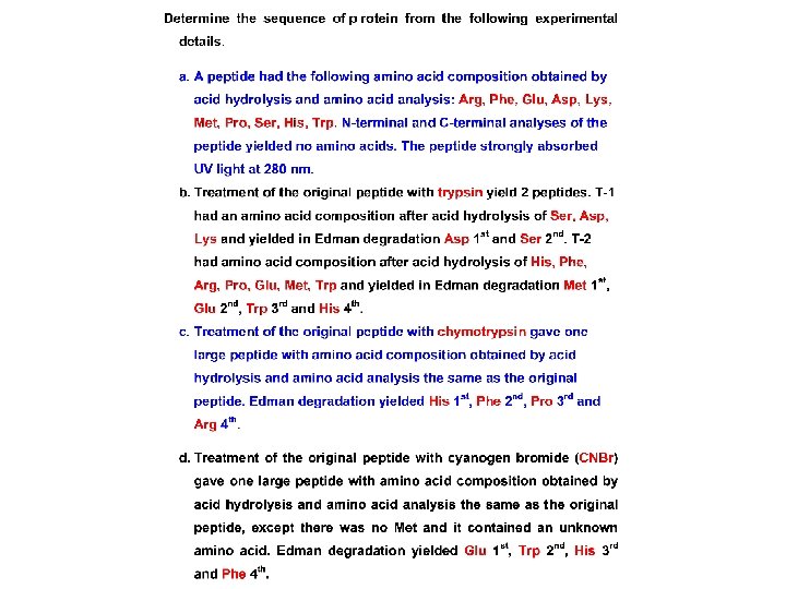
– Complete Sequence Analysis • The Sanger and Edman methods of analysis apply to short polypeptide sequences (up to about 60 amino acid residues by Edman degradation) • For large proteins and polypeptides, the sample is subjected to partial hydrolysis with dilute acid to give a random assortment of shorter polypeptides which are then analyzed – The smaller polypeptides are sequenced, and regions of overlap among them allow the entire polypeptide to be sequenced
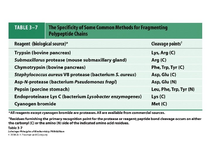
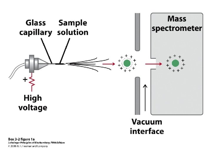
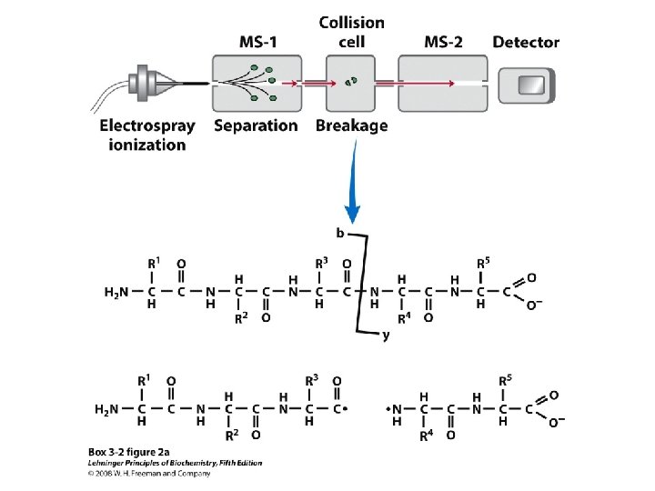
• Larger polypeptides can also be cleaved into smaller sequences using site-specific reagents and enzymes – The use of these agents gives more predictable fragments which can again be overlapped to obtain the sequence of the entire polypeptide – Cyanogen bromide (CNBr) cleaves peptide bonds only on the C-terminal side of methionine residues Mass spectrometry can be used to determine polypeptide and protein sequences
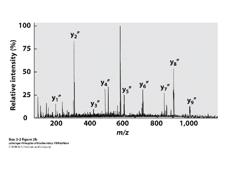
“Ladder sequencing” involves analyzing a polypeptide digest by mass spectrometry, wherein each polypeptide in the digest differs by one amino acid in length; the difference in mass between each adjacent peak indicates the amino acid that occupies that position in the sequence Mass spectra of polypeptide fragments from a protein can be compared with databases of known polypeptide sequences, thus leading to an identification of the protein or a part of its sequence by matching
The Blind Men and the Elephant by John Godfrey Saxe American poet John Godfrey Saxe (1816 -1887) based the following poem on a fable which was told in India many years ago. The Blind Men and the Elephant It was six men of Indostan To learning much inclined, Who went to see the Elephant (Though all of them were blind), That each by observation Might satisfy his mind The First approached the Elephant, And happening to fall Against his broad and sturdy side, At once began to bawl: “God bless me! but the Elephant Is very like a wall!” The Second, feeling of the tusk, Cried, “Ho! what have we here So very round and smooth and sharp? To me ’tis mighty clear This wonder of an Elephant Is very like a spear!” The Third approached the animal, And happening to take The squirming trunk within his hands, Thus boldly up and spake: “I see, ” quoth he, “the Elephant Is very like a snake!” The Fourth reached out an eager hand, And felt about the knee. “What most this wondrous beast is like Is mighty plain, ” quoth he; “ ‘Tis clear enough the Elephant Is very like a tree!” The Fifth, who chanced to touch the ear, Said: “E’en the blindest man Can tell what this resembles most; Deny the fact who can This marvel of an Elephant Is very like a fan!? The Sixth no sooner had begun About the beast to grope, Than, seizing on the swinging tail That fell within his scope, “I see, ” quoth he, “the Elephant Is very like a rope!” And so these men of Indostan Disputed loud and long, Each in his own opinion Exceeding stiff and strong, Though each was partly in the right, And all were in the wrong!
A Fan A Rope A Spear A Tree A Snake A Wall