Amino Acids Amino Acids Proteins are the most


 commonly found as constituents")


,")


![A. Derivation of the equation • By solving for the [H+] in the above](https://slidetodoc.com/presentation_image_h2/75c520914bccd3176cf61f12f55d0dc4/image-31.jpg "A. Derivation of the equation • By solving for the [H+] in the above")


 is the p. H at which")


- Slides: 48
Amino Acids
Amino Acids • Proteins are the most abundant and functionally diverse molecules in living systems. • Virtually every life process depends on proteins. • Proteins display an incredible diversity of functions, yet all share the common structural feature of being linear polymers of amino acids. • For example, enzymes and polypeptide hormones direct and regulate metabolism in the body, whereas contractile proteins in muscle permit movement. • In the bloodstream, proteins, such as hemoglobin and plasma albumin, shuttle molecules essential to life, whereas immuno globulins fight infectious bacteria and viruses.
Amino Acids • There are twenty Amino Acids (A. A) commonly found as constituents of mammalian proteins. • These are the only amino acids that are coded for by DNA, the genetic material in the cell.
General formula of Amino Acid • Each amino acid has a Carboxyl group, an amino group, and a distinctive side chain ("R-group") bonded to the α-carbon atom. • (except for proline)
General formula of Amino Acid • At physiologic p. H ( ~ p. H = 7. 4), the carboxyl group is dissociated, forming the negatively charged carboxylate ion(-COO-) and the amino group is protonated(-NH 3+).
General formula of amino acid • In proteins, almost all of these carboxyl and amino groups are combined in peptide linkage and, in general, are not available for chemical reaction except for hydrogen bond formation.
General formula of Amino Acid • The nature of the side chains dictates the role an amino acid plays in a protein. • It is, therefore, useful to classify the amino acids according to the properties of their side chains. • Side chains, whether they are § nonpolar (that is, have an even distribution of electrons) or § polar (that is, have an uneven distribution of electrons, such as acids and bases).
A. Amino acids with nonpolar side chains • Each of these amino acids has a nonpolar side chain that does not bind or give off protons or participate in hydrogen or ionic bonds. • The side chains of these amino acids can be thought of as "oily" or lipid like , a property that promotes hydrophobic interactions.
hydrophobic interactions
A. Amino acids with nonpolar side chains
1. Location of nonpolar amino acids in proteins • Proteins found in aqueous solutions (a polar environment), the side chains of the nonpolar amino acids tend to cluster together in the interior of the protein. • This phenomenon, known as the hydrophobic effect, is the result of the hydrophobicity of the nonpolar R-group, which act much like droplets of oil that clustered in an aqueous environment. • The nonpolar R-groups thus fill up the interior of the folded protein and help give it its three-dimensional shape.
1. Location of nonpolar amino acids in proteins • For proteins that are located in a hydrophobic environment, such as a membrane, the nonpolar R-groups are found on the outside surface of the protein, interacting with the lipid environment.
2. Proline • The side chain of proline and its α-amino group form a ring structure, and thus proline differs from other amino acids in that it contains an imino group, rather than an amino group.
B. Amino acids with uncharged polar side chains • These amino acids have zero net charge at neutral p. H, although the side chains of cysteine and tyrosine can lose a proton at an alkaline p. H. • Serine, threonine, and tyrosine each contain a polar hydroxyl group that can participate in hydrogen bond formation. • The side chains of asparagine and glutamine each contain a carbonyl group and an amide group, both of which can also participate in hydrogen bonds.
B. Amino acids with uncharged polar side chains
1 -Disulfide bond • The side chain of cysteine contains a sulfhydryl group (-SH), which is an important component of the active site of many enzymes. • In proteins, the -SH groups of two cysteines can become oxidized to form a dimer, cystine, which contains a covalent cross-link called a disulfide bond (-S-S-).
Disulfide bond:
2 - Side chains as sites of attachment for other compounds: • The polar hydroxyl group of Serine, threonine, and, rarely, tyrosine can serve as a site of attachment for structures such as a phosphate group. • In addition, the amide group of asparagine, as well as the hydroxyl group of serine or threonine, can serve as a site of attachment for oligosaccharide chains in glycoproteins.
C. Amino acids with acidic side chains • Aspartic acid and glutamic acid are proton donors. • At physiological p. H , the side chains of these amino acids are fully ionized, containing a negatively charged carboxylate group (-COO-). • They are, therefore, called Aspartate or Glutamate to emphasize that these amino acids are negatively charged at physiologic p. H.
C. Amino acids with acidic side chains
D. Amino acids with basic side chains • The side chains of the basic amino acids accept protons. • At physiologic PH the side chains of lysine and arginine are fully ionized and positively charged. • In contrast, histidine is weakly basic, and the free amino acid is largely uncharged at physiologic p. H. • However, when histidine is incorporated into a protein, its side chain can be either positively charged or neutral, depending on the ionic environment provided by the polypeptide chains of the protein.
D. Amino acids with basic side chains
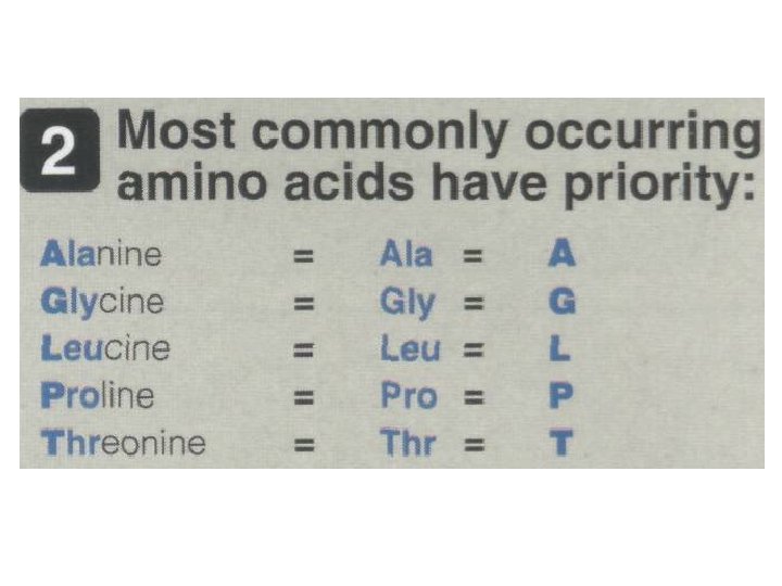
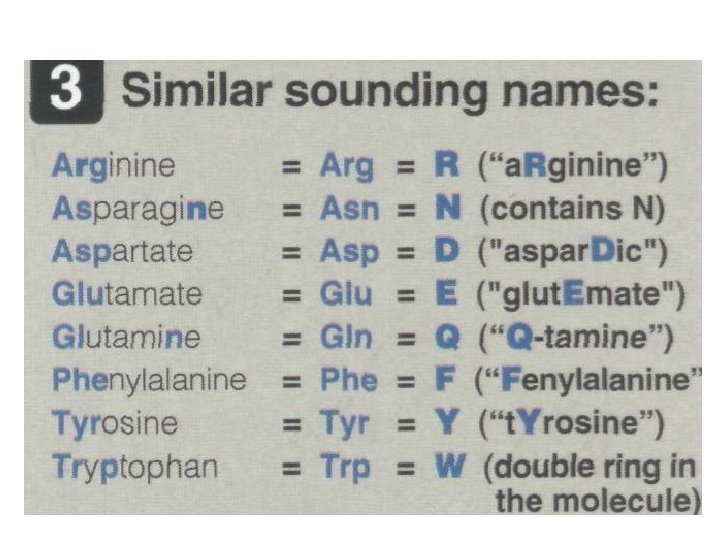
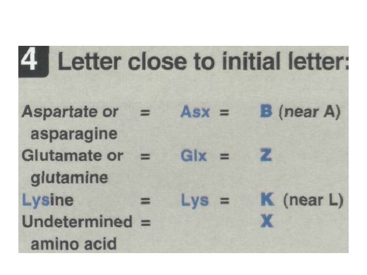
Abbreviations and symbols for the commonly occurring amino acids:
F. Optical properties of amino acids • The α-carbon of an amino acid is attached to four different chemical groups and is, therefore, a chiral or optically active carbon atom. • Glycine is the exception because its α-carbon has two hydrogen substituents and, therefore, is optically inactive. • Amino acids that have an asymmetric center at the α-carbon can exist in two forms, designated D and L, that are mirror images of each other. • The two forms in each pair are termed stereoisomers, opticalisomers, or enantiomers. • All amino acids found in proteins are of the L-configuration.
Acidic and basic properties of amino acids • Both free amino acids and some amino acids combined in peptide linkages can act as buffers. • Amino acids in aqueous solution contain weakly acidic αcarboxyl groups and weakly basic α-amino groups. • In addition, each of the acidic and basic amino acids contains an ionizable group in its side chain.
Acidic and basic properties of amino acids Acids may be defined as proton donors and bases as proton acceptors. Acids (or bases)described as “weak” ionize to only a limited extent. The concentration of protons in aqueous solution is expressed as p. H, where p. H = log 1/[H+] or –log [H+]. The quantitative relationship between the p. H of the solution and concentration of a weak acid (HA) and its con jugate base (A–) is described by the Henderson-Hasselbalch equation.
A. Derivation of the equation Consider the release of a proton by a weak acid represented by HA: HA H+ + A– Weak acid proton salt form acid or conjugate base The “salt” or conjugate base, A–, is the ionized form of a weak acid. By definition, the dissociation constant of the acid, Ka, is The larger the Ka, the stronger the acid, because most of the HA has dissociated into H+ and A–. The smaller the Ka, the less acid has dissociated and, therefore, the weaker the acid.
A. Derivation of the equation • By solving for the [H+] in the above equation, taking the logarithm of both sides of the equation, multiplying both sides of the equation by– 1, and substituting p. H = –log [H+] and p. Ka= – log Ka, we obtain the Henderson-Hasselbalch equation
B. Buffers • A buffer is a solution that resists change in p. H following the addition of an acid or base. • A buffer can be created by mixing a weak acid (HA) with its conjugate base(A-). • If an acid such as HCL is added to such a solution, A- can neutralize it, in the process being converted to HA. • If a base is added HA can neutralize it in the process being converted to A-
B. Buffers • Maximum buffering capacity occurs at a p. H equal to the p. Ka. • But a conjugate acid/base pair can still serve as an effective buffer when the p. H of a solution is within ± 1 p. H unit of the p. Ka. • If the amounts of HA and A– are equal, the p. H is equal to the p. Ka.
B. Buffers • A solution containing acetic acid (HA= CH 3 COOH and acetate (A-=CH 3 COO-) with a p. Ka of 4. 8 resists a change in p. H from p. H 3. 8 to 5. 8, with maximum buffering at p. H = 4. 8. • At p. H values less than the p. Ka the protonated acid form(CH 3 COOH) is the predominant species. • At p. H values greater than the p. Ka the deprotonated base form(CH 3 COO-) is the predominant species in solution.
Titration of an amino acid 1. Dissociation of the carboxyl group: • Alanine, for example, contains both an α-carboxyl and an αamino group. • At a low (acidic) p. H, both of these groups are protonated.
1. Dissociation of the carboxyl group: • As the p. H of the solution is raised, the -COOH group of (Form I) can dissociate by donating a proton to the medium. • The release of a proton results in the formation of the carboxylate group (-COO-). • This structure is shown as (Form II), which is the dipolar form of the molecule (zwitterion) (isoelectric form) (it has an overall net charge of zero).
2. Application of the Henderson-Hasselbalch equation • The dissociation constant of the carboxyl group of an amino acid is called K 1 rather than Ka because the molecule contains a second titratable group. • The Henderson-Hasselbalch equation can be used to analyze the dissociation of the carboxyl group of alanine in the same way as described for acetic acid. • where I is the fully protonated form of alanine, and II is the isoelectric form of alanine. • This equation can be rearranged and converted to its logarithmic form to yield:
3. Dissociation of the amino group • The second titratable group of alanine is the amino group (NH 3+). This is a much weaker acid than the -COOH group and, therefore, has a much smaller dissociation constant, K 2. • Release of a proton from the protonated amino group of Form II results in the fully deprotonated form of alanine, Form III.
4. p. Ks of alanine • Each titratable group has a p. Ka that is numerically equal to the p. H at which exactly one half of the protons have been removed from that group. • The p. Ka for the most acidic group(-COOH) is p. K 1 whereas the p. Ka for the next most acidic group(-NH 3+) is p. K 2.
5. Titration curve of alanine • The change in p. H that occurs during the addition of base to the fully protonated form of alanine (I) to produce the completely deprotonated form (III).
Buffer pairs: The -COOH/-COO- pair can serve as a buffer in the p. H region around Pk 1, and the -NH 3+/NH 2 pair can buffer in the region around p. K 2. When p. H = p. K: When the p. H is equal to p. K 1 (2. 3), equal amounts of Forms I and II of alanine exist in solution. When the p. H is equal to p. K 2 (9. 1), equal amounts of Forms II and III are present in solution.
c. Isoelectric point • The isoelectric point (PI) is the p. H at which an amino acid is electrically neutral, that is, in which the sum of the positive charges equals the sum of the negative charges. • For an amino acid, such as alanine, that has only two dissociable hydrogens (one from the α-carboxyl and one from the α-amino group), the PI is the average of p. Ka 1 and p. Ka 2 (PI=p. Ka+p. Ka/2) ==== [2. 3 + 9. 1]/2 = 5. 7
c. Isoelectric point • It corresponds to the p. H at which structure II (with a net charge of zero) predominates, and at which there also equal amounts of Form I (net charge of +1) and III (net charge of -1)
Net charge of amino acids at neutral p. H • At physiologic p. H, all amino acids have a negatively charged group (-COO-) and a positively charged group (NH 3+) both attached to the α-carbon. • [Note: Glutamate, aspartate, histidine, arginine, and lysine have additional potentially charged groups in their side chains. ] • Substances, such as amino acids, that can act either as an acid or a base are defined as amphoteric substances (amphoteric electrolytes).
Other applications of the Henderson-Hasselbalch equation • The Henderson-Hasselbalch equation can be used to calculate how the p. H of a physiologic solution responds to changes in the concentration of weak acid and/or its corresponding "salt" form. • For example, In the bicarbonate buffer system, the Henderson. Hasselbalch equation predicts how shifts in [HCO 3 -] and CO 2 influence p. H.
Summary • Each amino acid has an α-carboxyl group and a primary α -amino group (except for proline, which has a secondary amino group). • At physiologic p. H, the α-carboxyl group is dissociated, forming the negatively charged carboxylate ion (– COO–), and the α amino group is protonated (– NH 3+). • Each amino acid also contains one of 20 distinctive side chains attached to the α-carbon atom.
Summary • The chemical nature of this side chain determines the function of an amino acid in a protein, and provides the basis for classification of the amino acids as nonpolar, uncharged polar, acidic, or basic. • All free amino acids, plus charged amino acids in peptide chains, can serve as buffers. • The quantitative relationship between the p. H of a solution and the concentration of a weak acid (HA) and its conjugate base (A–) is described by the Henderson. Hasselbalch equation.
Summary • Buffering occurs within ± 1 p. H unit of the p. Ka, and is maximal when p. H = p. Ka, at which [A–] = [HA]. • The α-carbon of each amino acid (except glycine) is attached to four different chemical groups and is, therefore, a chiral or optically active carbon atom. • Only the L-form of amino acids is found in proteins synthesized by the human body.