
AMINO ACID METABOLISM Metabolic relationship of amino acids
AMINO ACID METABOLISM
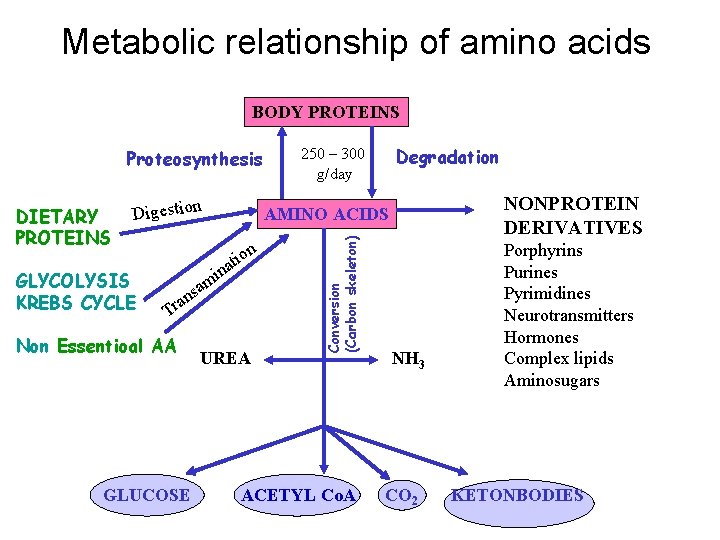
Metabolic relationship of amino acids BODY PROTEINS Proteosynthesis Digestion GLYCOLYSIS KREBS CYCLE on i t ina am s ran T Non Essentioal AA GLUCOSE NONPROTEIN DERIVATIVES AMINO ACIDS UREA Conversion (Carbon skeleton) DIETARY PROTEINS Degradation 250 – 300 g/day ACETYL Co. A NH 3 CO 2 Porphyrins Purines Pyrimidines Neurotransmitters Hormones Complex lipids Aminosugars KETONBODIES
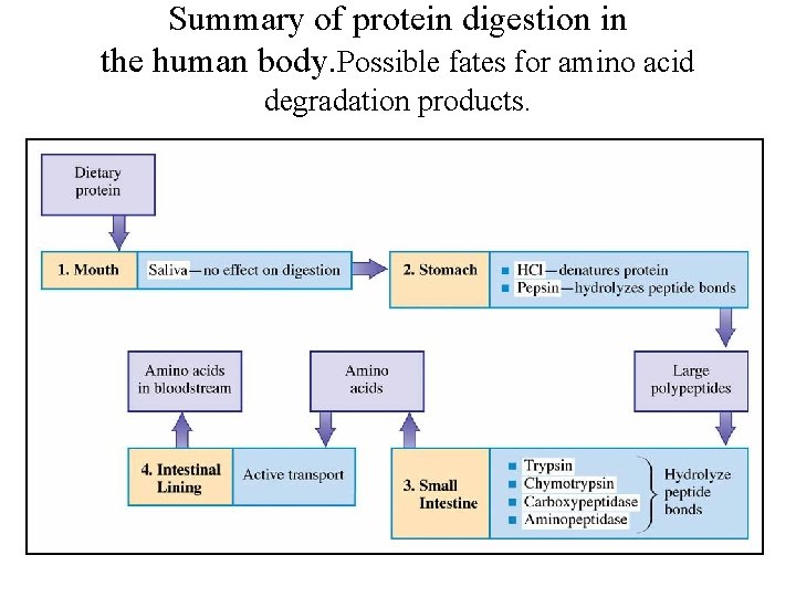
Summary of protein digestion in the human body. Possible fates for amino acid degradation products.
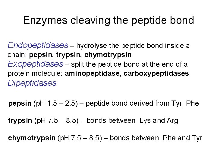
Enzymes cleaving the peptide bond Endopeptidases – hydrolyse the peptide bond inside a chain: pepsin, trypsin, chymotrypsin Exopeptidases – split the peptide bond at the end of a protein molecule: aminopeptidase, carboxypeptidases Dipeptidases pepsin (p. H 1. 5 – 2. 5) – peptide bond derived from Tyr, Phe trypsin (p. H 7. 5 – 8. 5) – bonds between Lys and Arg chymotrypsin (p. H 7. 5 – 8. 5) – bonds between Phe and Tyr

Gamma-glutamyl cycle
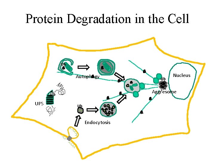
Protein Degradation in the Cell Ub Autophagy Nucleus Aggresome Ub UPS Ub Ub Endocytosis
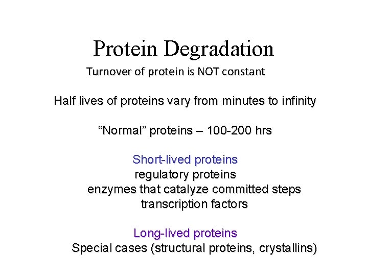
Protein Degradation Turnover of protein is NOT constant Half lives of proteins vary from minutes to infinity “Normal” proteins – 100 -200 hrs Short-lived proteins regulatory proteins enzymes that catalyze committed steps transcription factors Long-lived proteins Special cases (structural proteins, crystallins)
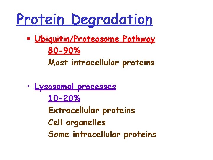
Protein Degradation § Ubiquitin/Proteasome Pathway 80 -90% Most intracellular proteins • Lysosomal processes 10 -20% Extracellular proteins Cell organelles Some intracellular proteins
How are proteins selected for degradation?
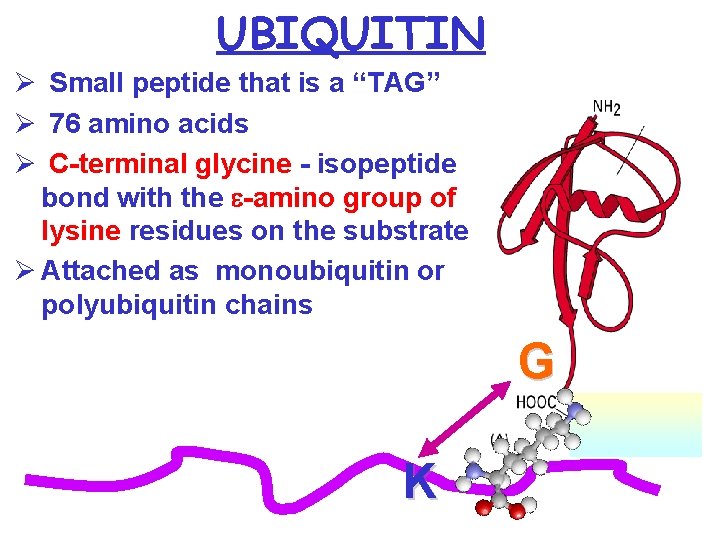
UBIQUITIN Ø Small peptide that is a “TAG” Ø 76 amino acids Ø C-terminal glycine - isopeptide bond with the e-amino group of lysine residues on the substrate Ø Attached as monoubiquitin or polyubiquitin chains G K
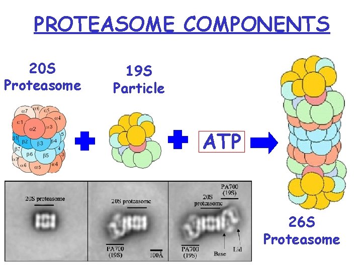
PROTEASOME COMPONENTS 20 S Proteasome 19 S Particle ATP 26 S Proteasome
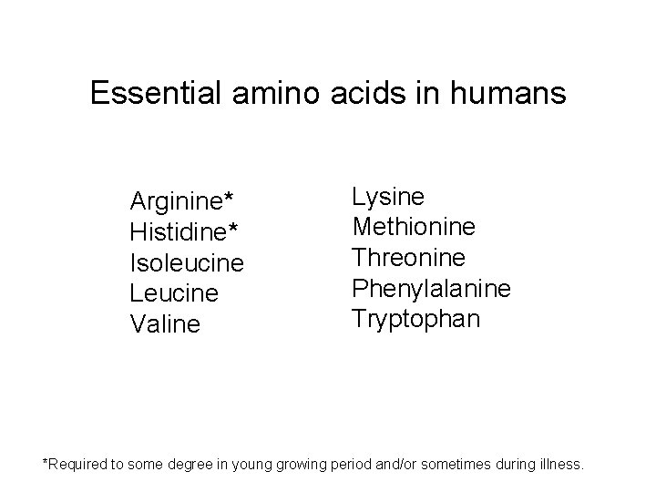
Essential amino acids in humans Arginine* Histidine* Isoleucine Leucine Valine Lysine Methionine Threonine Phenylalanine Tryptophan *Required to some degree in young growing period and/or sometimes during illness.
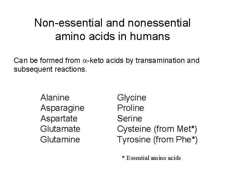
Non-essential and nonessential amino acids in humans Can be formed from -keto acids by transamination and subsequent reactions. Alanine Asparagine Aspartate Glutamine Glycine Proline Serine Cysteine (from Met*) Tyrosine (from Phe*) * Essential amino acids
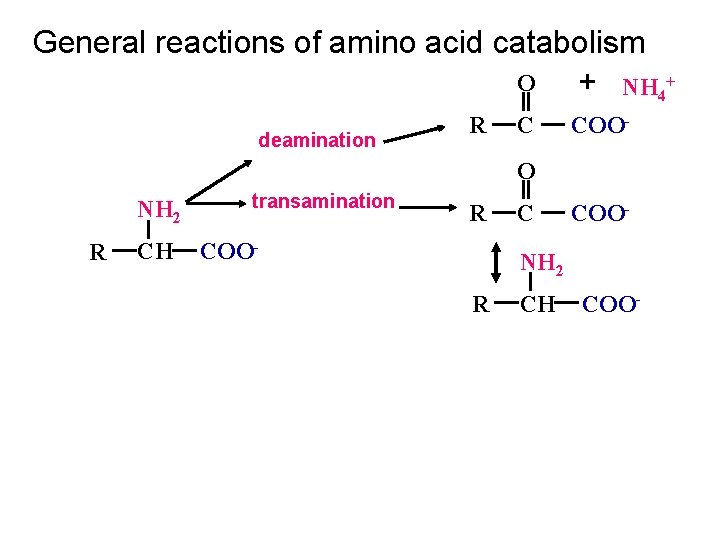
General reactions of amino acid catabolism O + NH 4+ deamination R C COO- O NH 2 R CH transamination R COO- C COO- NH 2 R CH COO-
Transamination reaction The first step in the catabolism of most amino acids is removal of a-amino groups by enzymes transaminases or aminotransferases All aminotransferases have the same prostethic group and the same reaction mechanism. The prostethic group is pyridoxal phosphate (PPL), the coenzyme form of pyridoxine (vitamin B 6)
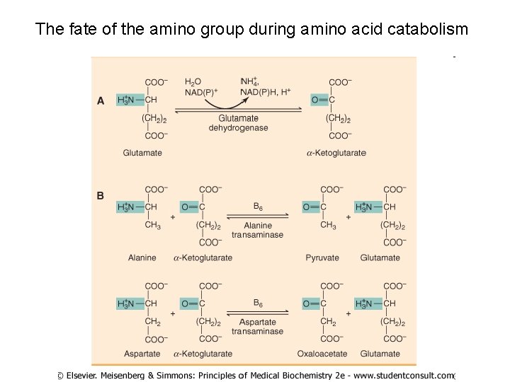
The fate of the amino group during amino acid catabolism
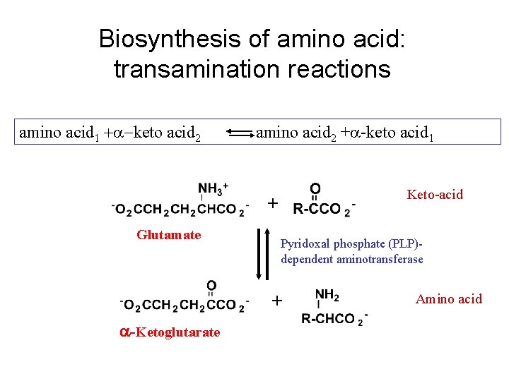
Biosynthesis of amino acid: transamination reactions amino acid 1 + -keto acid 2 amino acid 2 + -keto acid 1 Keto-acid + Glutamate Pyridoxal phosphate (PLP)dependent aminotransferase + -Ketoglutarate Amino acid
All amino acids except threonine, lysine, and proline can be transaminated Transaminases are differ in their specificity for L-amino acids. The enzymes are named for the amino group donor.
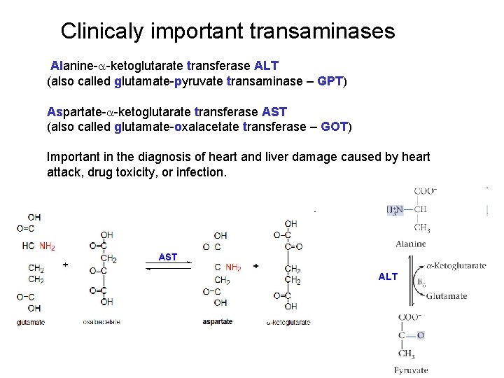
Clinicaly important transaminases Alanine- -ketoglutarate transferase ALT (also called glutamate-pyruvate transaminase – GPT) Aspartate- -ketoglutarate transferase AST (also called glutamate-oxalacetate transferase – GOT) Important in the diagnosis of heart and liver damage caused by heart attack, drug toxicity, or infection. ALT
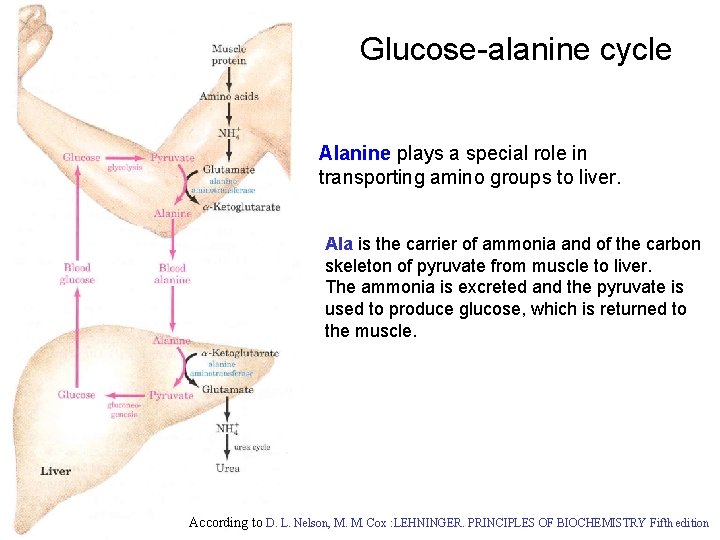
Glucose-alanine cycle Alanine plays a special role in transporting amino groups to liver. Ala is the carrier of ammonia and of the carbon skeleton of pyruvate from muscle to liver. The ammonia is excreted and the pyruvate is used to produce glucose, which is returned to the muscle. According to D. L. Nelson, M. M. Cox : LEHNINGER. PRINCIPLES OF BIOCHEMISTRY Fifth edition
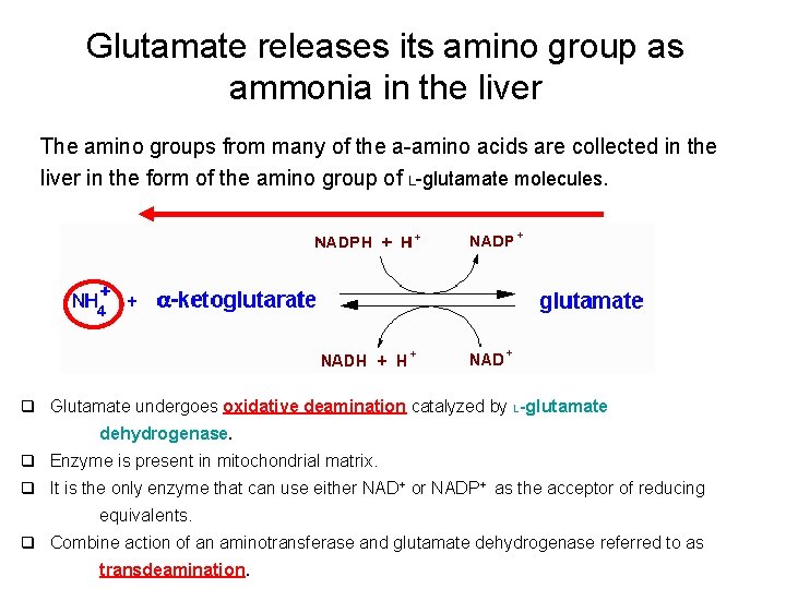
Glutamate releases its amino group as ammonia in the liver The amino groups from many of the a-amino acids are collected in the liver in the form of the amino group of L-glutamate molecules. q Glutamate undergoes oxidative deamination catalyzed by L-glutamate dehydrogenase. q Enzyme is present in mitochondrial matrix. q It is the only enzyme that can use either NAD+ or NADP+ as the acceptor of reducing equivalents. q Combine action of an aminotransferase and glutamate dehydrogenase referred to as transdeamination.
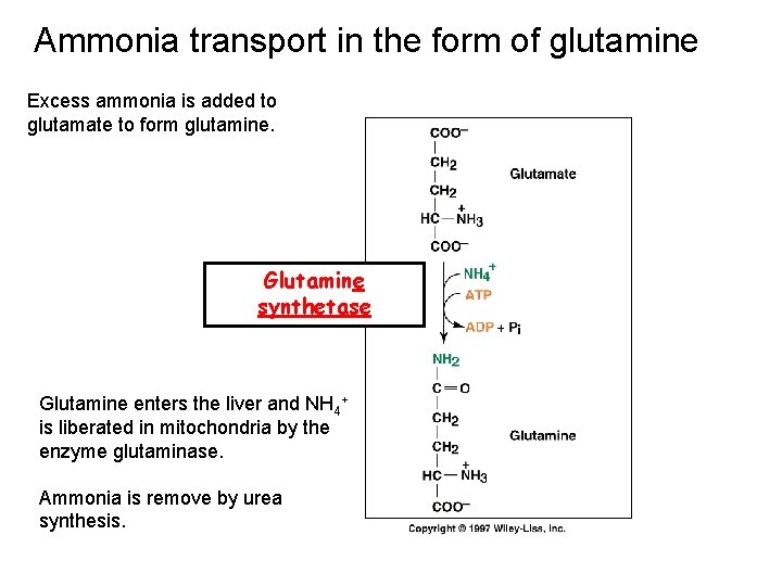
Ammonia transport in the form of glutamine Excess ammonia is added to glutamate to form glutamine. Glutamine synthetase Glutamine enters the liver and NH 4+ is liberated in mitochondria by the enzyme glutaminase. Ammonia is remove by urea synthesis.
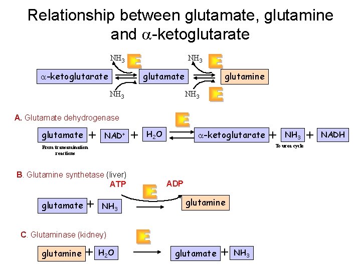
Relationship between glutamate, glutamine and -ketoglutarate NH 3 glutamate -ketoglutarate NH 3 glutamine NH 3 A. Glutamate dehydrogenase glutamate + NAD+ + H 2 O -ketoglutarate B. Glutamine synthetase (liver) ATP + NH 3 ADP glutamine C. Glutaminase (kidney) glutamine + NH 3 To urea cycle From transamination reactions glutamate + H 2 O glutamate + NH 3 + NADH
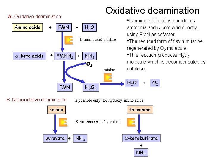
Oxidative deamination Amino acids + FMN + • L-amino acid oxidase produces H 2 O L-amino acid oxidase -keto acids + FMNH 2 + NH 3 O 2 FMN B. Nonoxidative deamination catalse H 2 O 2 ammonia and -keto acid directly, using FMN as cofactor. • The reduced form of flavin must be regenerated by O 2 molecule. • This reaction produces H 2 O 2 molecule which is decompensated by catalase. H 2 O + O 2 Is possible only for hydroxy amino acids serine threonine Serin-threonin dehydratase pyruvate + NH 3 -ketobutirate + NH 3
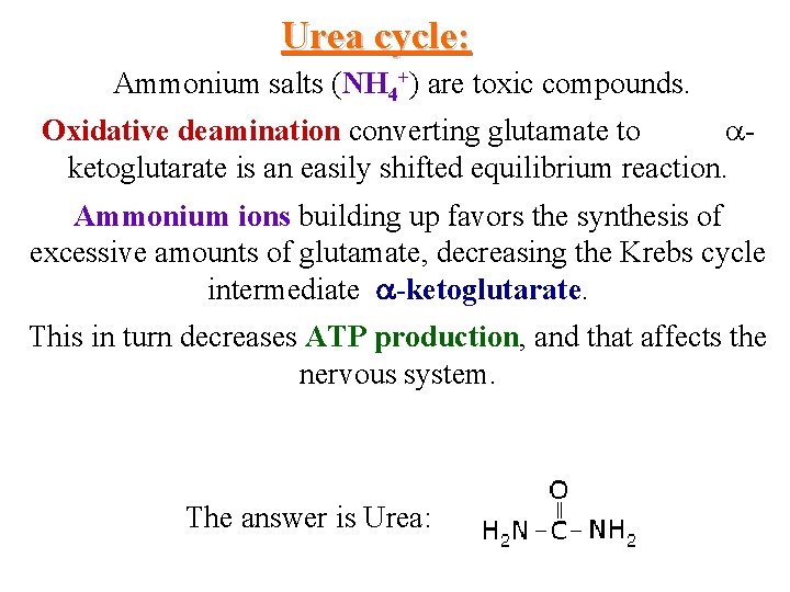
Urea cycle: Ammonium salts (NH 4+) are toxic compounds. Oxidative deamination converting glutamate to ketoglutarate is an easily shifted equilibrium reaction. Ammonium ions building up favors the synthesis of excessive amounts of glutamate, decreasing the Krebs cycle intermediate -ketoglutarate. This in turn decreases ATP production, and that affects the nervous system. The answer is Urea:
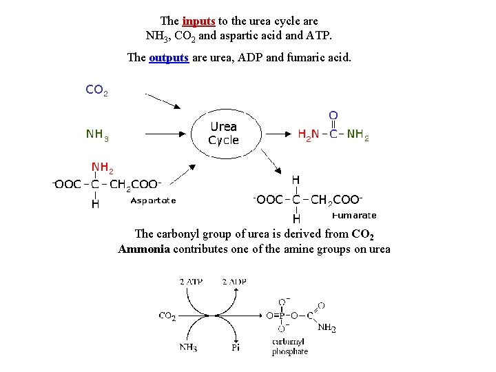
The inputs to the urea cycle are NH 3, CO 2 and aspartic acid and ATP. The outputs are urea, ADP and fumaric acid. The carbonyl group of urea is derived from CO 2 Ammonia contributes one of the amine groups on urea
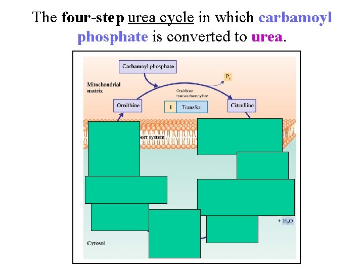
The four-step urea cycle in which carbamoyl phosphate is converted to urea.
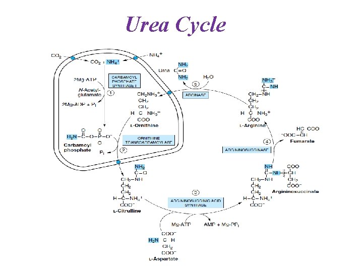
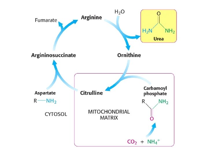
Urea Cycle
REGULATION OF THE UREA CYCLE Acute: N-acetylglutamate, allosteric effector, up regulates CPS I
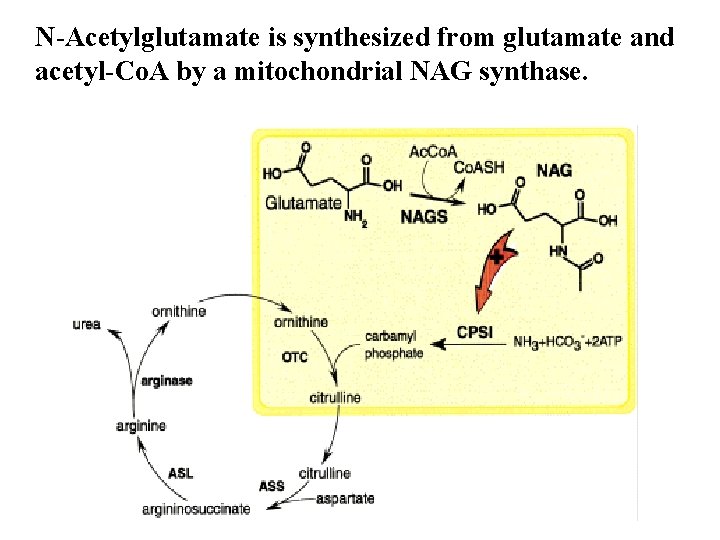
N-Acetylglutamate is synthesized from glutamate and acetyl-Co. A by a mitochondrial NAG synthase.
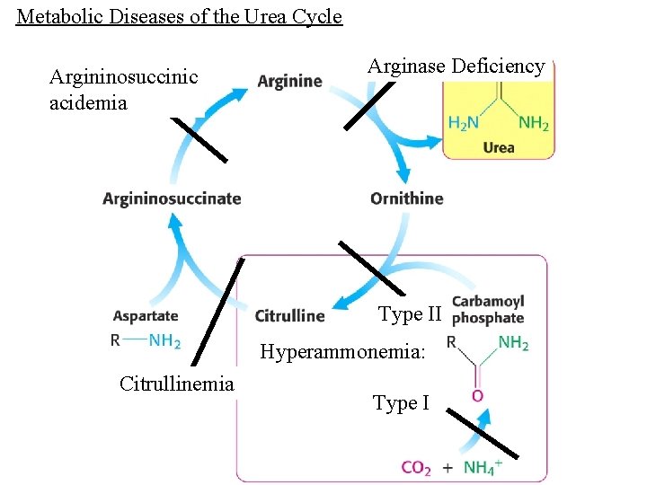
Metabolic Diseases of the Urea Cycle Argininosuccinic acidemia Arginase Deficiency Type II Hyperammonemia: Citrullinemia Type I
Metabolic Diseases of the Urea Cycle Disorders present in infants: Symptoms: Lethargy, swelling of the brain leads to mental retardation/brain damage Diagnosis: Low blood urea nitrogen (BUN) levels -high levels of ammonia in the blood elevated circulating glutamine -other metabolites that accumulate depend on the specific enzyme defect Treatment: Long term, dietary restriction. Low protein diet. Supplemented with Arginine
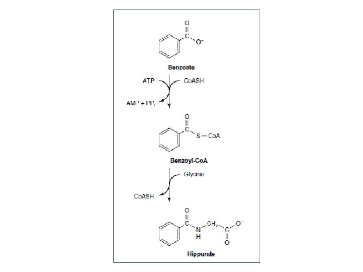
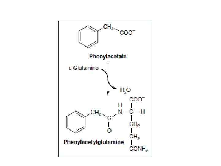
Excessive ammonia is toxic to the central nervous system. Alternative pathway therapy: Sodium benzoate to produce hippuric acid Sodium phenylacetate or phenyl-butyrate to produce phenylacetylglutamine
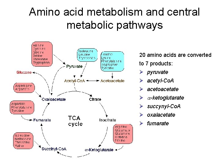
Amino acid metabolism and central metabolic pathways 20 amino acids are converted to 7 products: Ø pyruvate Ø acetyl-Co. A Ø acetoacetate Ø a-ketoglutarate Ø succynyl-Co. A Ø oxalacetate Ø fumarate
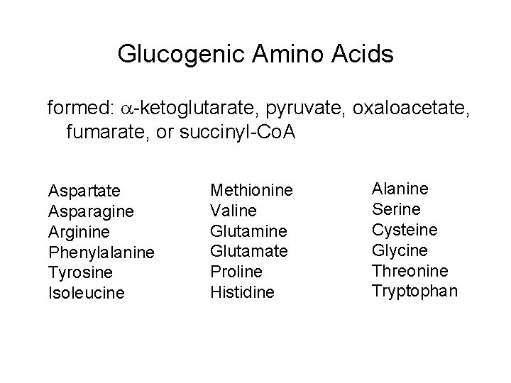
Glucogenic Amino Acids formed: -ketoglutarate, pyruvate, oxaloacetate, fumarate, or succinyl-Co. A Aspartate Asparagine Arginine Phenylalanine Tyrosine Isoleucine Methionine Valine Glutamate Proline Histidine Alanine Serine Cysteine Glycine Threonine Tryptophan
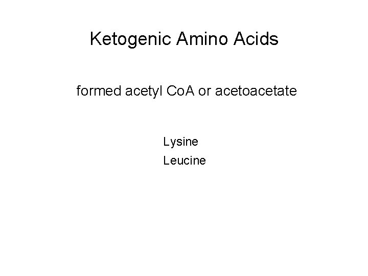
Ketogenic Amino Acids formed acetyl Co. A or acetoacetate Lysine Leucine
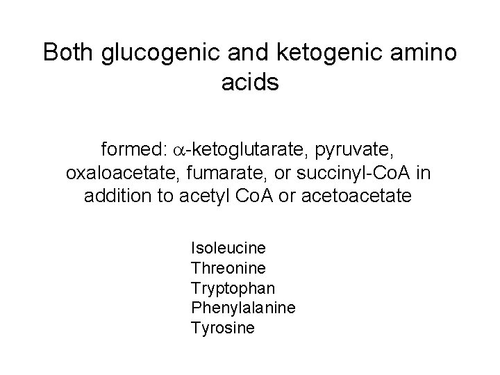
Both glucogenic and ketogenic amino acids formed: -ketoglutarate, pyruvate, oxaloacetate, fumarate, or succinyl-Co. A in addition to acetyl Co. A or acetoacetate Isoleucine Threonine Tryptophan Phenylalanine Tyrosine
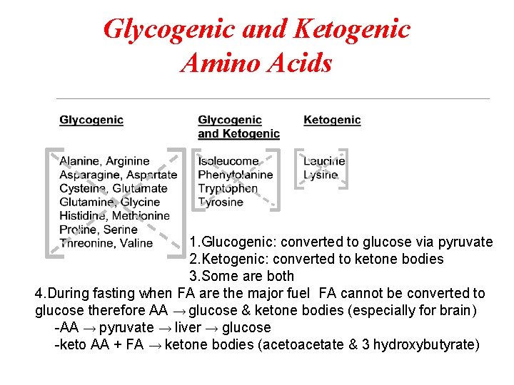
Glycogenic and Ketogenic Amino Acids 1. Glucogenic: converted to glucose via pyruvate 2. Ketogenic: converted to ketone bodies 3. Some are both 4. During fasting when FA are the major fuel FA cannot be converted to glucose therefore AA → glucose & ketone bodies (especially for brain) -AA → pyruvate → liver → glucose -keto AA + FA → ketone bodies (acetoacetate & 3 hydroxybutyrate)
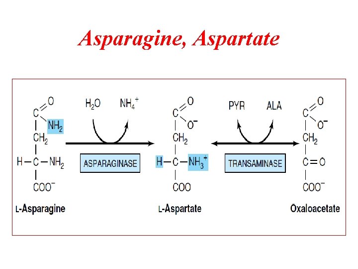
Asparagine, Aspartate
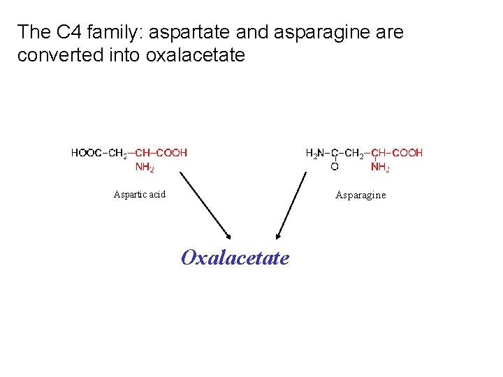
The C 4 family: aspartate and asparagine are converted into oxalacetate Asparagine Aspartic acid Oxalacetate
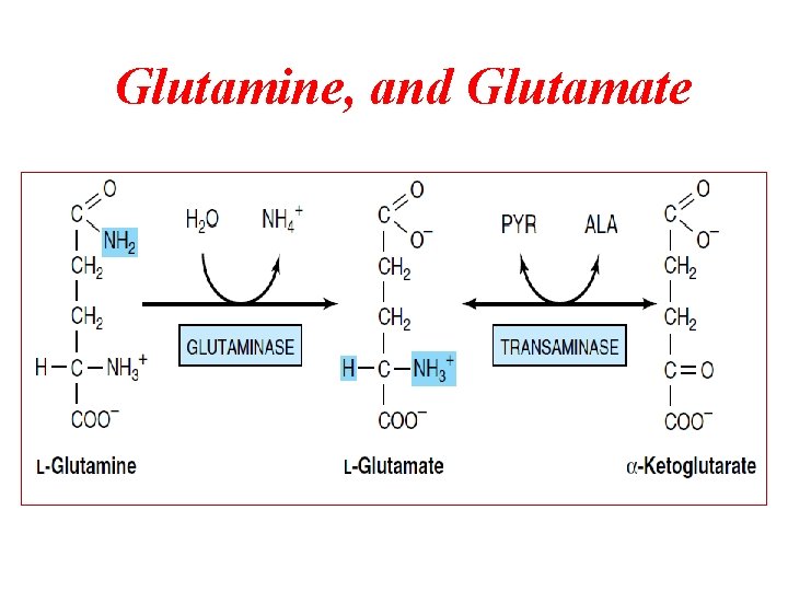
Glutamine, and Glutamate
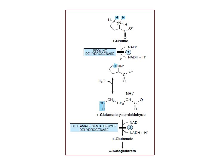
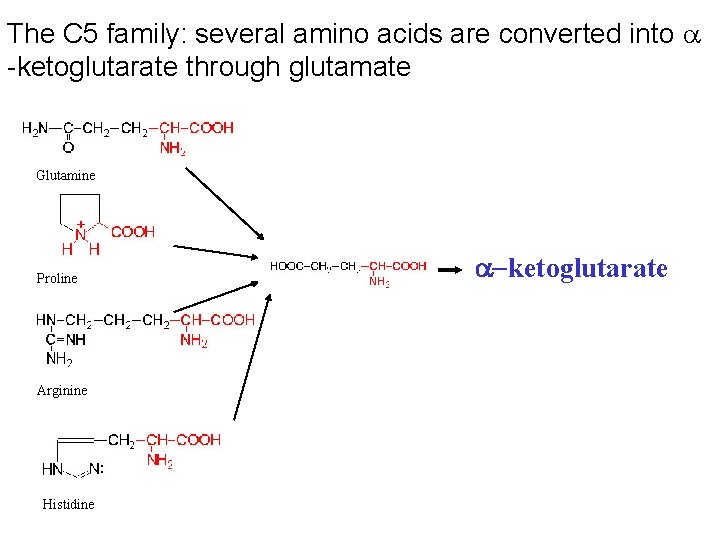
The C 5 family: several amino acids are converted into -ketoglutarate through glutamate Glutamine Proline Arginine Histidine -ketoglutarate
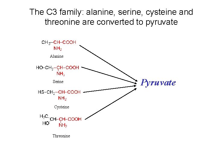
The C 3 family: alanine, serine, cysteine and threonine are converted to pyruvate Alanine Serine Cysteine Threonine Pyruvate
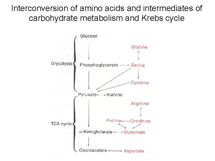
Interconversion of amino acids and intermediates of carbohydrate metabolism and Krebs cycle
Metabolism of some selected amino acids
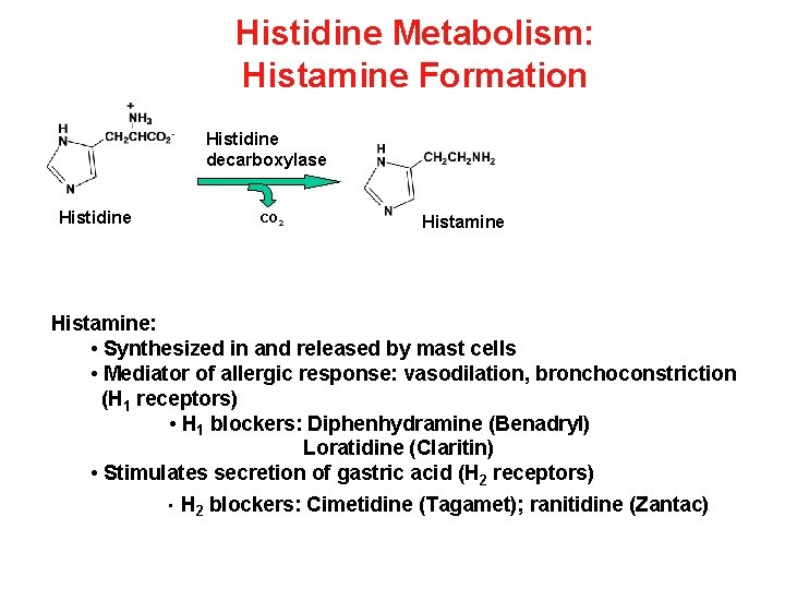
Histidine Metabolism: Histamine Formation Histidine decarboxylase Histidine CO 2 Histamine: • Synthesized in and released by mast cells • Mediator of allergic response: vasodilation, bronchoconstriction (H 1 receptors) • H 1 blockers: Diphenhydramine (Benadryl) Loratidine (Claritin) • Stimulates secretion of gastric acid (H 2 receptors) • H 2 blockers: Cimetidine (Tagamet); ranitidine (Zantac)
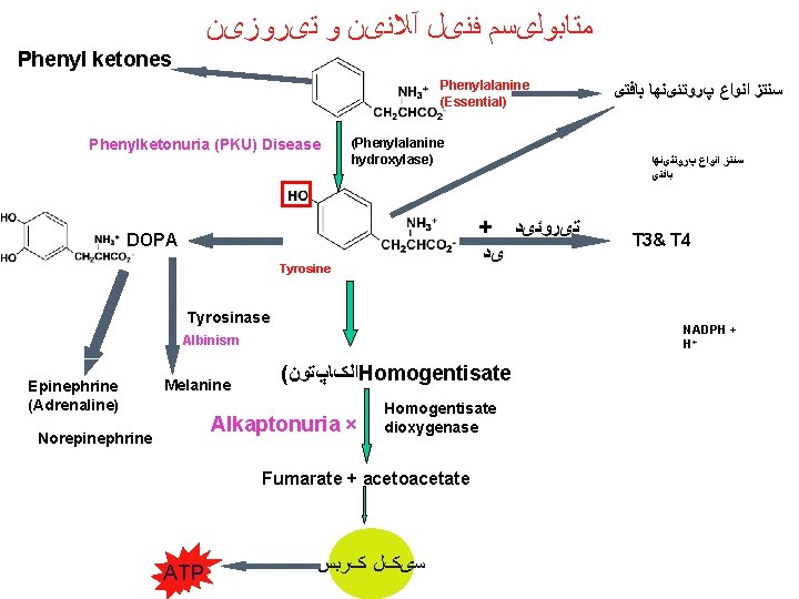
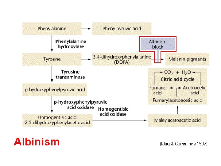
ﻣﺘﺎﺑﻮﻟیﺴﻢ ﻓﻨیﻞ آﻼﻧیﻦ ﻭ ﺗیﺮﻭﺯیﻦ Phenyl ketones Phenylalanine (Essential) Phenylketonuria (PKU) Disease (Phenylalanine hydroxylase) ﺳﻨﺘﺰ ﺍﻧﻮﺍﻉ پﺮﻭﺗﺌیﻨﻬﺎ ﺑﺎﻓﺘی + ﺗیﺮﻭﺋیﺪ DOPA Tyrosine Tyrosinase Melanine ( ﺍﻟکﺎپﺘﻮﻥ Homogentisate Alkaptonuria × Norepinephrine Homogentisate dioxygenase Fumarate + acetoacetate ATP T 3& T 4 NADPH + H+ Albinism Epinephrine (Adrenaline) ﺳﻨﺘﺰ ﺍﻧﻮﺍﻉ پﺮﻭﺗﺌیﻨﻬﺎ ﺑﺎﻓﺘی ﺳیکﻞ کﺮﺑﺲ
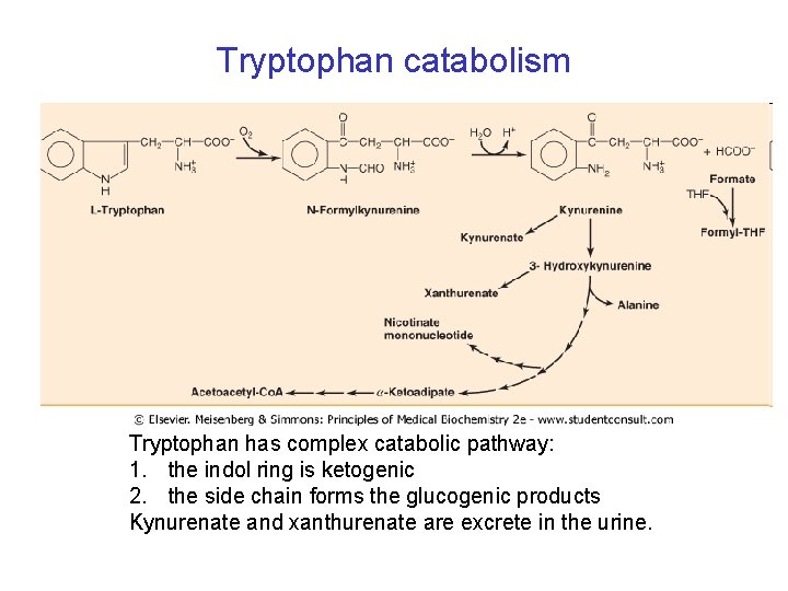
Tryptophan catabolism Tryptophan has complex catabolic pathway: 1. the indol ring is ketogenic 2. the side chain forms the glucogenic products Kynurenate and xanthurenate are excrete in the urine.
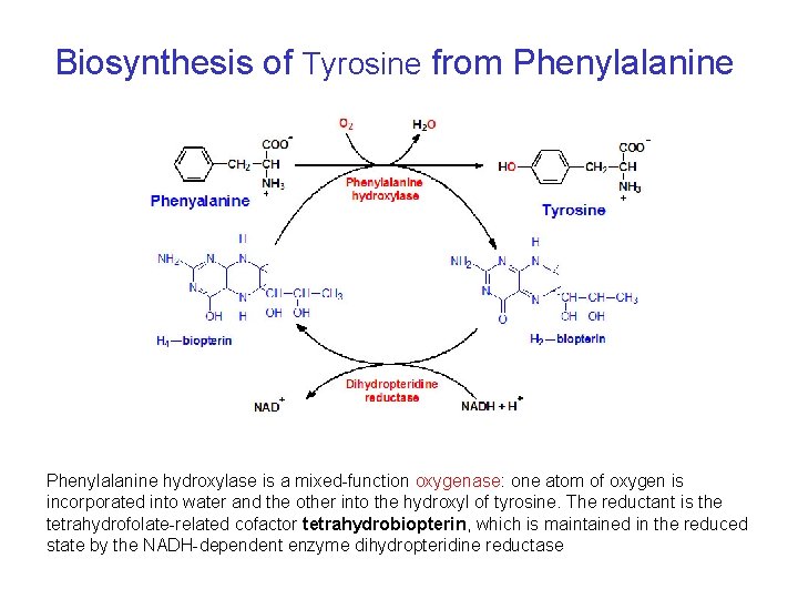
Biosynthesis of Tyrosine from Phenylalanine hydroxylase is a mixed-function oxygenase: one atom of oxygen is incorporated into water and the other into the hydroxyl of tyrosine. The reductant is the tetrahydrofolate-related cofactor tetrahydrobiopterin, which is maintained in the reduced state by the NADH-dependent enzyme dihydropteridine reductase
Phenylketonuria Hyperphenylalaninemia - complete deficiency of phenylalanine hydroxylase (plasma level of Phe raises from normal 0. 5 to 2 mg/d. L to more than 20 mg/d. L). The mental retardation is caused by the accumulation of phenylalanine, which becomes a major donor of amino groups in aminotransferase activity and depletes neural tissue of α-ketoglutarate. Absence of α-ketoglutarate in the brain shuts down the TCA cycle and the associated production of aerobic energy, which is essential to normal brain development. Newborns are routinelly tested for blood concentration of Phe. The diet with low-phenylalanine diet.
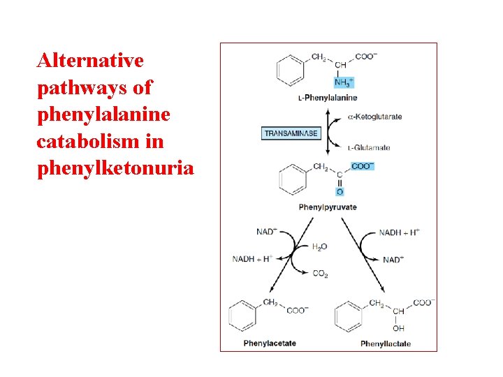
Alternative pathways of phenylalanine catabolism in phenylketonuria
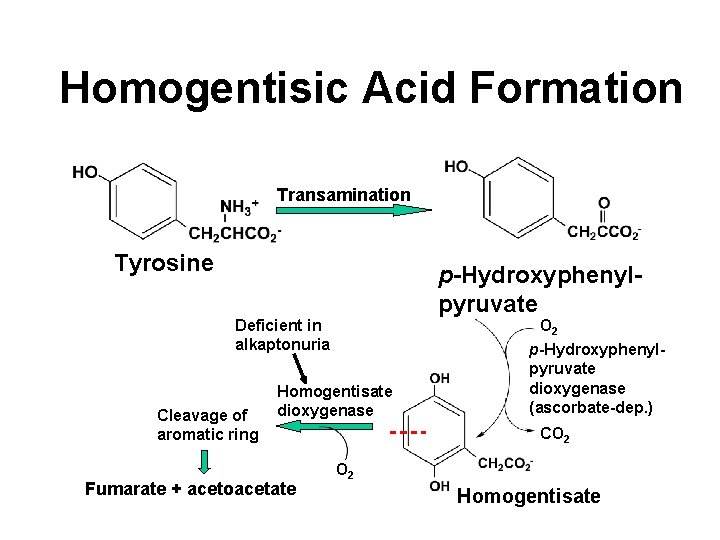
Homogentisic Acid Formation Transamination Tyrosine p-Hydroxyphenylpyruvate O 2 Deficient in alkaptonuria Cleavage of aromatic ring Homogentisate dioxygenase Fumarate + acetoacetate p-Hydroxyphenylpyruvate dioxygenase (ascorbate-dep. ) CO 2 Homogentisate
Alkaptonuria • First defect to which inborn error of metabolism applied – Sir Archibald Garrod in early 1900’s • Homogentisate appears in urine • Deposited in cartilage and elsewhere polymerization (black) • Deficiency of homogentisate dioxygenase • Urine turns dark on standing • Oxidation of homogentisic acid • Asymptomatic in childhood • Tendency toward arthritis in adulthood
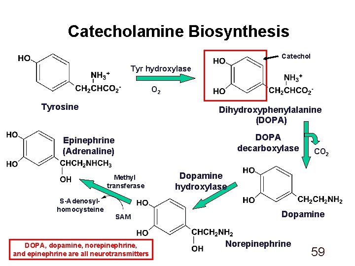
Catecholamine Biosynthesis Catechol Tyr hydroxylase O 2 Tyrosine Dihydroxyphenylalanine (DOPA) DOPA decarboxylase Epinephrine (Adrenaline) Methyl transferase S-Adenosylhomocysteine SAM DOPA, dopamine, norepinephrine, and epinephrine are all neurotransmitters CO 2 Dopamine hydroxylase Dopamine Norepinephrine 59
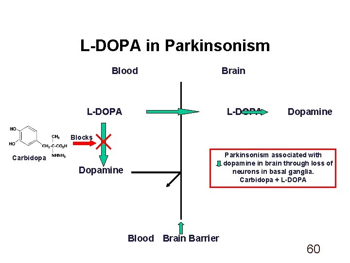
L-DOPA in Parkinsonism Blood L-DOPA Brain L-DOPA Dopamine Blocks Parkinsonism associated with dopamine in brain through loss of neurons in basal ganglia. Carbidopa + L-DOPA Carbidopa Dopamine Blood Brain Barrier 60
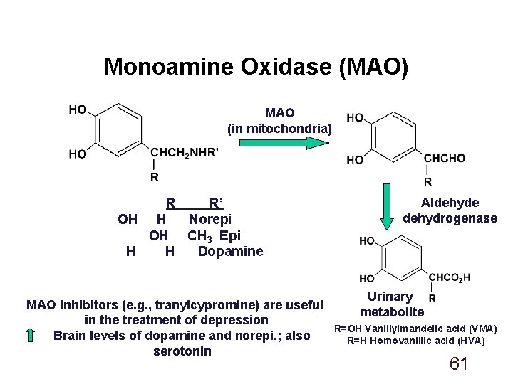
Monoamine Oxidase (MAO) MAO (in mitochondria) R R’ OH H Norepi OH CH 3 Epi H H Dopamine MAO inhibitors (e. g. , tranylcypromine) are useful in the treatment of depression Brain levels of dopamine and norepi. ; also serotonin Aldehyde dehydrogenase Urinary metabolite R=OH Vanillylmandelic acid (VMA) R=H Homovanillic acid (HVA) 61
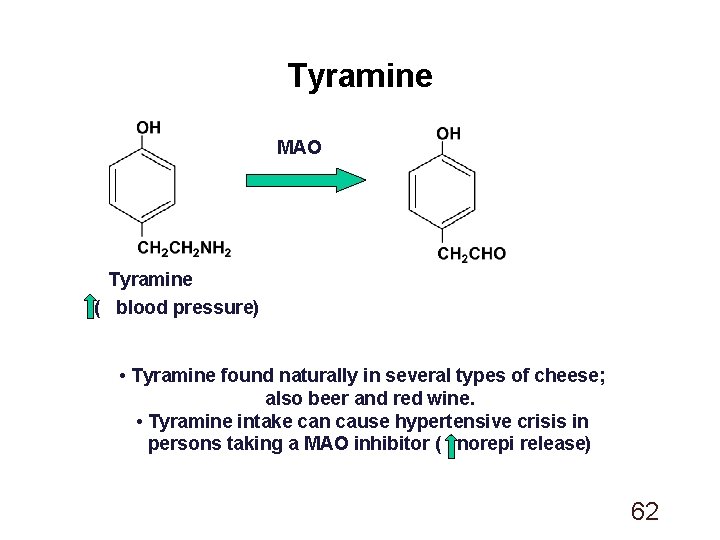
Tyramine MAO Tyramine ( blood pressure) • Tyramine found naturally in several types of cheese; also beer and red wine. • Tyramine intake can cause hypertensive crisis in persons taking a MAO inhibitor ( norepi release) 62
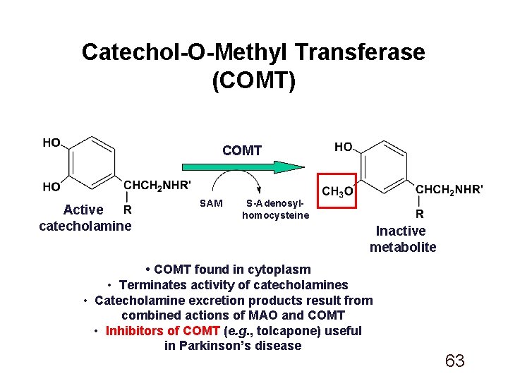
Catechol-O-Methyl Transferase (COMT) COMT Active catecholamine SAM S-Adenosylhomocysteine Inactive metabolite • COMT found in cytoplasm • Terminates activity of catecholamines • Catecholamine excretion products result from combined actions of MAO and COMT • Inhibitors of COMT (e. g. , tolcapone) useful in Parkinson’s disease 63
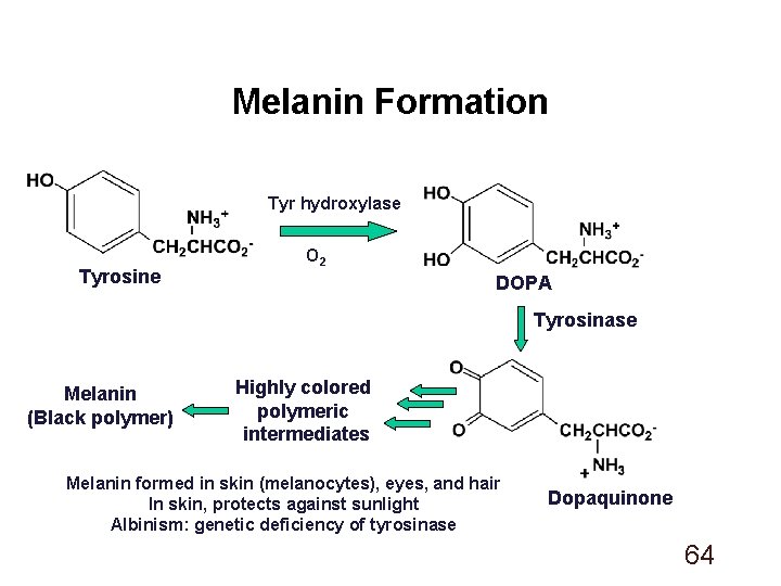
Melanin Formation Tyr hydroxylase Tyrosine O 2 DOPA Tyrosinase Melanin (Black polymer) Highly colored polymeric intermediates Melanin formed in skin (melanocytes), eyes, and hair In skin, protects against sunlight Albinism: genetic deficiency of tyrosinase Dopaquinone 64
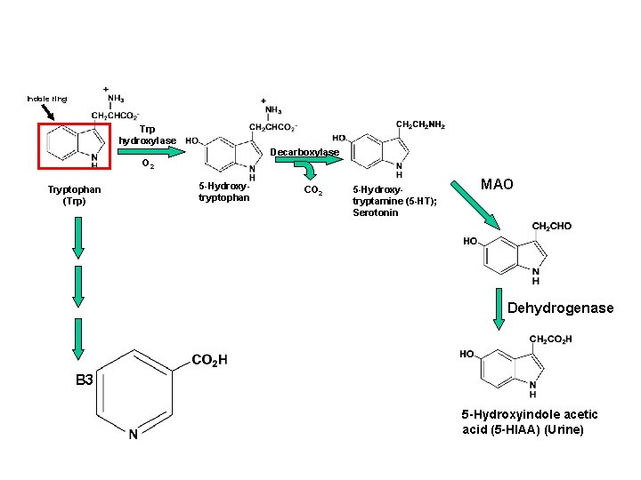
Indole ring Trp hydroxylase Decarboxylase O 2 Tryptophan (Trp) 5 -Hydroxytryptophan CO 2 5 -Hydroxytryptamine (5 -HT); Serotonin MAO Dehydrogenase B 3 5 -Hydroxyindole acetic acid (5 -HIAA) (Urine)
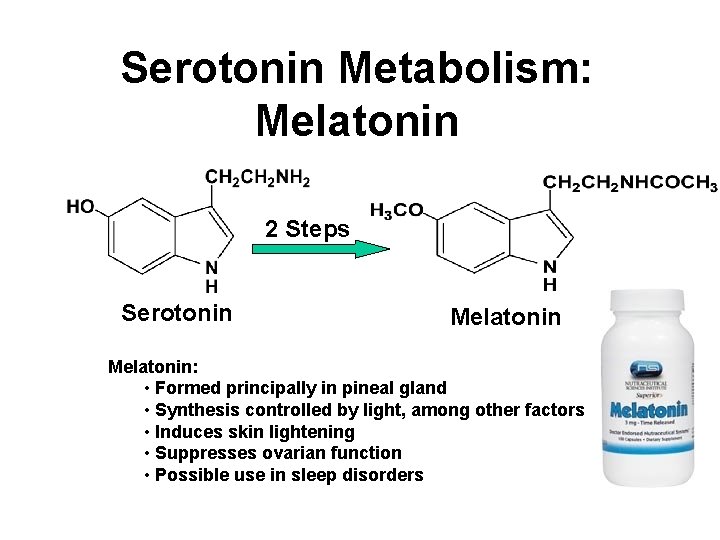
Serotonin Metabolism: Melatonin 2 Steps Serotonin Melatonin: • Formed principally in pineal gland • Synthesis controlled by light, among other factors • Induces skin lightening • Suppresses ovarian function • Possible use in sleep disorders
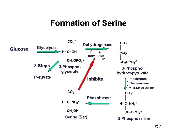
Formation of Serine Glucose Dehydrogenase Glycolysis NAD+ NADH + H+ 3 Steps 3 -Phosphoglycerate 3 -Phosphohydroxypyruvate Pyruvate Inhibits Glutamate Transaminase -Ketoglutarate Phosphatase Serine (Ser) 3 -Phosphoserine 67
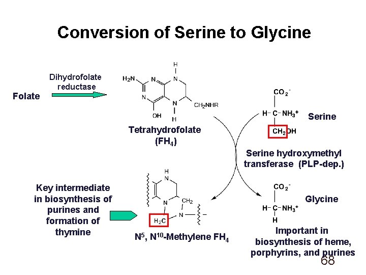
Conversion of Serine to Glycine Folate Dihydrofolate reductase Serine Tetrahydrofolate (FH 4) Serine hydroxymethyl transferase (PLP-dep. ) Key intermediate in biosynthesis of purines and formation of thymine Glycine N 5, N 10 -Methylene FH 4 Important in biosynthesis of heme, porphyrins, and purines 68
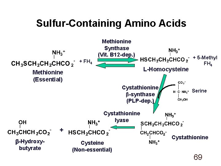
Sulfur-Containing Amino Acids Methionine Synthase (Vit. B 12 -dep. ) + FH 4 L-Homocysteine Methionine (Essential) Cystathionine b-synthase (PLP-dep. ) + 5 -Methyl FH 4 Serine Cystathionine lyase + b-Hydroxybutyrate Cysteine (Non-essential) Cystathionine 69
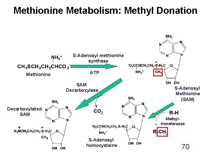
Methionine Metabolism: Methyl Donation S-Adenosyl methionine synthase Methionine ATP SAM Decarboxylase Decarboxylated SAM S-Adenosyl Methionine (SAM) CO 2 R-H Methyltransferases + R-CH 3 S-Adenosyl homocysteine 70
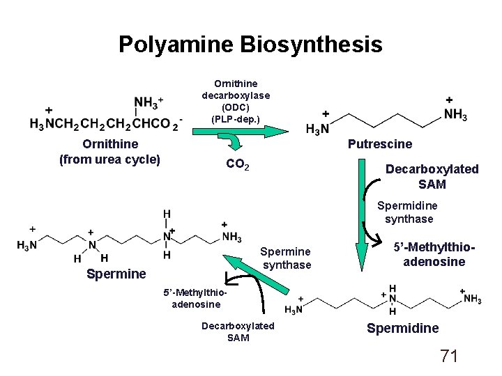
Polyamine Biosynthesis Ornithine decarboxylase (ODC) (PLP-dep. ) Ornithine (from urea cycle) Putrescine CO 2 Decarboxylated SAM Spermidine synthase Spermine 5’-Methylthioadenosine Decarboxylated SAM Spermidine 71
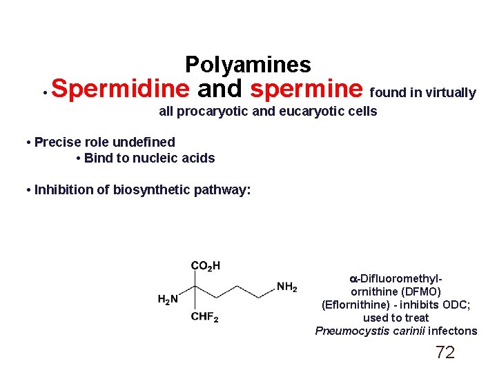
Polyamines • Spermidine and spermine found in virtually all procaryotic and eucaryotic cells • Precise role undefined • Bind to nucleic acids • Inhibition of biosynthetic pathway: -Difluoromethylornithine (DFMO) (Eflornithine) - inhibits ODC; used to treat Pneumocystis carinii infectons 72
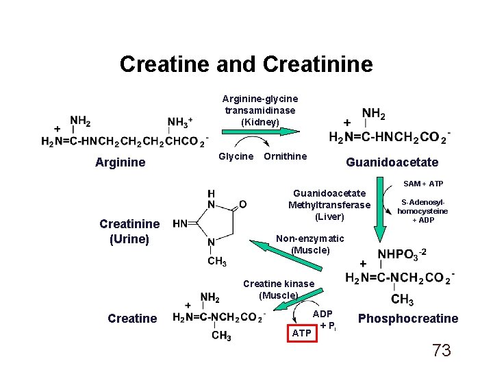
Creatine and Creatinine Arginine-glycine transamidinase (Kidney) Arginine Creatinine (Urine) Glycine Ornithine Guanidoacetate Methyltransferase (Liver) SAM + ATP S-Adenosylhomocysteine + ADP Non-enzymatic (Muscle) Creatine kinase (Muscle) Creatine ATP ADP + Pi Phosphocreatine 73
Creatine and Creatinine Creatine: • Dietary supplement • Used to improve athletic performance Creatinine: • Urinary excretion generally constant; proportional to muscle mass Creatinine Clearance Test: • Compares the level of creatinine in urine (24 hrs. ) with the creatinine level in the blood • Used to assess kidney function • Important determinant in dosing of several drugs in patients with impaired renal function 74
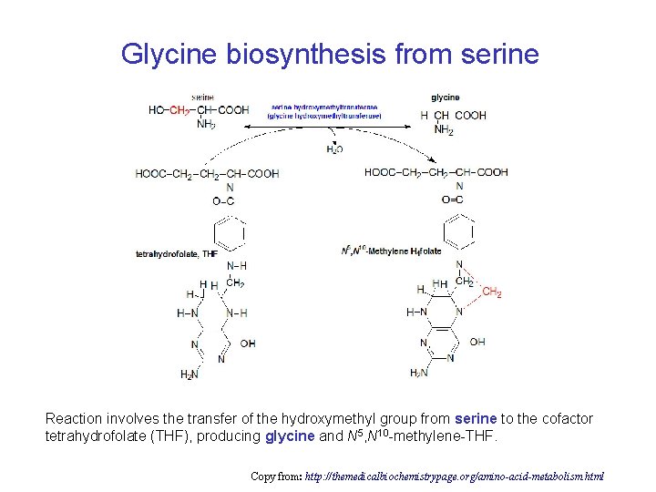
Glycine biosynthesis from serine Reaction involves the transfer of the hydroxymethyl group from serine to the cofactor tetrahydrofolate (THF), producing glycine and N 5, N 10 -methylene-THF. Copy from: http: //themedicalbiochemistrypage. org/amino-acid-metabolism. html
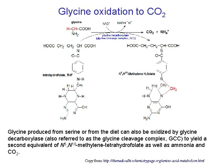
Glycine oxidation to CO 2 Glycine produced from serine or from the diet can also be oxidized by glycine decarboxylase (also referred to as the glycine cleavage complex, GCC) to yield a second equivalent of N 5, N 10 -methylene-tetrahydrofolate as well as ammonia and CO 2. Copy from: http: //themedicalbiochemistrypage. org/amino-acid-metabolism. html
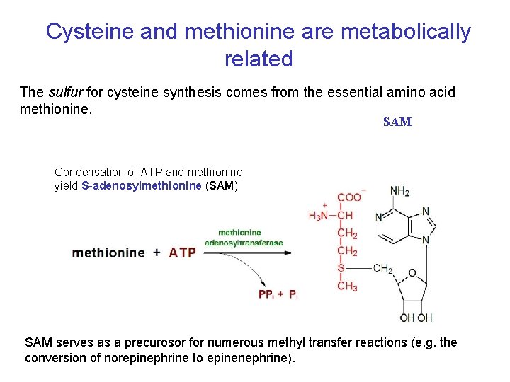
Cysteine and methionine are metabolically related The sulfur for cysteine synthesis comes from the essential amino acid methionine. SAM Condensation of ATP and methionine yield S-adenosylmethionine (SAM) SAM serves as a precurosor for numerous methyl transfer reactions (e. g. the conversion of norepinephrine to epinenephrine).
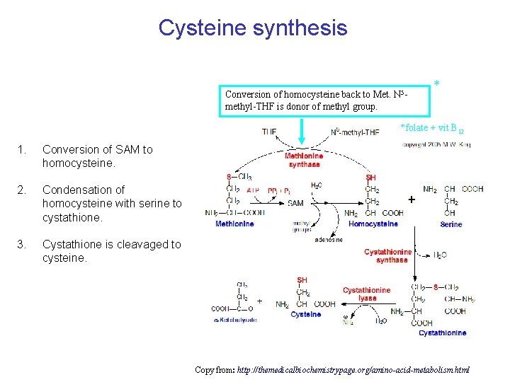
Cysteine synthesis Conversion of homocysteine back to Met. N 5 methyl-THF is donor of methyl group. * *folate + vit B 12 1. Conversion of SAM to homocysteine. 2. Condensation of homocysteine with serine to cystathione. 3. Cystathione is cleavaged to cysteine. Copy from: http: //themedicalbiochemistrypage. org/amino-acid-metabolism. html
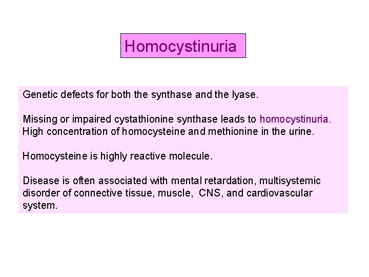
Homocystinuria Genetic defects for both the synthase and the lyase. Missing or impaired cystathionine synthase leads to homocystinuria. High concentration of homocysteine and methionine in the urine. Homocysteine is highly reactive molecule. Disease is often associated with mental retardation, multisystemic disorder of connective tissue, muscle, CNS, and cardiovascular system.
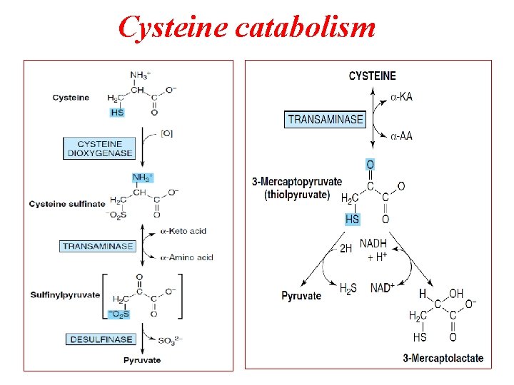
Cysteine catabolism
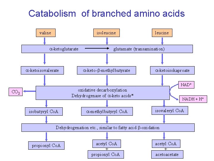
Catabolism of branched amino acids valine isoleucine -ketoglutarate -ketoisovalerate leucine glutamate (transamination) -keto-b-methylbutyrate -ketoisokaproate NAD+ oxidative decarboxylation Dehydrogenase of -keto acids* CO 2 isobutyryl Co. A -methylbutyryl Co. A NADH + H+ isovaleryl Co. A Dehydrogenation etc. , similar to fatty acid b-oxidation propionyl Co. A acetyl Co. A + acetoacetate
Branched-chain aminoaciduria Disease also called Maple Syrup Urine Disease (MSUD) (because of the characteristic odor of the urine in affected individuals). Deficiency in an enzyme, branched-chain α-keto acid dehydrogenase leads to an accumulation of three branchedchain amino acids and their corresponding branched-chain α-keto acids which are excreted in the urine. There is only one dehydrogenase enzyme for all three amino acids. Mental retardation in these cases is extensive.
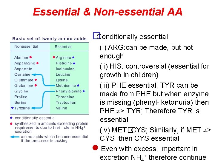
Essential & Non-essential AA �Conditionally essential (i) ARG: can be made, but not enough (ii) HIS: controversial (essential for growth in children) (iii) PHE essential, TYR can be made from PHE but when enzyme is missing (phenyl- ketonuria) then PHE > TYR; Therefore TYR is essential (iv) MET�CYS; Similarly, if MET > CYS then CYS essential Even with excess, important in excretion NH + therefore continue
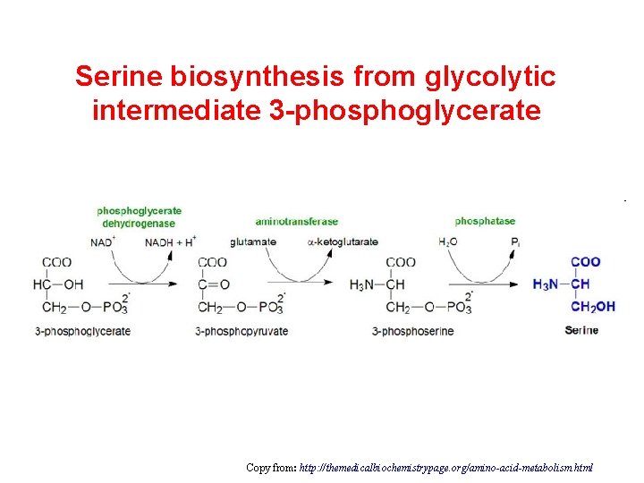
Serine biosynthesis from glycolytic intermediate 3 -phosphoglycerate Copy from: http: //themedicalbiochemistrypage. org/amino-acid-metabolism. html
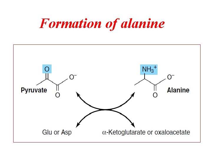
Formation of alanine
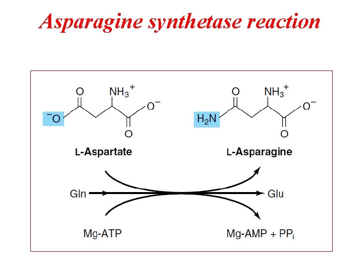
Asparagine synthetase reaction
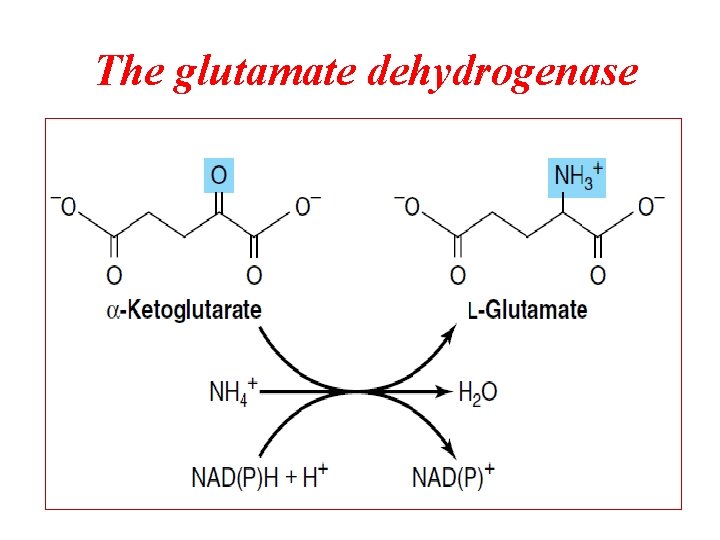
The glutamate dehydrogenase
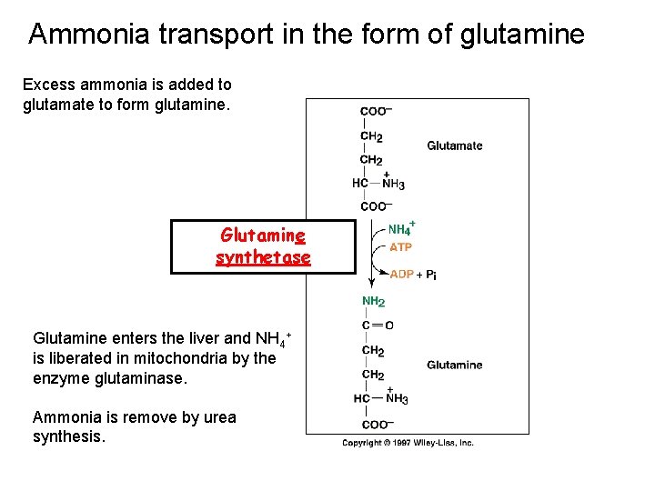
Ammonia transport in the form of glutamine Excess ammonia is added to glutamate to form glutamine. Glutamine synthetase Glutamine enters the liver and NH 4+ is liberated in mitochondria by the enzyme glutaminase. Ammonia is remove by urea synthesis.
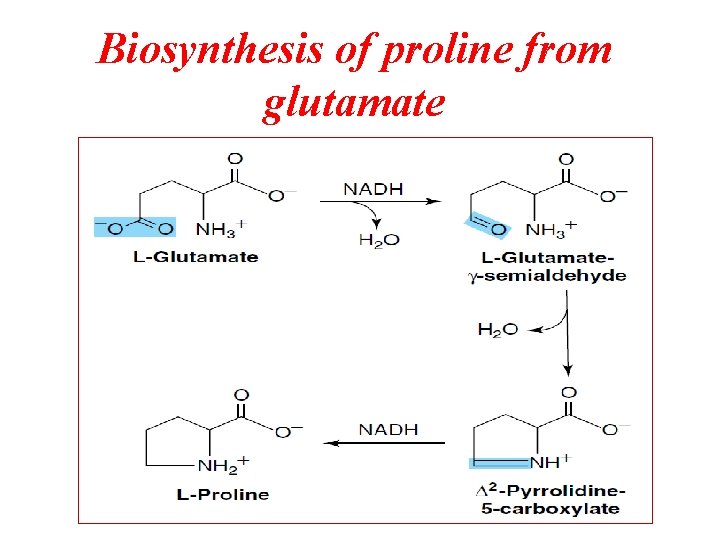
Biosynthesis of proline from glutamate
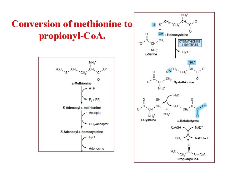
Conversion of methionine to propionyl-Co. A.
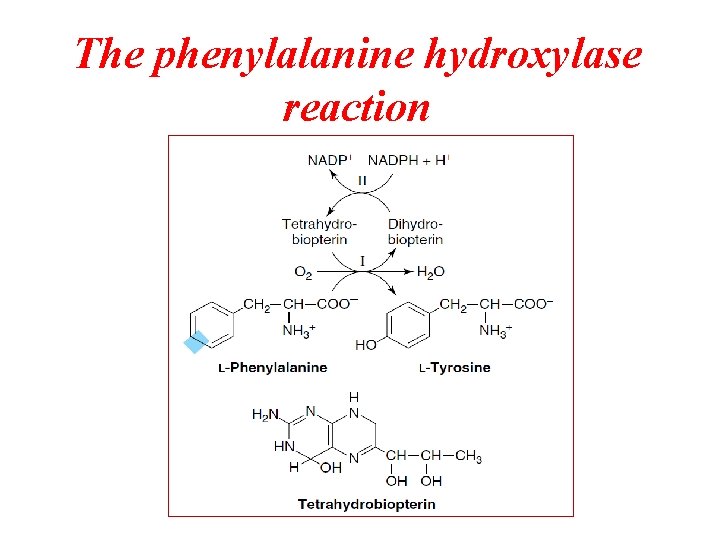
The phenylalanine hydroxylase reaction
Conversion of Amino Acids to Specialized Products
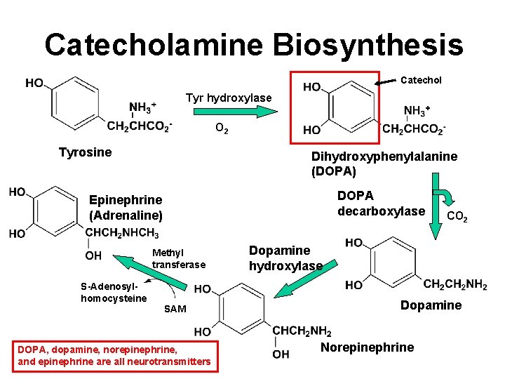
Catecholamine Biosynthesis Catechol Tyr hydroxylase O 2 Tyrosine Dihydroxyphenylalanine (DOPA) DOPA decarboxylase Epinephrine (Adrenaline) Methyl transferase S-Adenosylhomocysteine SAM DOPA, dopamine, norepinephrine, and epinephrine are all neurotransmitters CO 2 Dopamine hydroxylase Dopamine Norepinephrine
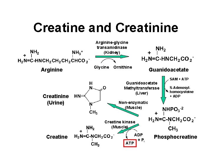
Creatine and Creatinine Arginine-glycine transamidinase (Kidney) Arginine Creatinine (Urine) Glycine Ornithine Guanidoacetate Methyltransferase (Liver) SAM + ATP S-Adenosylhomocysteine + ADP Non-enzymatic (Muscle) Creatine kinase (Muscle) Creatine ATP ADP + Pi Phosphocreatine
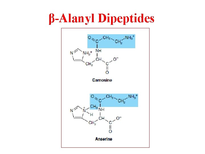
β-Alanyl Dipeptides
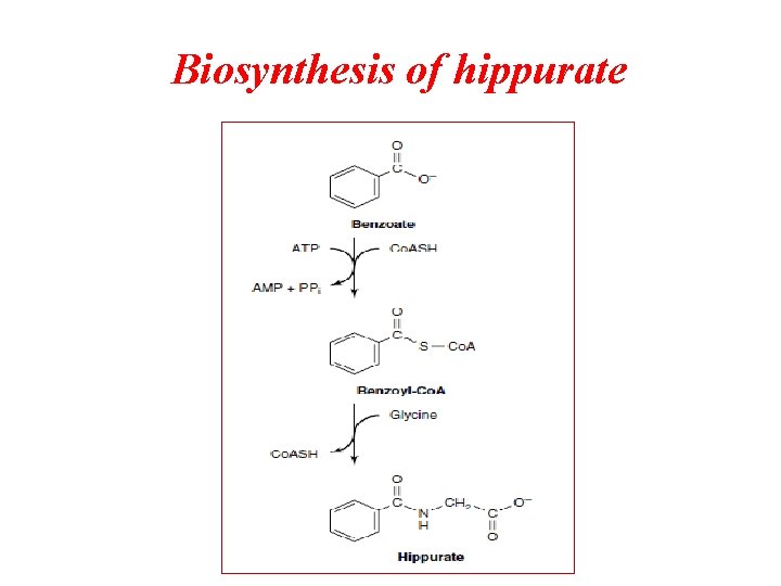
Biosynthesis of hippurate
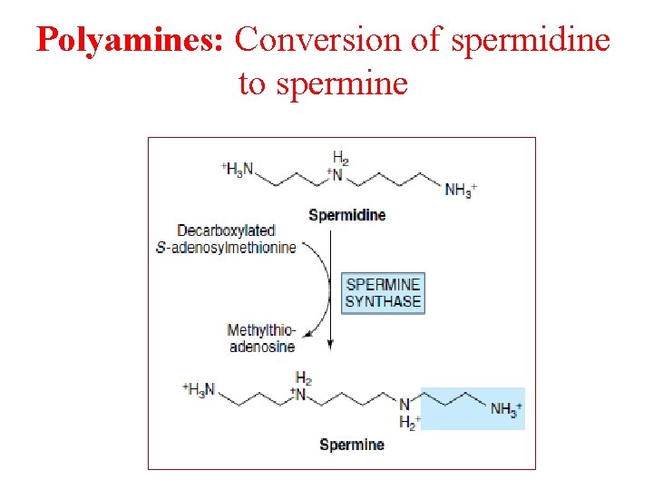
Polyamines: Conversion of spermidine to spermine
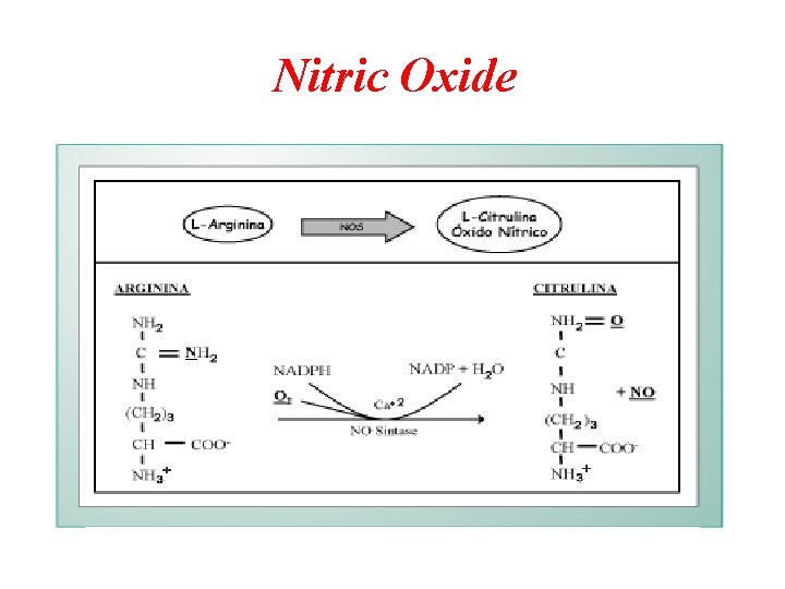
Nitric Oxide
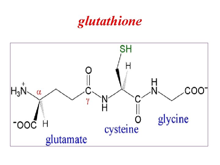
glutathione
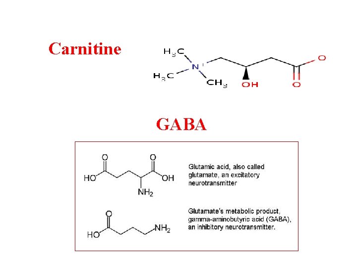
Carnitine GABA
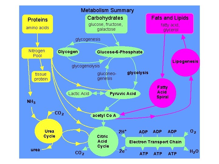
Protein Metabolism

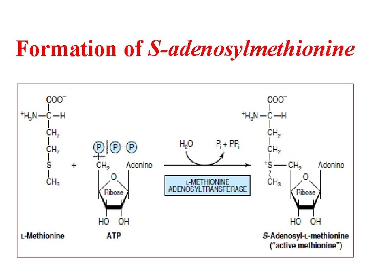
Formation of S-adenosylmethionine
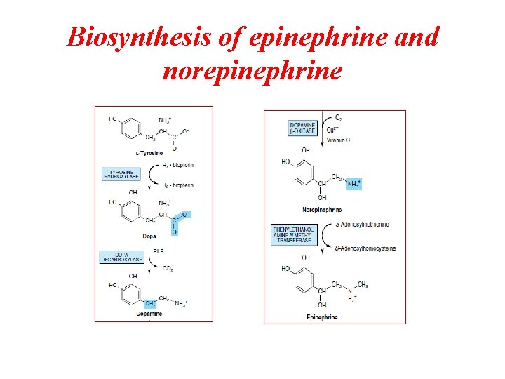
Biosynthesis of epinephrine and norepinephrine
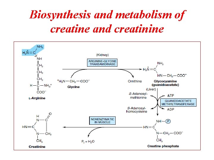
Biosynthesis and metabolism of creatine and creatinine
- Slides: 105