Amblyopia and Strabismus Latest Information on Patching Strabismus

Amblyopia and Strabismus: Latest Information on Patching, Strabismus and Research ACO PAEDIATRIC OPTOMETRY REFRESHER COURSE 2012 Lionel Kowal Melbourne

st 1 stop: Infantile onset esotropia �Lawrence Tychsen �St Louis MO

The Cause of Infantile Strabismus Lies Upstairs in the Cerebral Cortex, Not Downstairs in the Brainstem Tychsen, L Editorial Archives Ophthal Aug 2012 �Infantile-onset strabismus is a constellation (or syndrome) of ocular motor behaviors: eye misalignment; subnormal binocular fusion; a type of nystagmus; dissociated vertical & horizontal deviations. �Children at greatest risk are those who suffer cerebral lesions around the time of birth, esp PVL (=Peri Ventricular Leukomalacia, damage to the posterior-most fibers of the optic radiations, the binocular inputs to striate cortex). �These infants have a 30 - to 60 -fold greater risk of IOS c. f. with healthy

Can Ophthalmologists Repair the Brain in Infantile Esotropia? Early Surgery, Stereopsis, Monofixation Syndrome, and the Legacy of Marshall Parks Lawrence Tychsen J AAPOS 2005; 9: 510 -521 �Costenbader: landmark article published in the 1961 Transactions of the American Ophthalmologic Society, he defined infantile esotropia and reported that 1 in 5 children could develop gross stereopsis if surgically aligned by age 1 year.

1. Stereopsis emerges abruptly in human during the first 3 - 5 mo 2. Roughly equal proportions of normal and (prismatically aligned) esotropic infants possess the capacity for stereopsis 3. This capacity degenerates pathologically within a few months in uncorrected . esotropes

The graph of Figure 5 A shows that there is no obvious correlation between age of onset of misalignment and subsequent attainment of stereopsis. When the data are reanalyzed with strict attention to duration of misalignment, a strong correlation is evident: shorter durations mean better stereopsis. Excellent outcomes may be achievable in infants operated upon within 60 days of strabismus onset.

Is it really esotropia? . . or not Confounding issues: � 1. Telecanthus � 2. Positive/ negative angle kappa � 3. Cyclic esotropia - very rare, unless you think to ask: does it happen every 2 nd day?

Pseudo strabismus: is it? � 51 children � Av age, 1. 5 ± 0. 8 y; range, 3 -36 � Refractive accommodative ET mo developed in 16% of the children @ mean age of 2. 8 ± 1 y. � ET developed in 54% of the children with pseudoesotropia who were > + 1. 5 D c. f. 3% of those ≤ + 1. 50 D (P=0. 0001). � A positive family history of strabismus (P= 0. 193) and initial age at presentation with pseudoesotropia (P =0. 571) were not predisposing factors. � Development of refractive accommodative esotropia in children initially diagnosed with pseudoesotropia Kanwar Mohan & Ashok Sharma J AAPOS 2012; 16: 266 -268 Chandigarh

Pseudo strabismus: is it? 306 pts were diagnosed with pseudoesotropia with no significant refractive error on initial examination. � 201 had follow-up exam. � Av age @ initial examination was 13 mo (range, 2 -33), @ follow-up 33 mo (range, 4 -120). � 20 were later found to have strabismus (10%) 15 ET, 3 XT, 1 Duane, 1 Prader-Willi syndrome with ET. � 5 had significant refractive error & mild refractive amblyopia (2%). � Of children initially diagnosed with pseudoesotropia < age 3 who returned for follow- up, 12% later found to have strabismus or mild refractive amblyopia � � Incidence of strabismus and amblyopia in preverbal children previously diagnosed with pseudoesotropia Ariel L. Silbert, Noelle S. Matta, and David I. Silbert J AAPOS

Incidence of esotropia developing in subjects previously diagnosed with pseudoesotropia: a pilot study. Strabismus. 2012 Sep; 20(3): 124 -6. Repka MX. Wilmer Institute �Retrospective Anwar DS, Woreta FA, Weng CY, records review of children diagnosed with pseudo. ET prior to age 5 y, with check for strabismus after age 5 y. �N=31 �Median age at diagnosis of pseudo. ET was 1. 3 y. �Six (19%) children were subsequently diagnosed with esotropia. �Conclusion: Even when ‘world’s best’ examiners find no misalignment in young children, a small number will later be found with ET. �Serial examinations and parent education

Left-Sided Predominance in Pseudo -esotropia. Hesham N, Simon JW, Zobal-Ratner J. Binocul Vis Strabolog Q Simms Romano. 2012; 27(1): 39 -40. � Pseudo-ET is commonly encountered in pediatric ophthalmology. � In our practice, the left eye was reported by parents as being deviated more frequently than the right eye. � We attribute this laterality to the fact that most parents are right handed. They therefore tend to hold their children with their left arm, and to feed them with their right hands, in both cases viewing the left eye of the child in the adducted position.

Mayo longitudinal studies on ET �Accommodative ET �Non- Accommodative ET �Mohney et al �Ophthalmology March and June 2011

The Long-term Follow-up of Accommodative Esotropia AET in a Population-based Cohort of Children � 306 children with AET � 244 (80%) had fully AET (FAET) and 62 (20%) had partially AET (PAET). � Kaplan–Meier rate of discontinuing spectacles for strabismus in this population was 8% by 5 y after diagnosis, 20% by 10 y, and 37% by 20 y. � Children born prematurely or with a greater initial hyperopic refractive error were significantly less likely to become spectacle-free during the followup period. � During a median follow-up of 10 y (range, 0 – 28 y), 33 (13%) of the 244 subjects with FAET eventually underwent strabismus surgery. � Male gender and an earlier age at onset of FAET were associated with a higher likelihood of requiring surgery

Clinical Features Predictive of Successfully Weaning From Spectacles Those Children With Accommodative Esotropia Lambert, S et alii J AAPOS 2003; 7: 713 Beginning in 1995, children with fully accommodative esotropia with a baseline refractive error of +1. 50 to +5. 00 DS were gradually weaned from their hyperopic correction. � Spectacles were prescribed at a mean age of 4. 2 ± 1. 5 y, weaning was initiated at a mean age of 8± 1 y. � 12 of 20 children (60%) were successfully weaned from spectacles. � The spherical equivalent of the least hyperopic eye when spectacles were prescribed was +3± 1 D. � The clinical characteristic most clearly associated with successful weaning was the refractive error at the time glasses were prescribed. � Whereas 10 of 11 (91%) patients with < + 3 D were weaned from spectacles, only 2 of 9 (22%) patients with +3 to +5 D were successfully weaned from their spectacles (P =. 005).

Long-term Follow-up of Acquired Nonaccommodative Esotropia in a Population-based Cohort � 174 children were diagnosed during the 30 y period, yielding an incidence of 1 in 287 live births. � Median age at diagnosis was 4 y (range, 10 months to 18. 2 years), and 61% (107) were male. � 11% (8/75) of those queried were diplopic, none of the 174 was subsequently diagnosed with an intracranial lesion. � Mean follow-up of 11 y (range, 0 days to 37 years), 127 patients (73%) underwent strabismus surgery (mean, 1 surgery; range, 0 – 3 surgeries). Among the 127 patients who underwent surgery, the median final stereoacuity was 3000 seconds of arc, including 8 pts (6%) with ≥ 50 seconds of arc. � Patients who were older (>44 mo) at ANAET diagnosis and without amblyopia at their initial examination were more likely to achieve excellent final stereopsis.

Esotropia : Full +? Half +? �THE INFLUENCE OF REFRACTIVE ERROR MANAGEMENT ON THE NATURAL HISTORY AND TREATMENT OUTCOME OF ACCOMMODATIVE ESOTROPIA (AOS THESIS) BY BRADLEY CHARLES BLACK MD �Trans Am Ophthalmol Soc 2006; 104: 303 -321

THE INFLUENCE OF REFRACTIVE ERROR MANAGEMENT ON THE NATURAL HISTORY AND TREATMENT OUTCOME OF ACCOMMODATIVE ESOTROPIA AET � 285 patients with mean follow-up of 102 months. �After age 7, mean annual decrease in hypermetropia was 0. 24 D for pts wearing full cycloplegic refraction and for patients in whom hypermetropia was undercorrected by ≥ 1 D. �Age at diagnosis, oblique muscle dysfunction & abnormal distance-near relationship were associated with deterioration of AET. �Of 51 patients with an intermittent abnormal distance-near relationship, 19 (37%) had more + on cycloplegic refraction, and prescription of the increased + normalized the distance-near relationship.

THE INFLUENCE OF REFRACTIVE ERROR MANAGEMENT ON THE NATURAL HISTORY AND TREATMENT OUTCOME OF ACCOMMODATIVE ESOTROPIA � AET did not typically resolve. � Hypermetropic correction was discontinued for pts with enduring adequate alignment in 37 of the 285 pts (13%) at a mean age of 11. 6 y (range, 7 to 17). � For the 37 pts with resolution of AET, mean + on initial examination was 3. 18 DS compared with 4. 50 D for the group without resolution of AET(P <. 001). � If only patients ≥ 12 y @ last examination were included, 28 (20%) of 138 patients no longer required a hypermetropic correction to maintain adequate alignment. � Kaplan-Meier survival analysis predicts hypermetropic correction would be discontinued in 30% of the pat population 15 y after initiation of

THE INFLUENCE OF REFRACTIVE ERROR MANAGEMENT ON THE NATURAL HISTORY AND TREATMENT OUTCOME OF ACCOMMODATIVE ESOTROPIA �Conclusions: �The possibility that undercorrecting hypermetropia speeds its resolution is not supported by this study. �AET is usually stable, but younger age at diagnosis, oblique muscle dysfunction, and abnormal distance-near relationship are associated with deterioration. �Undercorrection of hypermetropia can cause an abnormal distance-near relationship, which in turn cause deterioration of AET. �Aggressive undercorrection of hypermetropia should be pursued carefully, because the risk may outweigh the potential advantages. .
: 624 -30.")
Genetics of ET & XT �Twin Res Hum Genet. 2012 Oct; 15(5): 624 -30. �Heritability of strabismus: genetic influence is specific to eso-deviation and independent of refractive error. �David Mackey & 15 others

Genetics of ET & XT �The heritability of an eso-deviation was 0. 64. The additive genetic correlation for eso-deviation and refractive error was 0. 13 and the bivariate heritability (i. e. , shared variance) was less than 1%, suggesting negligible shared genetic effect. �This study documents a substantial heritability of 64% for ET, yet no corresponding heritability for XT, suggesting that the genetic contribution to strabismus may be specific to ET.

Refraction, amblyopia, strabismus

Laser In Situ Keratomileusis for the Treatment of Refractive Accommodative Esotropia de Pagano & Pagano, Ophthalmology 2012; 119: 159 – 163 Argentina � 46 eyes / 23 pts with hyperopia and fully or partially refractive accommodative ET treated with LASIK 2000 - 2010. �Age 25± 13 y. �Mean hyperopia 3. 7± 1. 3 DS pre surgery and 0. 2± 0. 6 D after surgery (P<0. 001). �Mean ET without correction 21Δ before surgery, 4Δ after surgery (P<0. 001). �Refractive surgery is a promising option for the treatment of refractive accommodative ET

LASIK as an alternative line to treat noncompliant esotropic children. Saeed A, Ebsar Eye Center, Benha University, Benha, Egypt Clin Ophthalmol. 2011; 5: 1795 -801. Epub 2011 Dec 20. . � � � � � 20 eyes of 10 pts with accommodative ET. All patients were noncompliant with glasses and had refractive stability. They received brief GA and had bilateral LASIK to fully correct their hyperopic refractive errors aiming to achieve orthophoria. Preoperative and postoperative best corrected visual acuity, cycloplegic refraction, angle of squint, and any LASIK complications were recorded. Follow-up period was 9 mo. RESULTS: The age of patients ranged 5 -9 y and the hyperopic error range was +3. 5 D to +6. 75 D, with anisometropia 2 D or less. No patient had decreased best corrected visual acuity or loss of fusion ability. The postoperative refractive error ranged from -0. 75 D to +1. 5 D at the end of the study period. All patients achieved orthophoria. No significant intraoperative or postoperative complications were recorded. CONCLUSION: LASIK appears to be effective and relatively safe to treat accommodative ET children by reducing their hyperopic refractive error Larger studies with longer follow-up are necessary to determine its longterm effects.

Pediatric refractive surgery: Corneal and intraocular techniques and beyond #1 Evelyn A. Paysse, Lawrence Tychsen, and Erin Stahl, MD J AAPOS 2012; 16: 291 -297 The goal in pediatric refractive surgery: prevent blinding levels of refractive amblyopia The relevant measure of effectiveness in children who are completely noncompliant with spectacle or contact lens use is uncorrected VA. Refractive surgery has also been shown to have positive effects on children’s day-to-day visual function Paysse EA, et al. Developmental improvement in children with

Pediatric refractive surgery: Corneal and intraocular techniques and beyond Evelyn A. Paysse, MD, a Lawrence Tychsen, MD, b and Erin Stahl, MD J AAPOS 2012; 16: 291 -297 #2 There are 3 subsets in which conventional therapies for amblyopia are often ineffective: �Children with high magnitude isoametropia who are spectacle noncompliant or intolerant – often have neurobehavioral abnormalities related to genetic mutations, autism, cerebral palsy, or prematurity Children with high levels of uncorrected refractive error unnecessarily exist within a cocoon of visual isolation where visual stimuli are noxious and frightening - ‘visual autism’. This often leads to or compounds antisocial behavior, lack of interest, and behavioral difficulties. .

Pediatric refractive surgery: Corneal and intraocular techniques and beyond #3 Evelyn A. Paysse, MD, a Lawrence Tychsen, MD, b and Erin Stahl, MD J AAPOS 2012; 16: 291 -297 �Children with severe anisometropia who are non compliant or intolerant of spectacle and contact lens wear. �Children with high ametropia, either anisometropia or isoametropia, who have other special circumstances that preclude the use of refractive correction, such as craniofacial anomalies, ear deformities, or neck hypotonia

Esotropia surgery � 1. Moving target: marked convergence excess � 2. Poorer prognosis children

Advances in surgery 1. ET with very high convergence excess ≈ AC/A ratio �Medial rectus muscle pulley posterior fixation sutures in accommodative and partially accommodative esotropia with convergence excess � Logan Mitchell and Lionel Kowal � J AAPOS 2012; 16: 125 -130

MR pulley sutures

MR pulley suture

MR pulley suture

MR pulley suture

Long-Term Outcome of Medial Rectus Recession and Pulley Posterior Fixation in Esotropia With High AC/A Ratio. Wabulembo G, Demer JL. @ Jules Stein. Strabismus. 2012 Sep; 20(3): 115 -20. � Bi- Medial rectus (BMR) recession with pulley posterior fixation (PF) can be used to treat ET with a high AC/A ratio. N=21. � Mean follow-up was 3. 5 ± 2. 5 y. Mean age at surgery 4. 3 ± 1. 6 y. � Distance pre-operative ET 20Δ ± 10Δ, near pre-operative 37Δ ± 19Δ � Mean near-distance (N-D) disparity was 16Δ ± 12Δ. � Early mean postoperative ET was 1 ± 3Δ at distance and 3Δ ± 5Δ at near. � Mean late postoperative ET was 0. 1Δ ± 6Δ at distance and 1Δ ± 6Δ at near � At the final postoperative examination, mean N-D disparity was reduced to 0. 9Δ ± 3. 6Δ. � BMR-PF has a high long-term effectiveness, even in patients with amblyopia and autism.

Esotropia surgery in poorer prognosis children Retrospective analysis of 24 children with developmental delay who had ET surgery. Mean age 2. 8 ± 2. 5 y (range, 0. 8 -10 y). � Mean preoperative ET 50Δ ± 13Δ. Success: ET or XT <10Δ, � All had bilateral medial rectus muscle recessions, mean surgical dosage of 5. 1± 0. 7 mm per muscle, on average 0. 75 mm less than the standard amount. � The average postoperative follow-up was 5. 3± 3 y (range, 1 -13 years). � Surgical success was achieved in 9 /24 children (37. 5%) after one operation. � 10/24 were undercorrected, 5 /24 consecutive exotropia. 8/15 agreed to a second procedure. � The overall surgical success rate for all patients after all procedures was 63% after 1. 3 surgeries. CONCLUSIONS � Using reduced surgical doses in children with developmental delay, the initial success rate is ~40%, 5/24 become exotropic on long-term follow-up. � Better results may be achieved with additional procedures. Long-term results of esotropia surgery in children with developmental delay Zohar Habot-Wilner, Abraham Spierer, Irina S. Barequet, , and Tamara Wygnanski-Jaffe, J AAPOS 2012; 16: 32 -35

ET surgery in poorer prognosis children Strabismus surgery outcome in children and young adults with Down syndrome Yahalom et alii J AAPOS 2010; 14: 117 -119 Retrospective analysis of 14 consecutive cases of children with Down syndrome who had ET surgery. � Follow-up ≥ 6 mo (range, 0. 5 -16 y). � Mean preoperative angle 38Δ. � Surgical dose according to standard surgical tables. � Success (≤ 10Δ of orthophoria) in 12 of 14 children (86%). � The remaining 2 (14%) had residual esotropia. � CONCLUSIONS Good alignment outcomes were achieved in children with Down’s after strabismus surgery with standard surgical doses. � The tendency toward overcorrection reported in children with developmental and neurological disorders [& justification for using �

. Outcomes of strabismus surgery for esotropia in children with down syndrome compared with matched controls. Motley WW 3 rd, Melson AT, Gray ME, Salisbury SR. J Pediatr Ophthalmol Strabismus. 2012 Jul 1; 49(4): 211 -4. 16 pts with DS were matched with 16 control patients. Mean preoperative esotropia was 28Δ in the DS group & in the control group. No significant difference was found in surgical dosages between the two groups. 4 -mo and 24 -mo postoperative mean angles of esotropia were not different between groups. The 4 -month ET for the DS and control groups were 3. 1Δ and 2. 7 Δ. The 24 -mo ET angles for the DS and control groups were 7 and 6. 6 Δ Standard bilateral medial rectus recession surgical dosages need not be modified for individuals with DS

Repair of strabismus and binocular fusion in children with cerebral palsy: gross motor function classification scale. IOVS. 2011 Sep. Missouri USA �N=50. mean age 3. 5 y �Cong ET 54%, Cong XT 26% � 60+% of ET & XT achieved optimal (microtropic) alignment after an average of 2 surgical procedures. �% optimal alignment similar in children with mild vs severe CP. � 46% of children gained binocular fusion/stereopsis; quality of fusion gained was greater in children with mild CP (P < 0. 05). �Earlier surgery more likely to be

ALBINISM – NEW INFORMATION

Refractive profile in oculocutaneous albinism and its correlation with final visual outcome Yahalom et al Jerusalem Br J Ophthalmol 2012; 96: 537 -9 �OCA 1 (types A n= 53 white blond hair , B n= 51 blond hair , C n= 10 platinum hair) �OCA 2 (18 hair light blond to brown) �OCA 3 (Variable pigment) n=0 �OCA 4 (similar to #2) n=0 �All had acuity, cyclo ret, �genetic testing; some EMRs

Refractive profile in oculocutaneous albinism and its correlation with final visual outcome Yahalom et al Jerusalem Br J Ophthalmol 2012; 96: 537 -9 � 95% of whole cohort: Astigmatism mean 2. 1 DC +: �High + ≥ 5 DS 43% OCA 1 A �Nystagmus 90% of OCA 1 A&B, 80% of OCA 1 C & OCA 2 �A few myopes and a few high myopes

Refractive profile in oculocutaneous albinism and its correlation with final visual outcome Yahalom et al Jerusalem Br J Ophthalmol 2012; 96: 537 -9 �Conclusions �The poorest visual acuity was found in those with OCA 1 A, which was associated with the highest rate of high + (statistically significant different from other subgroups). �Astigmatism was the most common visually significant refractive error across all subtypes of albinism.

Positive Angle Kappa: A Sign of Albinism in Patients With CN MICHAEL C. BRODSKY, MD, AND KATHERINE J. FRAY, CO � +ve Kappa = pseudo exotropia � 21 consecutive pts with albinism & CN and 12 consecutive pts with idiopathic congenital nystagmus, ICN. � Abnormal +ve angle kappa in both eyes was noted in 15/21 (71%) patients with albinism versus 2/10 (20%) patients with ICN). � Abnormal +ve angle kappa in at least one eye was noted in 20/21 (95%) patients with albinism versus 4/12 (33%) patients ICN � The abnormal positive angle kappa persisted under conditions of binocular fixation producing the clinical appearance of exotropia with nystagmus, but no fixation shift was present on alternate cover testing. � No consistent difference in VA or nystagmus intensity between the 2 eyes was noted in patients with asymmetrical or monocular +ve angle kappa.

Amblyopia �Is it a unilateral disease? �Treating older kids

Amblyopia is it a bilateral disease? � VA in the fellow eye of 112 amblyopes was compared with that of age-matched healthy subjects. � 112 : strabismic, 14; anisometropic, 51; combined 47 � Baseline VA in fellow eye differed significantly from that of age-matched controls up to age 8. � Av log. MAR acuity reached 0. 0 at age 5 in controls vs age 9 in fellow eyes. � 21% developed temporary occlusion amblyopia. � Full-time patching had no additional benefit when compared with part- time patching

Effect of Age on Response to Amblyopia Treatment in Children PEDIG Jnl Club Arch Ophth July 2011 �Meta-analysis of 996 PEDIG cases �there was a decrease in treatment response with increasing age that was most evident for children with more severe amblyopia. �we found an association between a greater improvement in amblyopic eye visual acuity and a less hyperopic amblyopic eye �less improvement in amblyopic eye visual acuity with a history of prior amblyopia treatment than without in

Effect of Age on Response to Amblyopia Treatment in Children PEDIG Jnl Club Arch Ophth July 2011 �Conclusions: Amblyopia is more responsive to treatment among children <7 y. Although the average treatment response is smaller in children 7 to <13 y, some children show a marked response to treatment.

Aniseikonia and other ‘new’ causes of diplopia

Old Diplopia Qs �Does the 2 nd image go away when you close either eye? �Is it to the L / R / above / below? �Does the L / R / higher / lower one go away when you close the L / R eye? �Is one tilted? Which one? Tilted in / out? �Is there a position where the doubling is gone? . . is worst?

‘New’ diplopia Qs – under-recognised SENSORY & MOTOR BARRIERS TO FUSION Is the image seen by the R: � Larger / smaller than image seen by L � Same shape as L Are the horizontal and vertical lines on the E as they should be � Paler / darker than L � Tilted [torsion]

Case 1: Reducing anisometropia “sensible” cataract surgery � 56 yo Dr for R phaco/IOL �Pre-op refractions (SE) �R -8 D L -2. 5 D �Post-op refractions (SE) �R +0. 25 D (6/8) L -2. 5 D (6/6) & CONSTANT DIPLOPIA �PCT = XT 8 ∆, LHT 8 ∆ Presumably this was all asymptomatic phoria before cataract surgery

Knapp’s Rule �Axial anisometropia corrected in the spectacle plane doesn’t usually cause aniseikonia

56 yo Dr Caught “Knapping”? * � If Axial anisometropia is converted to Lenticular anisometropia, then aniseikonia is to be expected � Aniseikonia impairs motor & sensory fusion and will predispose to diplopia [esp if there is also a (hitherto) trivial motor phoria] � Axial lengths : R 29. 48 mm � Now has 13% R macropsia L 26. 75 mm Likely to have been anticipated by pre-op CL testing � Galilean system has resolved diplopia by minimising RE image : + CL [start +1. 50, with equivalent - to spectacle lens] � Opposite optical arrangement to LE � Trial / error, or use Aniseikonia Inspector © *Thank you Logan Mitchell

Measuring aniseikonia � Free Space Estimation LK: experience ++, preference + � Space Eikonometer (Stereoscopic method) LK: no experience � Awaya Test (Direct Comparison Method) LK: experience ++, preference ± � Size [Thick] lenses LK: experience +, preference ++ MEASUREMENT ARTEFACT – each technique can give a different answer

DETECTING & MEASURING ANISEIKONIA 1 • Look @ 6/60 E • Which one is bigger? BDΔR, R sees higher image • Does it look like an ‘E’ should? [metamorphopsia] • Is the ‘E’ tilted? [detect torsion] • If a bar of the ‘E’ is worth 20%, how much bigger is it? Also check with BD prism in front of other eye - prisms can also cause magnification
 Use R-G glasses. Find the pair")
MEASURING ANISEIKONIA 2: AWAYA’S NEW ANISEIKONIA TEST (NAT) Use R-G glasses. Find the pair of semi-circles where the difference in size compensates for the patient’s aniseikonia

Differences in Tests of Aniseikonia Glen Mc. Cormack, * Eli Peli, t and Patrick Stone* � We compared of NAT to the Space Eikonometer in 3 experiments: (1) IOVS, Vol. 33, No. 6, validity May 1992 � � � aniseikonia was induced by calibrated size lenses in a double- blind study of 15 normals; (2) habitual aniseikonia was measured with both instruments in 4 pts; and (3) 8 normals retested with a computer-video simulation of NAT. The NAT underestimated induced aniseikonia by a factor of 3 in normals and underestimated habitual aniseikonia in 4 pts. The Space Eikonometer correctly measured the magnitude of induced aniseikonia in normals. The simulation test did not show underesti- mation in the 8 normals. We could not attribute NAT's underestimation of aniseikonia to the red/green anaglyph method, printing error, psychophysical method, or the direct-comparison test format. We speculate that NAT induces a different sensory fusion response to aniseikonia than do the other tests, and that this altered sensory fusion response diminishes measured aniseikonia…. NAT is not a valid measure of aniseikonia. LK: does not exclude it’s continuing use in the one patient: measures ‘NAT aniseikonia’ not ‘True aniseikonia’

MEASURING ANISEIKONIA 3 most ‘real life’ measurement: SIZE LENSES up to ± 13% Recommended: repeatable & leads to precise optical solution

56 yo Dr 2 very important Qs 1. How much anisometropia is it safe to surgically reduce to try produce glasses independence? �No data 2. How much anisometropia is it safe to surgically introduce in order to give monovision MV? �Some data

Surgical / permanent MV ≠ intermittent / temporary MV 1 � 3 month MV [early PRK days] : 1/50 pts asymptomatic reduction in fusional reserve White J. Excimer laser photorefractive keratectomy: the effect on binocular function. In Spiritus M ( Ed): Transactions, 24 th Meeting, European Strabismological Association. Buren: Acolus Press, 1997; 252 – 56

Surgical / permanent MV ≠ intermittent / temporary MV 2 118 RS patients. 48 planned MV. ‘Abnormal binocular vision’ (ABV) in 11/48 (22%), ≥ 1 of �Intermittent / persistent diplopia �Visual confusion �‘Binocular blur requiring occlusion to focus comfortably’. � 70 pts did not have MV, 2 had ABV (3%). Average anisometropia in � 13 pts with ABV: 1. 90 DS � 105 pts with normal BV: 0. 50 DS

Surgical / permanent MV ≠ intermittent / temporary MV 3 � 3 pts with MV IOLs who developed ET with diplopia ≥ 2 y after IOLs �Rx: Reverse the MV Pollard et al Am J Ophthal 2011 This paper also contained examples of CL MV causing delayed diplopia

How much anisometropia is it safe to: 1. reduce? ? � 1. 2. introduce Evidence based: Reduce: no evidence Introduce: RS cohort: 1. 9 DS too much; ~20% have ABV

How much anisometropia is it safe to: 2. introduce ? � 2. Eminence based: . . introduce / reduce as little as possible. � Anisometropia in RS: ‘mini- MV’ 0. 5 to 1. 5 DS… others up to 2. 75 DS � No universally accepted criteria for IOL-MV. Common: Full distance Rx to dominant eye. Ocular Dominance: hole- in- card to VEP. Some ‘cross MV’. � Every time you reduce or introduce anisometropia …. there is an unknown [? ] low % of problem patients, and the % probably increases with time after surgery.

Modern macular treatments preserve acuity but do not prevent metamorphopsia & aniseikonia Can be occult until vision improving surgery

Diplopia Secondary to Aniseikonia Associated With Macular Disease Arch Ophthalmol. 1999; 117: 896 -899 � 7 patients : diplopia & epiretinal membranes (6/7) or vitreomacular traction (1/7). � All had aniseikonia, 5% - 18% [Awaya]. � 5 : the image in involved eye was larger, 2 smaller. � All had concomitant small-angle strabismus and at least initially did not fuse when the deviation was offset with a prism. � Variable response to optical management & retinal surgery. � Concomitant small angle strabismus and the inability to fuse with prisms may lead the clinician to the incorrect diagnosis of central disruption of fusion. Surgical intervention does not necessarily improve the aniseikonia. Nancy M. Benegas, MD; James Egbert, MD; W. Keith Engel, MD; Burton J. Kushner, MD

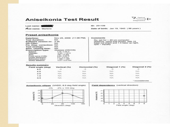
ANISEIKONIA INSPECTOR �Designed by Gerard De Wit �Direct comparison method of the perceived images �Patient fixates centrally whilst the images flash for 0. 5 sec (static aniseikonia) �Field angle is set by the size of the rectangles

Aniseikonia Inspector Testing

Minimising Aniseikonia
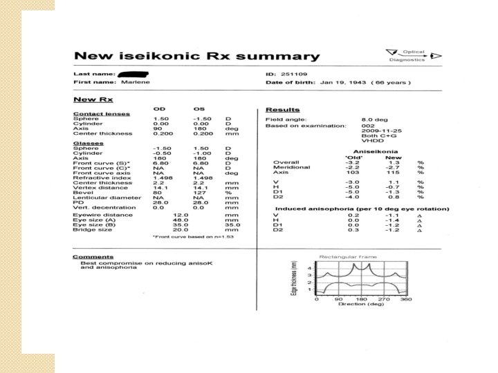

Aniseikona and motor fusion �In most patients, aniseikonia precipitates small angle strabismus due to loss of sensory fusion which in turn impairs motor fusion �Compensation for the strabismus with prisms along with lenses modified to compensate for the aniseikonia works in most optically corrected patients

Case 2: : A newly recognised mechanism for small angle diplopia in the elderly: Saggy eye muscles � 82 y o Intermittent Horizontal diplopia, mainly on le gaze, since cataract surgery 4 y ago � R 6/9, L 6/6 Horizontal Deviation: 0 0 6 ET 12 ET 6 ET Small L hypo in primary � Prescribed glasses: 8Δ BO, 2Δ BU LE single vision

Restricted depression on L a. Bduction

‘better’ SR – LR tissue sling some atrophy of LSR – LLR tissue sling Sagging of LLR pulley Not directly related to cataract surgery, but happens in same age group and will be attributed by patients to cataract surgery

LR-SR inter-muscular sling Degeneration of the LR-SR sling may occur in elderly Inferior displacement of the LR Pulley. LR is now a less capable a. Bductor, & now has an infraduction vector as well ET & Hypotropia Demer JL et alii “Heavy Eye” Syndrome in the Absence of High Myopia: A Connective Tissue Degeneration in Elderly Strabismic Patients J AAPOS. 2009 February; 13(1): 36– 44.

High risk #1: Beware correcting / ‘improving’ anisometropia �Spectacles compensate for most cases of aniseikonia 2° to axial anisometropia BETTER than do IOLs or corneal refractive surgery �Converting R: -12, L: -4 to -2 DS OU runs a real risk of PRODUCING aniseikonia, ABV & permanent troublesome diplopia esp if there is a small hitherto asymptomatic & unrecognised phoria �NO prospective studies to guide us how to handle anisometropic pts having IOLs

High risk #2: Beware of monovision There are insufficient prospective studies that can tell us which pts are safe for IOL MV You need to tell MV pts that there is a small risk [? %] of problems that seem to be fixable by reversing the MV. Sometimes these problems can present 2 -3 y after surgery. CL testing probably <100% predictive

High risk #3 : Beware macular membranes �Metamorphopsia / aniseikonia can be beyond the ability of optical devices to resolve �Cataract surgery can cause permanent diplopia in these pts

Is the disc swollen. . or is it drusen? IOVS 2011

Differentiating Optic Disc Edema From Optic Nerve Head Drusen on Optical Coherence Tomography Lenworth N. Johnson et al KEY: Lumpy/ bumpy appearance Arch Ophthalmol. 2009; 127(1): 45 -49

Thank you & good luck When assessing your results, you need to get the whole picture


Long-Term Outcome of Medial Rectus Recession and Pulley Posterior Fixation in Esotropia With High AC/A Ratio. Wabulembo G, Demer JL. @ Jules Stein. Strabismus. 2012 Sep; 20(3): 115 -20. � Medial rectus (MR) recession with pulley posterior fixation (PF) can be used to treat esotropia (ET) with a high AC/A ratio � N=21 LK 26 � Distance and near pre-operative ET averaged LK 23 19. 6Δ ± 10. 5Δ and 36. 9Δ ± 18. 9Δ, respectively. Mean neardistance (N-D) disparity was LK 26. 4 16. 4Δ ± 12. 3Δ. � The MR recession averaged 4. 4 ± 0. 9 mm. � Early mean postoperative ET was LK 0. 5 XT 1. 3 ± 3. 3Δ at distance and 2. 8Δ ± 5. 2Δ at near. � Mean late postoperative ET was 0. 1Δ ± 5. 8Δ and 1. 0Δ ± 6. 2Δ at distance and near, respectively. � At the final postoperative examination, mean N-D disparity was reduced to 0. 9Δ ± 3. 6Δ. LK 4. 5
- Slides: 86