Alveolar Process Gingiva Alveolar bone composed of a

Alveolar Process Gingiva
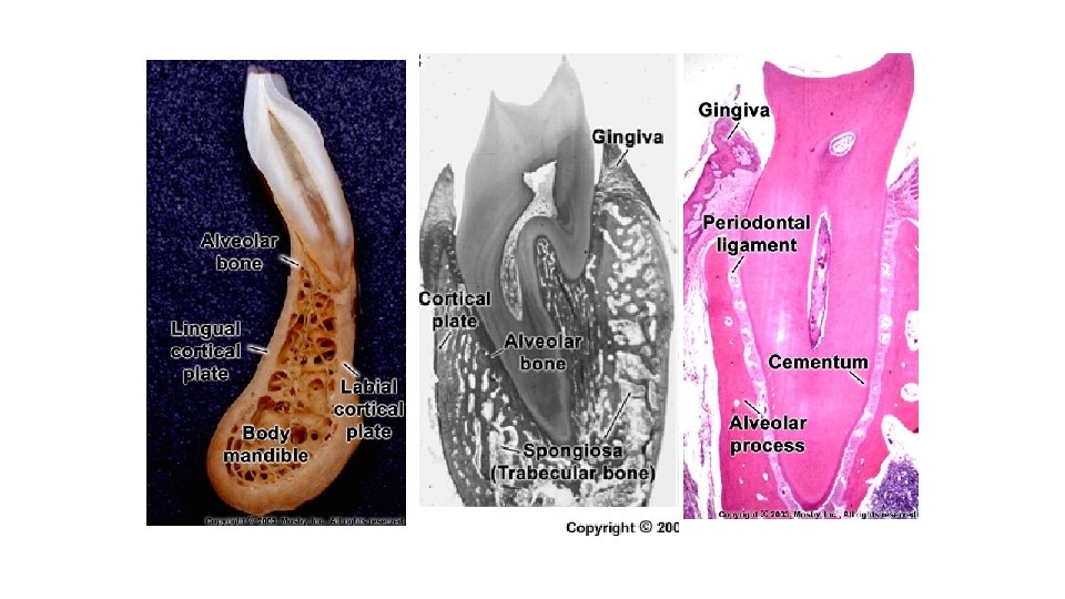
 outer cortical plates b) a central spongiosa c) bone")
Alveolar bone composed of a) outer cortical plates b) a central spongiosa c) bone lining the alveolus (bundle bone)

Alveolar bone proper: lines the tooth. Contains Sharpey’s fibers embedded into it Because alveolar process is regularly penetrated by collagen fiber bundles, it is also called bundle bone. It appears more radiodense(radio-opaqe) than surrounding supporting bone in X-rays called lamina dura

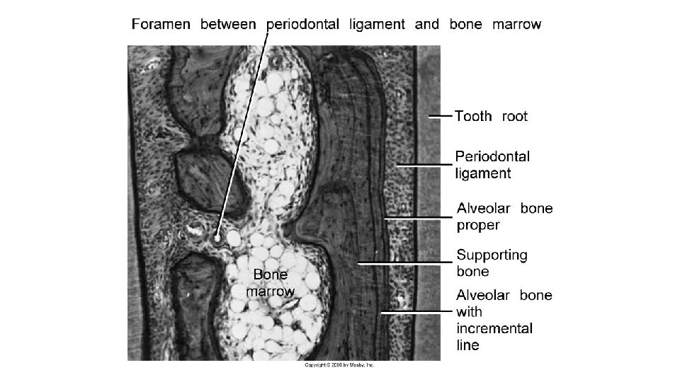
Bundle Bone It is perforated by many foramina that transmit nerves and vessels (cribriform plate). Radiographically, the bundle bone is the lamina dura. The lining of the alveolus is fairly smooth in the young but rougher in the adults. Radiodense because increased mineral content around fiber bundles Lamina Dura
 Extends both on")
Supporting Compact Bone Similar to compact bone anywhere else (Haversian bone) Extends both on the lingual (palatal) and buccal side Contains haversian and Volkman’s canals (they both form a continuous channel of nutrient canals)

Bundle bone and Trabecular bone Arrows: Sharpey’s fiber

The alveolar crest is found 1. 5 -2. 0 mm below the level of the CEJ.

Clinical considerations Resorption and regeneration of alveolar bone This process can occur during orthodontic movement of teeth. Bone is resorbed on the side of pressure and opposed on the site of tension.

Periodontal Ligament *PDL is the soft specialized connective tissue situated between cementum and alveolar bone proper *Ranges in thickness between 0. 15 and 0. 38 mm and is thinnest in the middle portion of the root *The width decreases with age *PDL Tissue with high turnover rate *Contains fibers, cells and intercellular substance

Embryogenesis The PDL forms from the dental follicle root development begins

FUNCTIONS OF PERIODONTIUM Tooth support Shock absorber: Withstanding the forces of mastication Sensory receptor necessary for proper positioning of the jaw Nutritive: blood vessels provide the essential nutrients to the vitality of the PDL
")
CELLULAR COMPOSITION The cells of periodontal ligament are categorized as: 1. Synthetic Cells a) Osteoblasts b) Fibroblasts c) Cementoblasts 2. Resorptive Cells a) Osteoclasts b) Cementoclasts c) Fibroblasts 3. Progenitor Cells 4. Epithelial Cell rests of malassez 5. Connective Tissue cells a) Mast cells b) Macrophages 14
 Osteoblasts(formative cell of bone) b) Osteoclasts (critical for periodontal disease and tooth")
Cells a) Osteoblasts(formative cell of bone) b) Osteoclasts (critical for periodontal disease and tooth movement) c) Fibroblasts (Most abundant, PRODUCTION OF VARIOUS TYPES OF FIBERS ) d) Epithelial cells (remnants of Hertwig’s epithelial root sheath-epithelial cell rests of Malassez) e) Macrophages (important defense cells) f) Undifferentiated cells (perivascular location) h) Cementoblasts(formative cell of cementum) i) Cementoclasts (only in pathologic conditions)

CEMENTOBLASTS • Cementoblasts synthesize collagen and protein polysaccharides, which make up the organic matrix of cementum. • After some cementum has been laid down, its mineralization begins with the help of calcium and phosphate ions. • Sharpey’s fibers 16

RESORPTIVE CELLS OSTEOCLASTS • Resorb bone. • The surface of an osteoclasts which is in contact with bone has a ruffled border. • Resorption occurs in two stages: • The mineral is removed at bone margins and then exposed organic matrix disintegrates. The osteoclasts demineralise the inorganic part as well as disintegrates the organic matrix. 17

18

FIBROBLASTS • Fibroblasts are capable of both synthesis and resorption. • They exhibit lysosomes, which contain collagen fragments undergoing digestion. • The presence of collagen resorbing fibroblasts in a normal functioning periodontal ligament indicates resorption of fibers occurring during remodeling of periodontal ligament. 19

PROGENITOR CELLS a • Progenitor cells are the undifferentiated mesenchymal cells, which have the capacity to undergo mitotic division and replace the differentiated cells dying at the end of their life span. • These cells are located in perivascular region and have cytoplasm. • When cell division occurs, one of the daughter cells differentiate into functional type of connective tissue cells. The other remaining cells retain their capacity to divide. 20

EPITHELIAL CELL RESTS OF MALASSEZ • These cells are the remnants of the epithelium of Hertwig’s Epithelial Root Sheath and are found close to cementum. 21

Periodontal ligament showing epithelial cell rests of malassez, indicated by arrows. 22

MAST CELLS • Mast cells are small round or oval. These cells are characterized by numerous cytoplasm, which mask its small, indistinct nucleus. • The diameter of mast cells is about 12 to 15 microns. • The granules contain heparin and histamine. The release of histamine into the extracellular compartment causes proliferation of the endothelial and mesenchymal cells. • Degranulate in response to antigen- antibody formation on their surface. 23

MACROPHAGES • Macrophages are derived from blood monocytes and are present near the blood vessels. • These cells have a horse-shoe shaped or kidney shaped nucleus with peripheral chromatin and cytoplasm contain phagocytosed material. • Macrophages help in phagocytosing dead cells and secreting growth factor, which help to regulate the proliferation of adjacent fibroblasts. 24

Epithelial Cell Rests of Malassez

PDL fibers - Collagen fibers: I, III and XII. Groups of fibers that are continually remodeled. (Principal fiber bundles of the PDL). The average diameter of individual fibers are smaller than other areas of the body, due to the shorter half-life of PDL fibers (so they have less time for fibrillar assembly) - Oxytalan fibers: variant of elastic fibers, perpendicular to teeth, adjacent to capillaries

• The damaged periodontal fibers are replaced and remodeled by newly formed fibers. • The “RENEWAL CAPABILITY” is an important characteristic of periodontal ligament. 27

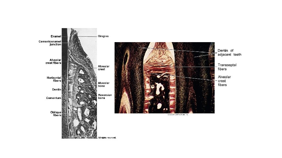
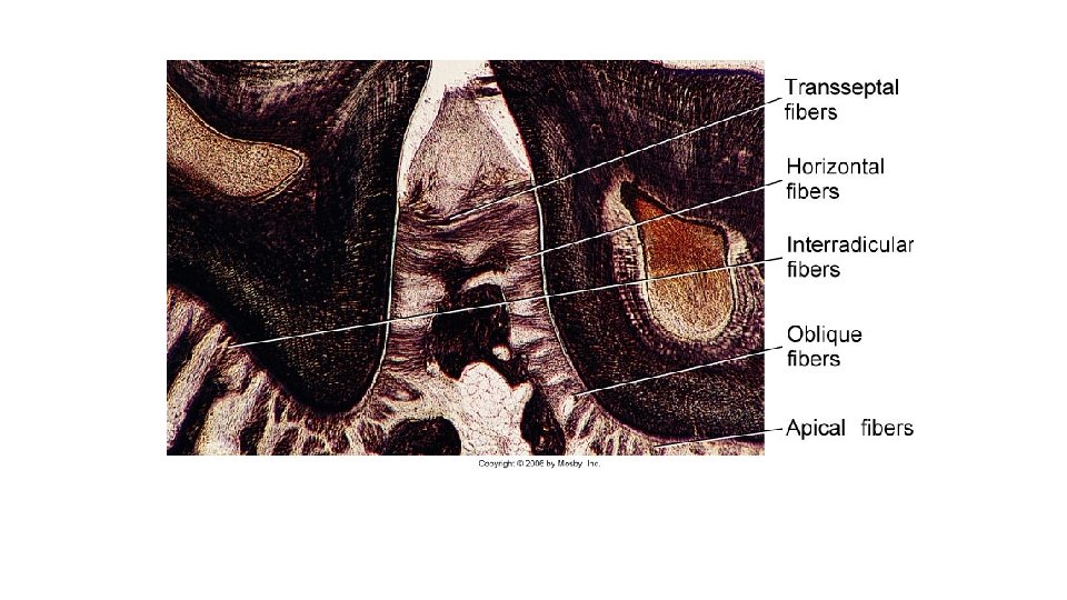
Principal Fibers Run between tooth and bone. Can be classified as dentoalveolar and gingival group Dentoalveolar group a. Alveolar crest group (ACG): below CE junction, downward, outward b. Horizontal group: apical to ACG, right angle to the root surface c. Oblique group: most numerous, oblique direction and attaches coronally to bone d. Apical group: around the apex, base of socket e. Interradicular group: multirooted teeth Runs from cementum and bone , forming the crest of the interradicular septum At each end, fibers embedded in bone and cementum: Sharpey’s fiber
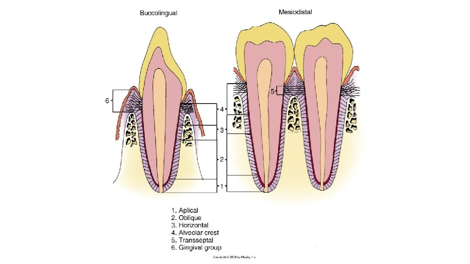

Gingival ligament fibers: the principal fibers in the gingival area are referred to as gingival fibers. Not strictly related to periodontium. Present in the lamina propria of the gingiva. a. Dentogingival: most numerous; cervical cementum to f/a gingiva b. Alveologingival: bone of the alveolar crest to f/a gingiva c. Circular: around neck of teeth, free gingiva d. Dentoperiosteal: runs apically from the cementum over the outer cortical plate to alv. process or vestibule (muscle) or floor of mouth e. Transseptal: cementum between adjacent teeth, over the alveolar crest
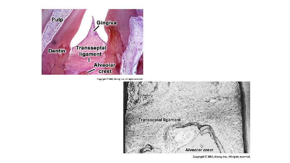

Transeptal Alveolar crest Horizontal Oblique
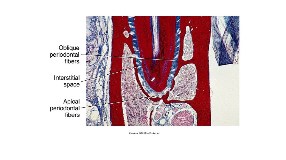

Interstitial Space Present between each bundle of ligament fibers Contains blood vessels and nerves

Blood supply to PDL BLOOD VESSELS Main blood supply is from superior and inferior alveolar arteries. The blood vessels are derived from the following: 1. BRANCHES FROM Vessels supplying the pulp. APICAL VESSELS 2. BRANCHES FROM INTRA-ALVEOLAR VESSELS: 3. BRANCHES FROM GINGIVAL VESSELS

39
- Slides: 39