AlQadisiya Universitybiotechnology College Embryology second stage 2017 Alaa
. Alaa kamil Abualla lab. 2 : FERTILIZATION")
Al-Qadisiya University/biotechnology College Embryology second stage. (2017). Alaa kamil Abualla lab. 2 : FERTILIZATION

FERTILIZATION • The embryo development begins with a sperm fertilizing an egg to become a zygote which undergoes many cleavages to develop into a ball of cells called a morula and development to blastula which precedes the formation of the gastrula in which the germ layers of the embryo form. • Fertilization is the fusion of gametes, occurs in the ampulla region of the uterine tube to initiate the development of a new individual organism. In animals, the process involves the fusion of an ovum with a sperm, which first creates a zygote and then leads to the development of an embryo. • • Only 1% of the sperms deposited in the vagina enter the cervix, yet movement of sperm from cervix to uterine tube occurs by muscular contractions of uterus and uterine tube & by their own propulsion, and this sperms reach to the uterine tube during 2 -7 hrs.

FERTILIZATION

zygote the zygote is a eukaryotic cell formed by a fertilization event between two gametes, the zygote's genome is a combination of the DNA in each gamete, and contains all of the genetic information necessary to form a new individual.

Cleavage follows fertilization: The cleavage is a series of rapid mitotic divisions (without cell growth). At the end of the cleavage stage, cells making up the blastula move about and surface proteins help cells recognize each other. The gastrula is formed, which consists of 3 “germ layers” Endoderm, Mesoderm and Ectoderm
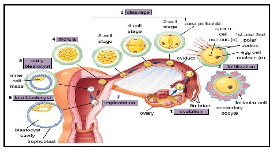

CLEAVAGE 1. Morula is a mass formed in 3 -4 days post fertilization, consist from 8 cell in a spherical shape if untouched and allowed to remain implanted, will eventually develop into a blastocyst. Its produced by a series of cleavage divisions of the early embryo, starting with the single-celled zygote. Once the embryo has divided into 16 cells.

Morula

Morula

Morula
 has a cavity inside the")
2. Blastocyst The blastocyst (4 -5 days post fertilization) has a cavity inside the zona pellucida along with an inner cell mass. It’s a hollow sphere of cells, surrounding an inner fluid-filled cavity called the blastula formed during an early stage of embryonic development in animals. The inner cell mass which subsequenn trophoblast to forms the embryo. The outer layer of the blastocyst consists of cells collectively called the which rise to the placenta, this layer surrounds the inner cell mass and a fluid-filled cavity known as the blastocoele.

Blastocyst

Blastocyst

3. Implantation Is the very early stage of pregnancy at which the conceptus adheres to the wall of the uterus. At this stage of prenatal development, the conceptus is a blastocyst. It is by this adhesion that the foetus receives oxygen and nutrients from the mother to be able to grow. • In humans the endometrium of the uterus is usually termed the "implantation window" and lasts about 4 days.

Implantation The implantation window is characterized by changes to the endometrium cells, which aid in the absorption of the uterine fluid. These changes are collectively known as the plasma membrane transformation and bring the blastocyst nearer to the endometrium and immobilize it.
 • During the process of")
4. Gastrulation: three Germ Layers Formed (day 12: ) • During the process of gastrulation, a special type of cells a hole on the surface of the blastula which is called the dorsal lip of the blastopore. Once this lip has been established, the bottle cells will extend inward and migrate along the inner wall of the blastula. The once superficial cells of the animal pole are destined to become the cells of the middle germ layer called the mesoderm. • These processes allow for the prospective mesoderm cells to be placed between the ectoderm and the endoderm.

1. Endoderm • The endoderm is formed from cells that migrate toward the center of the developing gastrula. They are initially found as flattened cells, but eventually begin to pile on top of each other in a columnar arrangement. The function of the endoderm is to provide an epithelial lining for the body’s two major tubes: the digestive and respiratory tube. • It forms the epithelial lining of multiple systems. • Endocrine glands , organs and Urinary system

2. Mesoderm • The mesoderm is forms blood cells, the muscles in a process known as myogenesis, and forms part of the gonads (the rest being the gametes). • All muscle, bone, and connective tissue • Entire vascular and lymphatic system, including blood • Urogenital organs (kidneys, gonads, and reproductive ducts) • The dermis (middle layer of the skin)

3. Ectoderm • The ectoderm is one of the three primary germ layers in the very early embryo, the ectoderm as the most exterior (or distal) layer. It originates from the outer layer of germ cells. • The entire nervous system (spine, peripheral nerves and brain), • Posterior pituitary • Adrenal medulla • Cornea and lens • Epidermis of skin and its derivatives (i. e. hair, nails, sweat glands, sensory receptors) • tooth enamel and the epidermis • It also forms the lining of mouth, anus, nostrils, sweat glands, hair and nails.
- Slides: 19