ALLIED FUNGI SLIME MOLDS Slime molds share the


 and extra")
 Kingdom – Protozoa Phyllum")


- Slides: 16
ALLIED FUNGI SLIME MOLDS
Slime molds share the following features with fungi: �They are achlorophyllous, true nucleated heterotrophs. �Slime molds inhabit the moist decaying lignocellulosic substratum like fallen leaves, twigs, wooden logs, decaying animal dung, sometimes on the surface of garden plants tree trunks, and act as decomposers of complex polysaccharides into simple sugars like fungi. �Slime molds somatic structure is coenocytic as in the coenocytic fungi. �Slime molds plasmodium (vegetative thallus) absorbs and assimilates its nutrition in the soluble form by direct contact of the plasma membrane with substratum. �Slime molds possess reserve food material as fat droplets and glycogen particles. �They reproduce asexually by amoeboid cells(motile or non motile) which during favourable conditions divide mitotically several times and parennate as microcysts or spherules during unfavourable conditions and remain viable for very long period in the soil. �They produce sexual meiotic spores in the fruiting bodies. The Spores are thin walled, hyaline surrounded by definite cell wall of cellulosic in nature but not chitinous as in other fungi and mode of dispersal is like fungi by wind, water and by insectal movements. �The pattern of life cycle slime molds follow is same like fungi. �Slime molds produce sclerotia during unfavourable conditions same like fungi.
Slime molds differ from fungi: 1. Mode of nutrition – Ingestion (Phagocystosis) and extra cellular degradation and absorption. 2. Absence of definite cell wall around somatic structures. 3. Plasmodium shows cytoplasmic streaming.
CLASSIFICATION Webster J. and Weber R. W. S. 2007(Molecular phylogenetic) Kingdom – Protozoa Phyllum – Myxomycota Class – Acrasiomycetes - Dictyosteliomycetes - Protosteliomycetes - Myxomycetes
Occurrence Habit: Slime molds are cosmopolitan and are mostly saprophytic in nature. Habitat: Slime molds prefer mostly cool, shady and moist places for their growth and are found in almost all kinds of environment i. e. Tropics, Temperate, in alpine zones near melting snow peaks and even in deserts. They live on decaying lignocellulosic substrates like moist fallen leave, twigs, wooden logs in the forests, sometimes on the surface of garden plants, tree trunks, bark, on the body of dead cacti and decaying animal dung etc. These are mostly studied, located and collected by mycologists during fungal collections. Lamproderma and Lepidoderma etc. are seen at snow peaks.
�They can be used as an edible protein source in future. The young fruiting bodies of Enteridium, Lycoperdon and Fuligo septica are eaten in Mexico. �The exenic cultures of slime molds are used in cell and molecular biology e. g. Physarum polycephalum. and Didymium iridis cultures are used to study the genetic variability in nature and in understanding the process of ageing and how it can be controlled both by the nucleus and cytoplasm (cell longevity). �Some antibiotics have been isolated from. Physarum gyrosum which can be used against yeasts, gram (+) and (-) bacteria. �Slime molds wherever they grow utilize all kinds of spores of microbes as food and don’t allow any microbe to colonise that substratum are considered as a purest form of protoplasm found in nature. It is frequently employed in research to understand the chemical composition of protoplasm.
Economic Importance Slime molds in general have little direct economic importance. The colourful plasmodium and fruiting bodies of slime molds are beautiful and the delicately constructed intricate designs have attracted the photographers and artists to exhibit them in the form of paintings, sceneries and photographs e. g. the creamish yellow coloured fruiting bodies of Physorum polycephalum and the grayish black linear hair like fruting bodies of Stemonitis sp growing on woods. �Ecologically these are important in the food web as food for insects. �They can be used as an edible protein source in future. The young fruiting bodies of Enteridium, Lycoperdon and Fuligo septica are eaten in Mexico.
�The exenic cultures of slime molds are used in cell and molecular biology e. g. Physarum polycephalum. and Didymium iridis cultures are used to study the genetic variability in nature and in understanding the process of ageing and how it can be controlled both by the nucleus and cytoplasm (cell longevity). �Some antibiotics have been isolated from. Physarum gyrosum which can be used against yeasts, gram (+) and (-) bacteria. �Slime molds wherever they grow utilize all kinds of spores of microbes as food and don’t allow any microbe to colonise that substratum are considered as a purest form of protoplasm found in nature. It is frequently employed in research to understand the chemical composition of protoplasm. �These are used as an ideal experimental tools in research laboratories to understand the various processes in living beings i. e. mitotic cycle, morphogenesis, physiology, the chemical changes which govern reproduction, the structure and movement of protoplasm etc. e. g. Physarum polycephalum is used in cancer research and Dictystelium discoideum (Acrasiomycete) in morphogenesis. �Myxomycetes are used in space programms to understand the behaviour of living protoplasm in microgravity environment e. g. the plasmodium of Physarum polycephalum have been successfully maintained by the scientists under space
Types of Plasmodia There are three general types of plasmodia seen in myxomycetes (Alexopoulos 1960): 1. Protoplasmodium Figure: Diagrammatic representation of a typical protoplasmodium.
It is a small, irregular hyaline lump like homogeneous mess of protoplasm and remains microscopic throughout its existence. It is a primitive type of plasmodium and does not form any veins or branches, and exhibits a very slow irregular streaming of protoplasm instead of rapid, rhythmic, reversible streaming of the other plasmodial types. Protoplamodium gives rise to only one single sporangium during fruiting e. g. members of Echinosteliales, Echinostelium minutum, licea sp, Clastoderma debaryanum etc. Figure: Licea biforis protoplasmodium and fruiting body
Figure: Licea biforis protoplasmodium and fruiting body
2. Aphanoplasmodium: Plasmodium is initially an irregular lump like hyaline mess but soon gets differentiated into long branches and forms a network of very fine, transparent strands. The protoplasm is not very granular and the plasmodium is devoid of slime sheath. The veins or branches are not conspicuously differentiated into gelified and fluid regions, the streaming protoplasm is confined by a very delicate membrane. Streaming is rapid and rhythmically reversible. This type of plasmodium is a characteristic of stemonitomycetidae. AB Figure: Diagrammatic representation (A) and photograph (B) of aphanoplasmodium.
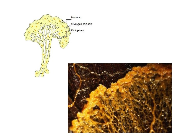
3. Phaneroplasmodium: This plasmodium initially resembles a protoplasmodium which later grows larger and becomes massive. The protoplasm is very granular and easily visible. The gelified and fluid parts of the veins are easily differentiated and the rhythmic, reversible streaming is very conspicuous. The Plasmodium is surrounded by plasma membrane and a gelatinous layer in which calcium and silica granules are deposited. This plasmodium is creamish yellow macroscopic seen flourishing on the leaves and stem of ornamentals in the garden or on the decaying wooden logs in the forests. Plasmodium in the initial phase of development is like protoplasmic type which later grows larger granular and becomes more massive and visible by naked eye. The gellified and fluid portions of the veins are easily differentiated and the rhythmic, reversible streaming is very conspicuous which is probably due to the interaction of cations with cytoskeleton elements lining the veins. The plasmodium deposits the excretory or undigested food particles on the side of the branches or veins exhibiting the plasmodial tracks on the substratum. It is seen in order Physarales (Didymium, Physarum etc. ) AB Figure: A. Diagrammatic representation of phaneroplasmodium; B. Photograph showing phaneroplasmodium and young fruiting bodies
Figure: Life cycle of Stemonitis exhibited by photographs; A. aphanoplasmodium; B. Mature fruiting body; C, D. magnified view of capilitium and columella ; E. Spores magnified; F. Mature fruiting bodies
This type of sporangial development is characteristic of members of stemonitales i. e. known as epihypothallic type. The plasmodium is laid down on the substratum, becomes concentrated into one or more sphaerical masses. Inside these sphaerical masses it begins to deposit a stalk on the hypothallus. The stalk begins to grow and becomes longer, the protoplasm crawls upward and continues to deposit the materials to the tip of the stalk until the total length is attained. Now the protoplasm inside the fruiting body secrets a thin wall (peridium) around it and deposit the capillitial threads intra protoplasmically which get extended from the columella towards the surface of the developing sporangium. The sporangial protoplasm now cleaves into spores where nuclei undergo meiosis and eventually spore tetrads (n) are formed. Slime Molds