
Alexander Disease and GFAP By Austin Pier What
Alexander Disease and GFAP By: Austin Pier
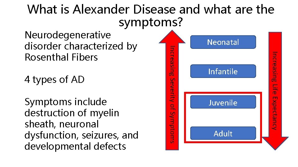
What is Alexander Disease and what are the symptoms? Symptoms include destruction of myelin sheath, neuronal dysfunction, seizures, and developmental defects Neonatal Infantile Juvenile Adult Increasing Life Expectancy 4 types of AD Increasing Severity of Symptoms Neurodegenerative disorder characterized by Rosenthal Fibers
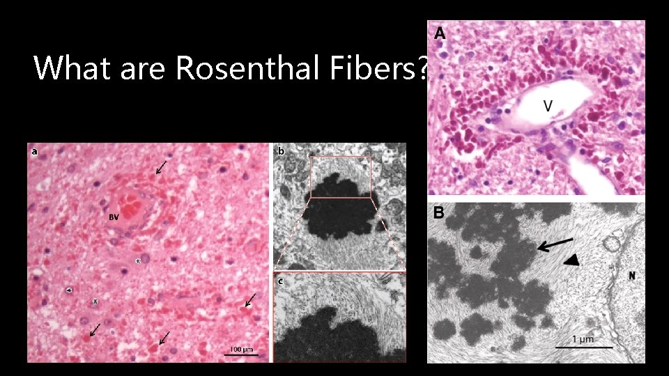
What are Rosenthal Fibers?
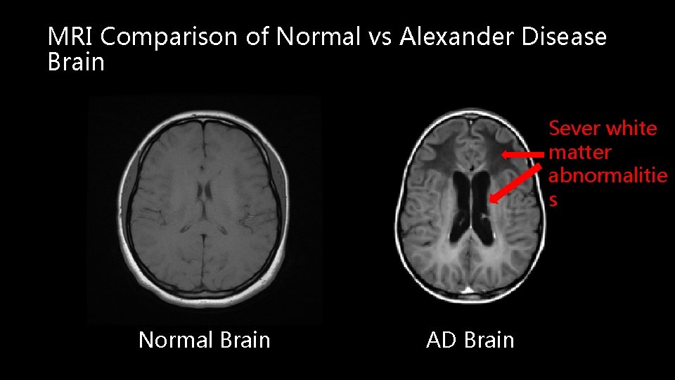
MRI Comparison of Normal vs Alexander Disease Brain Sever white matter abnormalitie s Normal Brain AD Brain
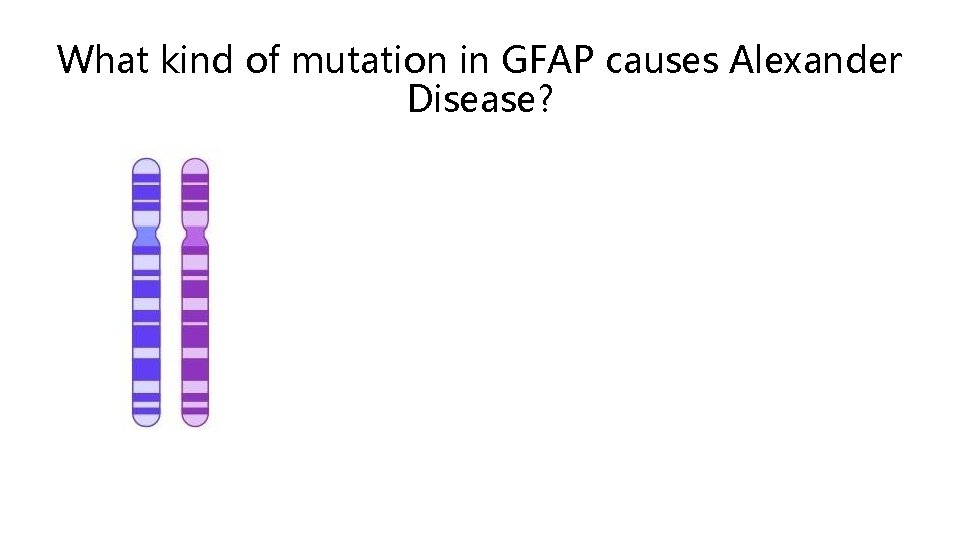
What kind of mutation in GFAP causes Alexander Disease?
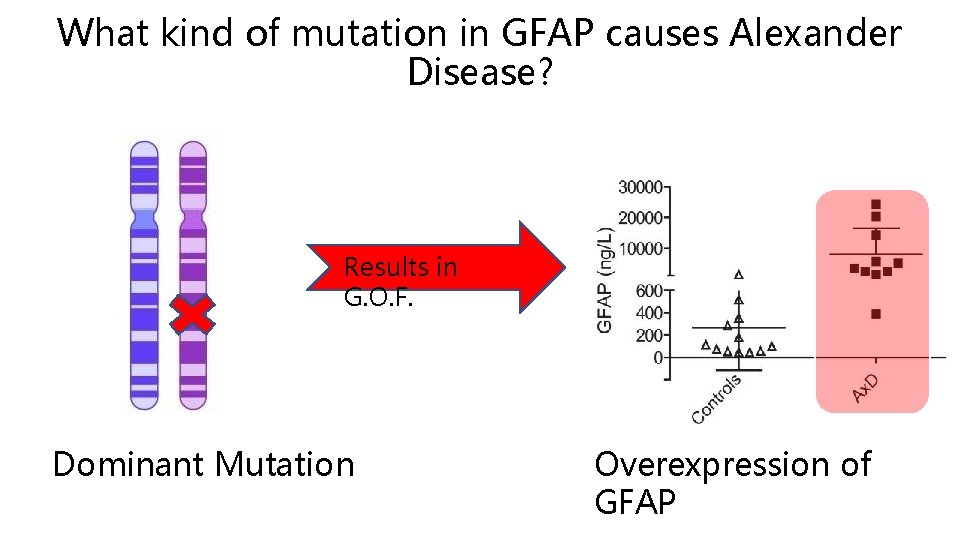
What kind of mutation in GFAP causes Alexander Disease? Results in G. O. F. Dominant Mutation Overexpression of GFAP
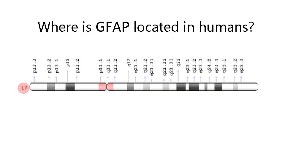
Where is GFAP located in humans? GFAP is located at q 21. 31 on chromosome 17
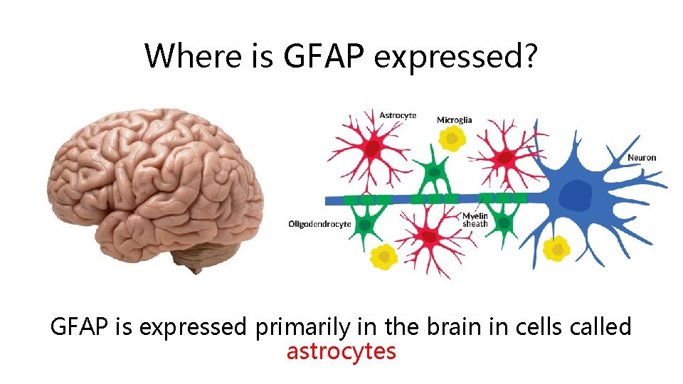
Where is GFAP expressed? GFAP is expressed primarily in the brain in cells called astrocytes
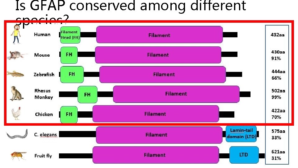
Is GFAP conserved among different species?
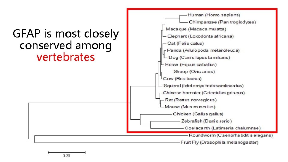
GFAP is most closely conserved among vertebrates
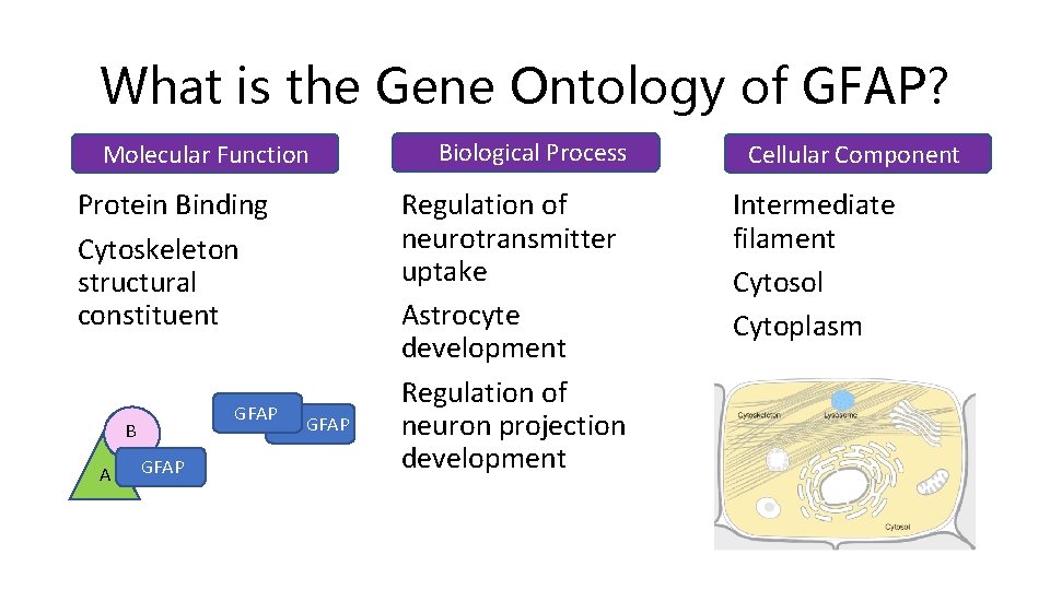
What is the Gene Ontology of GFAP? Molecular Function Protein Binding Cytoskeleton structural constituent GFAP B A GFAP Biological Process Regulation of neurotransmitter uptake Astrocyte development Regulation of neuron projection development Cellular Component Intermediate filament Cytosol Cytoplasm
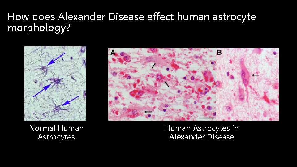
How does Alexander Disease effect human astrocyte morphology? Normal Human Astrocytes in Alexander Disease
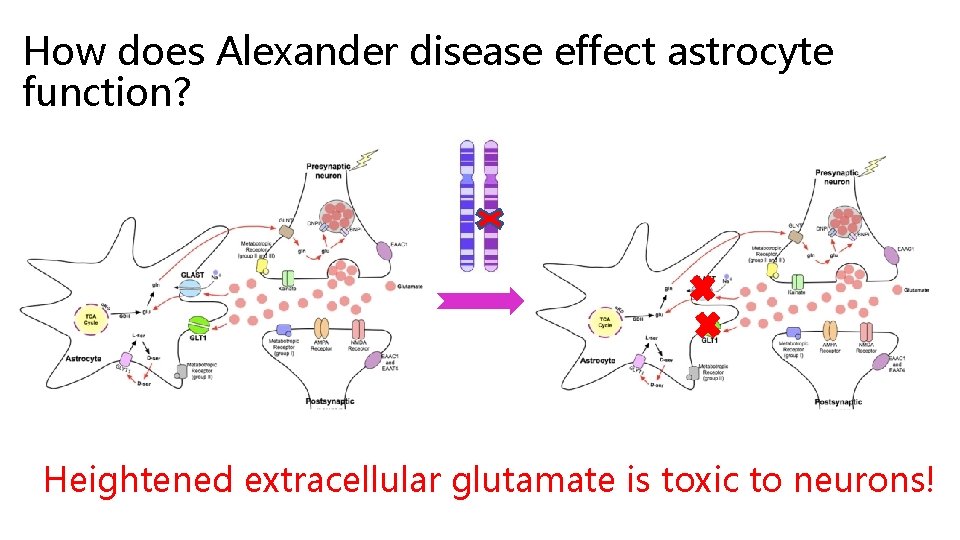
How does Alexander disease effect astrocyte function? Heightened extracellular glutamate is toxic to neurons!
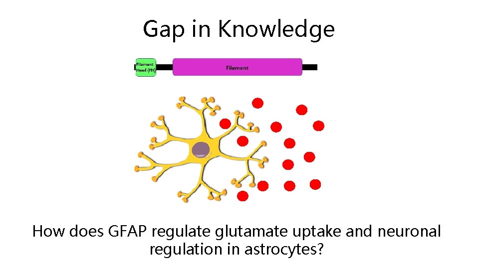
Gap in Knowledge How does GFAP regulate glutamate uptake and neuronal regulation in astrocytes?
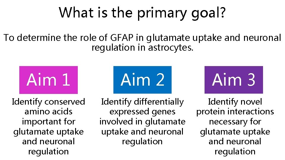
What is the primary goal? To determine the role of GFAP in glutamate uptake and neuronal regulation in astrocytes. Aim 1 Aim 2 Aim 3 Identify conserved amino acids important for glutamate uptake and neuronal regulation Identify differentially expressed genes involved in glutamate uptake and neuronal regulation Identify novel protein interactions necessary for glutamate uptake and neuronal regulation
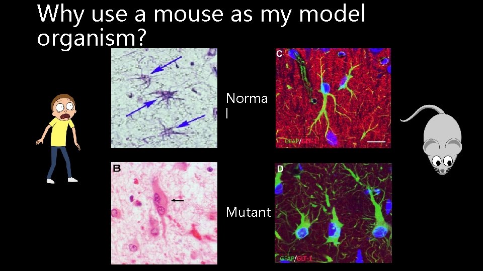
Why use a mouse as my model organism? Norma l Mutant
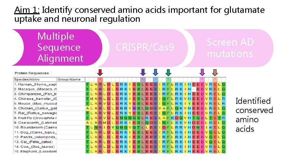
Aim 1: Identify conserved amino acids important for glutamate uptake and neuronal regulation Multiple Sequence Alignment CRISPR/Cas 9 Screen AD mutations Identified conserved amino acids
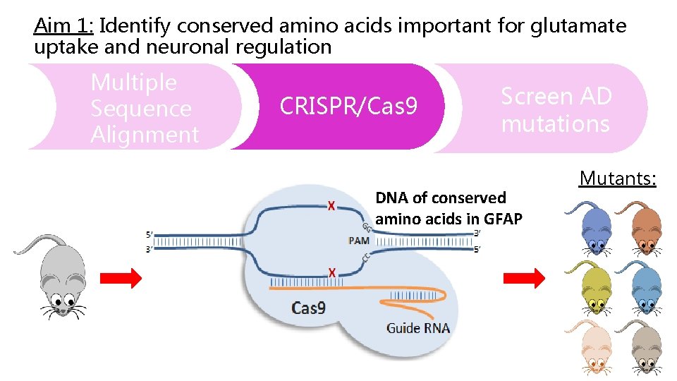
Aim 1: Identify conserved amino acids important for glutamate uptake and neuronal regulation Multiple Sequence Alignment CRISPR/Cas 9 Screen AD mutations DNA of conserved amino acids in GFAP Mutants:
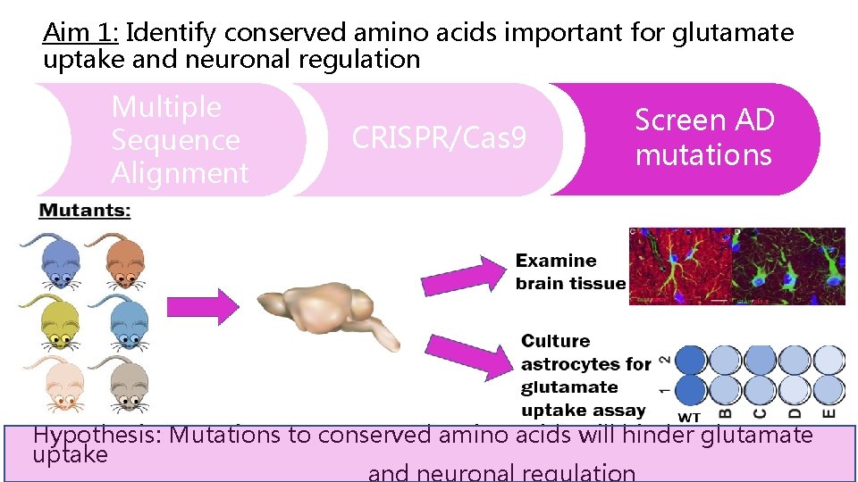
Aim 1: Identify conserved amino acids important for glutamate uptake and neuronal regulation Multiple Sequence Alignment CRISPR/Cas 9 Screen AD mutations Hypothesis: Mutations to conserved amino acids will hinder glutamate uptake and neuronal regulation
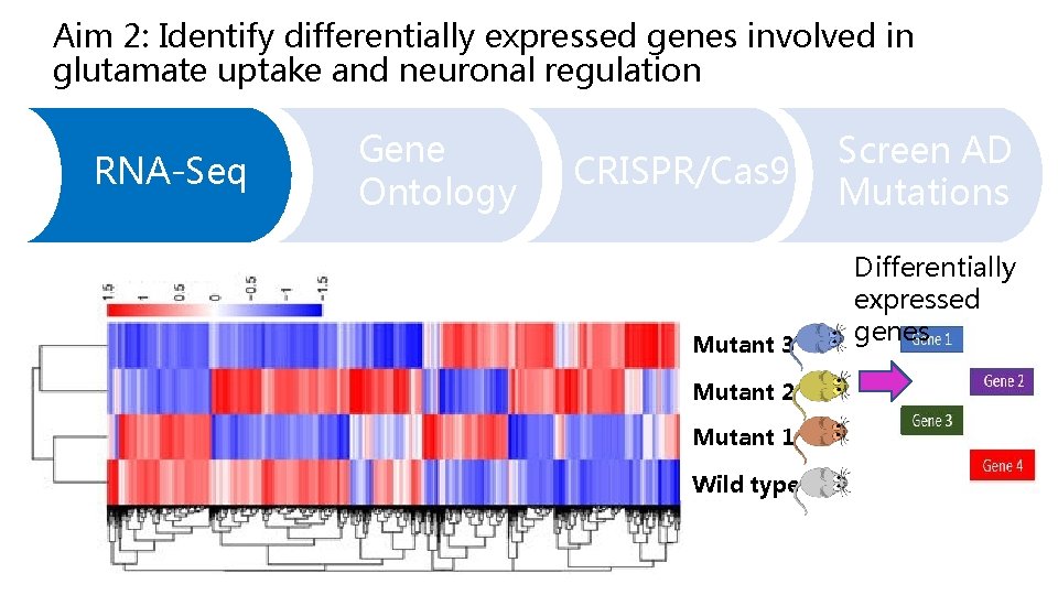
Aim 2: Identify differentially expressed genes involved in glutamate uptake and neuronal regulation RNA-Seq Gene Ontology CRISPR/Cas 9 Mutant 3 Mutant 2 Mutant 1 Wild type Screen AD Mutations Differentially expressed genes
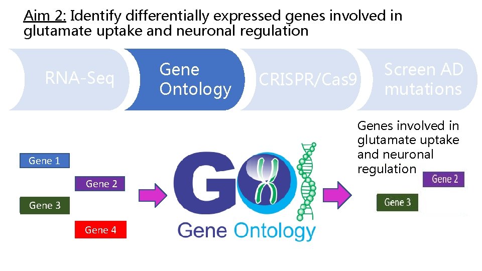
Aim 2: Identify differentially expressed genes involved in glutamate uptake and neuronal regulation RNA-Seq Gene Ontology CRISPR/Cas 9 Screen AD mutations Genes involved in glutamate uptake and neuronal regulation Gene 1 Gene 2 Gene 3 Gene 4
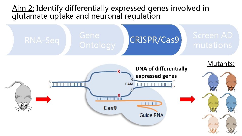
Aim 2: Identify differentially expressed genes involved in glutamate uptake and neuronal regulation RNA-Seq Gene Ontology CRISPR/Cas 9 DNA of differentially expressed genes Screen AD mutations Mutants:
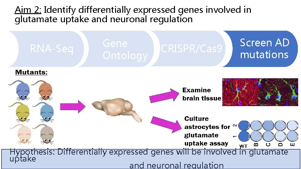
Aim 2: Identify differentially expressed genes involved in glutamate uptake and neuronal regulation RNA-Seq Gene Ontology CRISPR/Cas 9 Screen AD mutations Hypothesis: Differentially expressed genes will be involved in glutamate uptake and neuronal regulation
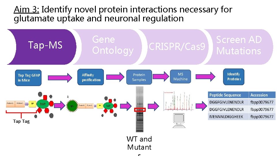
Aim 3: Identify novel protein interactions necessary for glutamate uptake and neuronal regulation Tap-MS Tap Tag GFAP in Mice Gene Ontology CRISPR/Cas 9 Screen AD Mutations Identify Proteins Affinity purification Tap Tag WT and Mutant
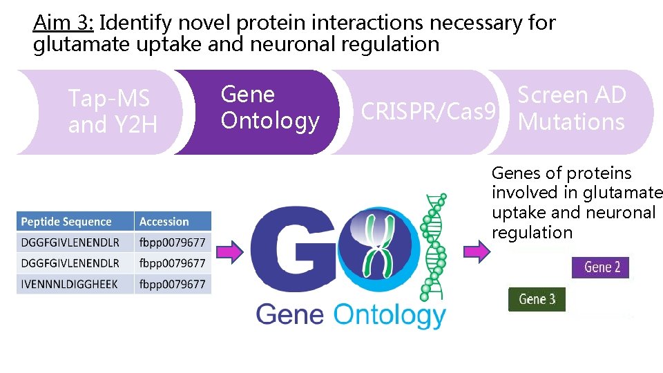
Aim 3: Identify novel protein interactions necessary for glutamate uptake and neuronal regulation Tap-MS and Y 2 H Gene Ontology Screen AD CRISPR/Cas 9 Mutations Genes of proteins involved in glutamate uptake and neuronal regulation
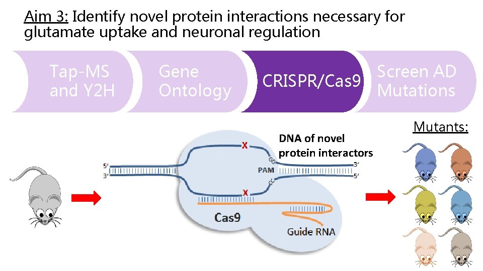
Aim 3: Identify novel protein interactions necessary for glutamate uptake and neuronal regulation Tap-MS and Y 2 H Gene Ontology Screen AD CRISPR/Cas 9 Mutations DNA of novel protein interactors Mutants:
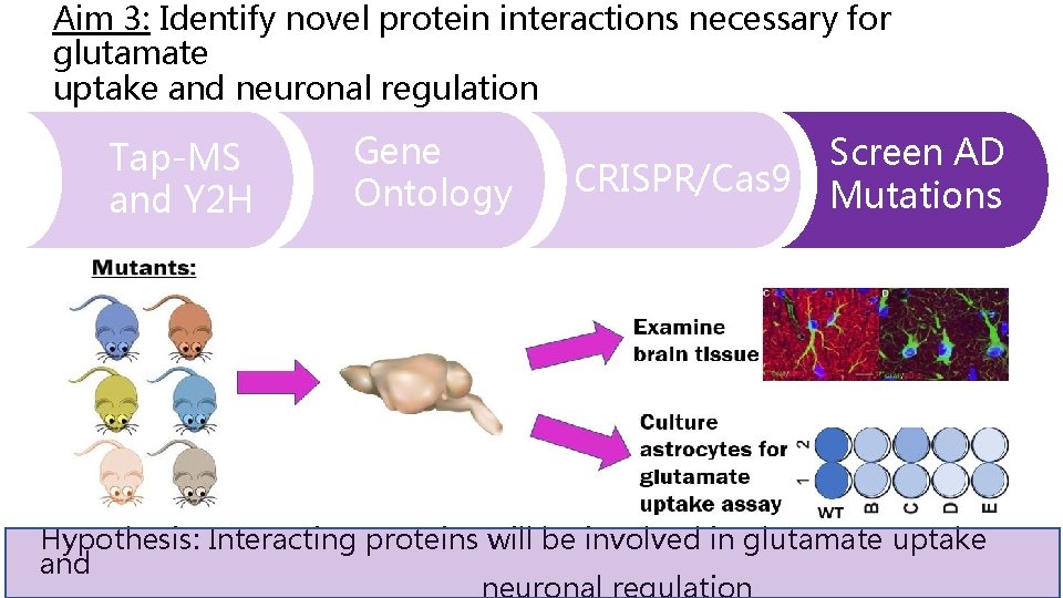
Aim 3: Identify novel protein interactions necessary for glutamate uptake and neuronal regulation Tap-MS and Y 2 H Gene Ontology CRISPR/Cas 9 Screen AD Mutations Hypothesis: Interacting proteins will be involved in glutamate uptake and neuronal regulation
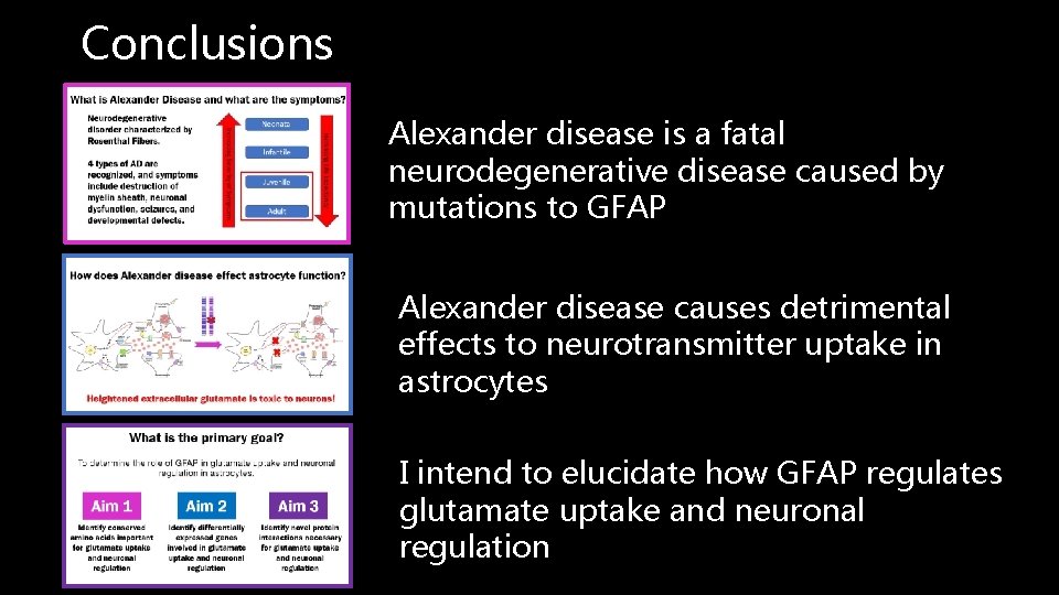
Conclusions Alexander disease is a fatal neurodegenerative disease caused by mutations to GFAP Alexander disease causes detrimental effects to neurotransmitter uptake in astrocytes I intend to elucidate how GFAP regulates glutamate uptake and neuronal regulation
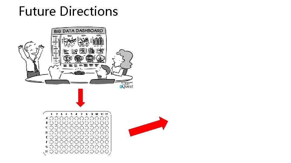
Future Directions
Questions?
Work Cited: [3] Martidis, A. , Yee, R. D. , Azzarelli, B. , & Biller, J. (1999). Neuro-ophthalmic, radiographic, and pathologic manifestations of adult-onset Alexander disease. Archives of Ophthalmology (Chicago, Ill. : 1960), 117(2), 265– 267. [4] Namekawa, M. , Takiyama, Y. , Aoki, Y. , Takayashiki, N. , Sakoe, K. , Shimazaki, H. , … Nakano, I. (2002). Identification of GFAP gene mutation in hereditary adult-onset Alexander’s disease. Annals of Neurology, 52(6), 779– 785. https: //doi. org/10. 1002/ana. 10375 [5] Sosunov, A. A. , Mc. Khann, G. M. , & Goldman, J. E. (2017). The origin of Rosenthal fibers and their contributions to astrocyte pathology in Alexander disease. Acta Neuropathologica Communications, 5, 27. https: //doi. org/10. 1186/s 40478 -0170425 -9 Spanagel, R. , Pendyala, G. , Abarca, C. , Zghoul, T. , Sanchis-Segura, C. , Magnone, M. C. , … Albrecht, U. (2005). ERRATUM: The clock gene Per 2 influences the glutamatergic system and modulates alcohol consumption. Nature Medicine, 11(2), 233. https: //doi. org/10. 1038/nm 0205 -233 c [6] Springer, S. , Erlewein, R. , Naegele, T. , Becker, I. , Auer, D. , Grodd, W. , & Krägeloh-Mann, I. (2000). Alexander disease--classification revisited and isolation of a neonatal form. Neuropediatrics, 31(2), 86– 92. https: //doi. org/10. 1055/s-2000 -7479 [7] Srivastava, S. , & Naidu, S. (1993). Alexander Disease. In M. P. Adam, H. H. Ardinger, R. A. Pagon, S. E. Wallace, L. J. Bean, K. Stephens, & A. Amemiya (Eds. ), Gene. Reviews®. Seattle (WA): University of Washington, Seattle. Retrieved from http: //www. ncbi. nlm. nih. gov/books/NBK 1172/ Basic Local Alignment Search Tool. (n. d. ). Retrieved March 14, 2018, from http: //blast. ncbi. nlm. nih. gov/Blast. cgi [2] Pearson, W. R. (2013). An Introduction to Sequence Similarity (“Homology”) Searching. Current Protocols in Bioinformatics / Editoral Board, Andreas D. Baxevanis. . . [et Al. ] , 0 3. https: //doi. org/10. 1002/0471250953. bi 0301 s 42 [3] Skop, A. (2018). Lab 2: Homology & Phylogeny. Retrieved March 14, 2018, from http: //genetics 564. weebly. com/homology--phylogeny. html [3] Sievers F, Wilm A, Dineen D, Gibson T, Karplus K, Li W, Lopez R, Mc. William H, Remmert M, Söding J, Thompson J, Higgins D, Fast, scalable generation of high-quality protein multiple sequence alignments using Clustal Omega, MSB 7 (2011) 1 -6. PMID: 21988835 [4] Skop, A. (2018). Lab 2: Homology & Phylogeny. Retrieved March 14, 2018, from http: //genetics 564. weebly. com/homology--phylogeny. html [2] Skop, A. (2018). Lab 3: Protein domain discovery & DNA motifs. Retrieved April 12, 2016, from http: //genetics 564. weebly. com/motif--domain-discovery. html Olabarria, M. , & Goldman, J. E. (2017). Disorders of Astrocytes: Alexander Disease as a Model. Annual Review of Pathology: Mechanisms of Disease , 12(1), 131– 152. https: //doi. org/10. 1146/annurev-pathol-052016 -100218 https: //www. google. com/search? safe=off&tbm=isch&q=neuron+background&chips=q: neuron+background, g_3: desktop&sa=X&ved=0 ah. UKEwix_amhoq. La. Ah. UJ 7 Z 8 KHYUWAqw. Q 4 l. YILCg. A&biw=1229&bih=587&dpr=1. 56#imgrc=96 n 9 i. Qnb. Wt 5 YJM https: //ghr. nlm. nih. gov/gene/GFAP#location https: //www. google. com/search? q=astrocytes+images&safe=off&tbm=isch&tbs=rimg: Cdz 171 Pk. BWNw. Iji 49 o 7 u. Ra 1 kc. GFp 7 vp-7 V_1 Dbob 9 j. YHi. K 4 lpy 12 Ax. S 5 wl 3 B 4 Tr. FOKbx. P 1 xwd. C_1 npxj. Sy. GZYIl-Lps. Co. SCbj 2 ju 5 Fr. WRw. EV 3_1 aqq. C 3 Y_1 c. Kh. IJYWnun 7 t. X 8 MRhoz. Y 9 ojgvssq. Egluhv 2 Nge. Iri. RGZYism. Mb. Hmjio. SCWn. LXYDFLn. CXEcx. HQgk. JCBz_1 Kh. IJc. Hh. Os. U 4 pv. E 8 Rsw 0 NE 3 euh. W 4 q. Egn. XHB 0 L-en. GNBHrkv. Pa. Pe. WK 4 Co. SCb. IZlgi. X 4 umw. EYQgw. O_1 HRa. Y&tbo=u&sa=X&ved=2 ah. UKEwjt 5 LGO 4 KPa. Ah. Xrv 1 QKHX 4 s. CMw. Q 9 C 96 BAg. AEBs&biw=616&bih=572&dpr=1. 56#imgrc=1 jo. Ptn. IOIe-Lb. M https: //www. google. com/search? q=myelin+sheath+degeneration&safe=off&source=lnms&tbm=isch&sa=X&ved=0 ah. UKEwitqa. PNpq. Ta. Ah. UNCXw. KHfe. UBC 0 Q_AUICig. B&biw=614&bih=572#imgrc=qk. Esr. FYDau. G 7 DM: https: //radiopaedia. org/cases/normal-brain-mri-6 https: //www-annualreviews-org. ezproxy. library. wisc. edu/na 101/home/literatum/publisher/ar/journals/content/pathmechdis/2017/pathmechdis. 2017. 12. issue-1/annurev-pathol-052016 -100218/20170125/images/large/pm 120131. f 2. jpeg https: //www. nature. com/articles/s 41598 -017 -03024 -0/figures/3 https: //www. google. com/search? safe=off&hl=en&tbm=isch&sa=1&ei=p. Hjn. Wvq. YJu. SYjw. Sd 3 p. VA&q=mouse+clipart+transparent+background&oq=mouse+clipart&gs_l=psy-ab. 1. 5. 0 l 10. 1936. 4556. 0. 5933. 16. 13. 1. 2. 2. 0. 357. 1561. 0 j 7 j 1 j 1. 10. 0. . . 1 c. 1. 64. psyab. . 3. 12. 1574. 0. . 0 i 67 k 1. 100. Hnx. GOTV 8 px. E#imgrc=Cw. H 62 d. JUds 5 UQM: http: //www. uniprot. org/uniprot/P 14136#function
- Slides: 31