
Alevel Biology 3 6 4 3 Control of
A-level Biology 3. 6. 4. 3 Control of blood water potential To be used alongside AQA A-level Biology Control of blood water potential teaching notes 1 of 20 Version 1. 0 Copyright © 2016 AQA and its licensors. All rights reserved.
What might happen if. . . • The water potential of the blood became too low (more negative)? • The water potential of the blood became too high (less negative)? 2 of 20 Version 1. 0 Copyright © 2016 AQA and its licensors. All rights reserved.
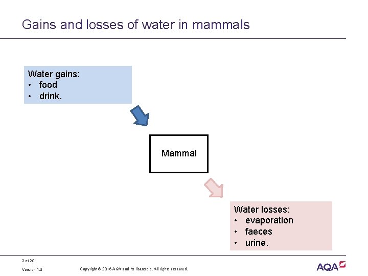
Gains and losses of water in mammals Water gains: • food • drink. Mammal Water losses: • evaporation • faeces • urine. 3 of 20 Version 1. 0 Copyright © 2016 AQA and its licensors. All rights reserved.
Diuresis - terminology • Excessive water loss in the urine is known as diuresis. • An antidote to diuresis is called an antidiuretic. • Mammals produce a hormone called antidiuretic hormone (ADH) that reduces water loss in the urine. • ADH is produced in the hypothalamus then secreted to and stored in the posterior pituitary gland. • When the water potential of the blood decreases (becomes more negative), osmoreceptors in the hypothalamus detect this and send impulses to the posterior pituitary stimulating it to release ADH. 4 of 20 Version 1. 0 Copyright © 2016 AQA and its licensors. All rights reserved.
Formation of urine: an overview • In a way similar to the formation of tissue fluid, small molecules are forced out of capillary networks within the kidneys into microscopic kidney tubules. • Useful substances are reabsorbed back into the blood from these kidney tubules. • ADH regulates the reabsorption of water from the kidney tubules back into the blood. 5 of 20 Version 1. 0 Copyright © 2016 AQA and its licensors. All rights reserved.
Control of water loss in the urine • Hypothalamus • contains osmoreceptors • produces ADH, which it secretes into the posterior pituitary body. • Posterior pituitary body • releases ADH into the blood when stimulated to do so by osmoreceptors in the hypothalamus. • ADH • stimulates reabsorption of water by collecting ducts in the kidney. 6 of 20 Version 1. 0 Copyright © 2016 AQA and its licensors. All rights reserved.
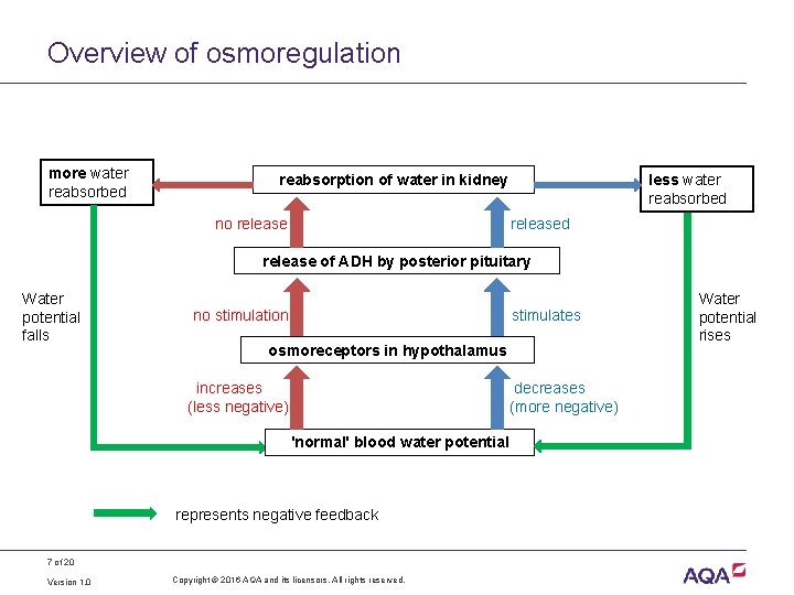
Overview of osmoregulation more water reabsorbed reabsorption of water in kidney no release less water reabsorbed release of ADH by posterior pituitary Water potential falls no stimulation stimulates osmoreceptors in hypothalamus increases (less negative) decreases (more negative) ‘normal’ blood water potential represents negative feedback 7 of 20 Version 1. 0 Copyright © 2016 AQA and its licensors. All rights reserved. Water potential rises
The role of the kidney • The functional units within the kidneys are microscopic tubules, called nephrons. • Each nephron starts as a blind ended Bowman’s capsule supplied by a knot of capillaries, called a glomerulus. The nephron then becomes, in turn; • a coiled region called the proximal convoluted tubule • a hairpin bend called the loop of Henle • a second coiled region called the distal convoluted tubule before joining a collecting duct. 8 of 20 Version 1. 0 Copyright © 2016 AQA and its licensors. All rights reserved.
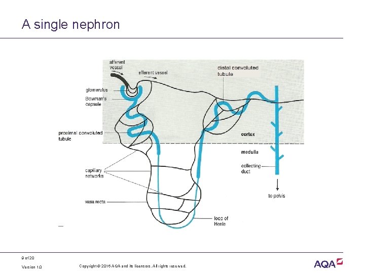
A single nephron 9 of 20 Version 1. 0 Copyright © 2016 AQA and its licensors. All rights reserved.
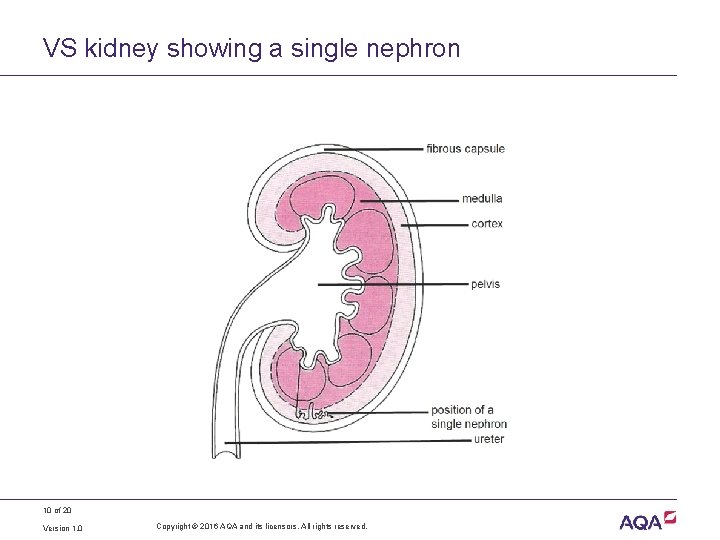
VS kidney showing a single nephron 10 of 20 Version 1. 0 Copyright © 2016 AQA and its licensors. All rights reserved.
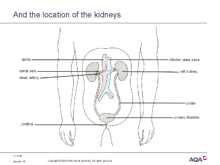
And the location of the kidneys 11 of 20 Version 1. 0 Copyright © 2016 AQA and its licensors. All rights reserved.
Formation of glomerular filtrate Ultrafiltration of blood • The high blood pressure in each glomerulus forces ions, water and other small molecules from the capillaries into the lumen of the surrounding Bowman’s capsule. This is the glomerular filtrate. • The blood pressure is high enough to do this partly because the capillary leaving each glomerulus (efferent) is narrower than that entering (afferent). • Filtration occurs through the microscopic pores between the cells lining each capillary and each capsule and the basement membrane between them. 12 of 20 Version 1. 0 Copyright © 2016 AQA and its licensors. All rights reserved.
Selective reabsorption in the proximal convoluted tubule • Many of the ions, small molecules and water molecules are reabsorbed from the ultrafiltrate back into the capillaries surrounding the proximal convoluted tubule. • Glucose molecules are reabsorbed by active transport. • Water molecules are reabsorbed by osmosis. 13 of 20 Version 1. 0 Copyright © 2016 AQA and its licensors. All rights reserved.
Explain the advantage of the following • The wall of each proximal convoluted tubules is one cell thick. • Each cell in the wall of a proximal convoluted tubule: • has many folds (microvilli) on its surface in contact with the glomerular filtrate • has co-transport proteins in its surface membrane • has cytoplasm packed with mitochondria. 14 of 20 Version 1. 0 Copyright © 2016 AQA and its licensors. All rights reserved.
The role of the loop of Henle • The loop of Henle maintains a high concentration of sodium chloride (Na. Cl) in the tissues of the medulla of the kidney. • As a result, water moves by osmosis from the filtrate in the collecting ducts and is taken up by the vasa recta capillaries. 15 of 20 Version 1. 0 Copyright © 2016 AQA and its licensors. All rights reserved.
The loop of Henle uses a counter-current mechanism The second half of the loop (the ascending limb) actively transports sodium ions (Na+) and chloride ions (Cl–) from the filtrate into the medulla. However: • its wall is impermeable to water • the first half of the loop (the descending limb) is permeable to Na+ and Cl– and so takes up these ions • as a result, a high concentration of Na+ and Cl– ions develops in the medulla as the filtrate moves down the loop of Henle. 16 of 20 Version 1. 0 Copyright © 2016 AQA and its licensors. All rights reserved.
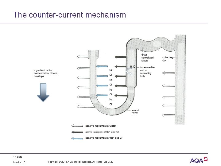
The counter-current mechanism 17 of 20 Version 1. 0 Copyright © 2016 AQA and its licensors. All rights reserved.
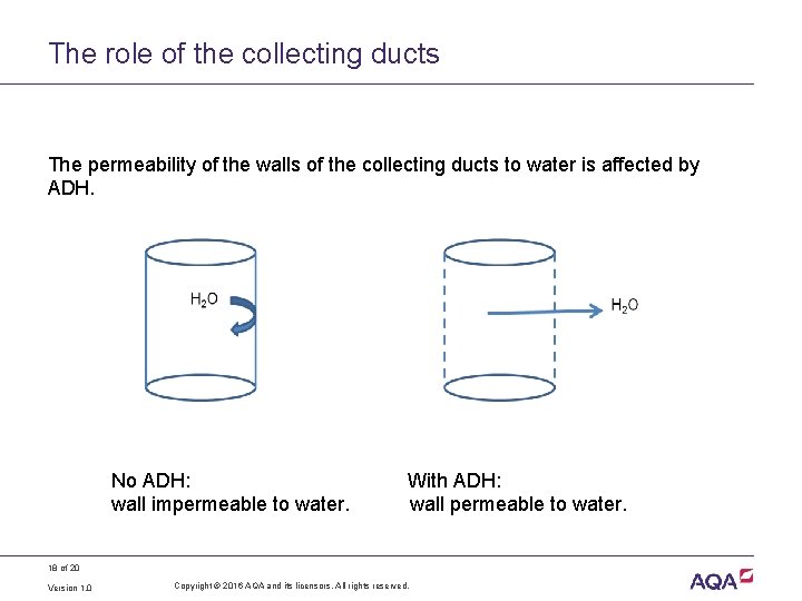
The role of the collecting ducts The permeability of the walls of the collecting ducts to water is affected by ADH. No ADH: wall impermeable to water. With ADH: wall permeable to water. 18 of 20 Version 1. 0 Copyright © 2016 AQA and its licensors. All rights reserved.
Reabsorption of water • In the presence of ADH, the walls of the collecting duct are permeable to water. • Water molecules move by osmosis from the filtrate in the collecting ducts into the tissue of the medulla. • The vasa recta capillaries in the medulla absorb the water back into the circulation. • The volume of the filtrate (and therefore of the urine) becomes less and its concentration increases. 19 of 20 Version 1. 0 Copyright © 2016 AQA and its licensors. All rights reserved.
Acknowledgements Diagrams on slides 9, 10, 11 and 17 Rowland, M. , 1992, Biology, Thomas Nelson & Sons Ltd 20 of 20 Version 1. 0 Copyright © 2016 AQA and its licensors. All rights reserved.
- Slides: 20