
Alberts Bray Hopkin Johnson Lewis Raff Roberts Walter
Alberts • Bray • Hopkin • Johnson • Lewis • Raff • Roberts • Walter Essential Cell Biology FOURTH EDITION Chapter 16 Cell Signaling Copyright © Garland Science 2014
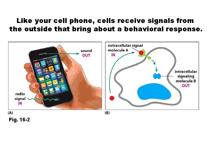
Like your cell phone, cells receive signals from the outside that bring about a behavioral response. Fig. 16 -2
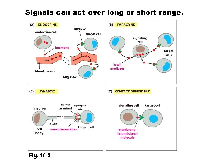
Signals can act over long or short range. Fig. 16 -3
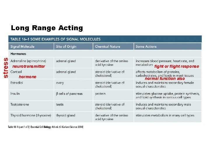
stress Long Range Acting neurotransmitter hormone fight or flight response normal function also
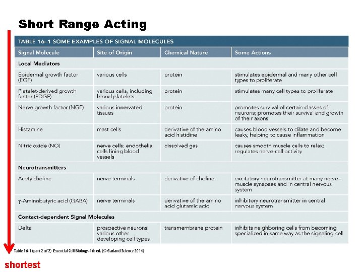
Short Range Acting shortest
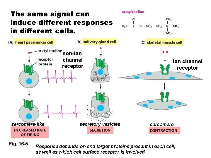
The same signal can induce different responses in different cells. non-ion channel receptor sarcomere-like Fig. 16 -5 secretory vesicles ion channel receptor sarcomere Response depends on end target proteins present in each cell, as well as which cell surface receptor is involved.
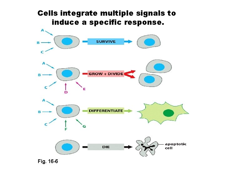
Cells integrate multiple signals to induce a specific response. Fig. 16 -6
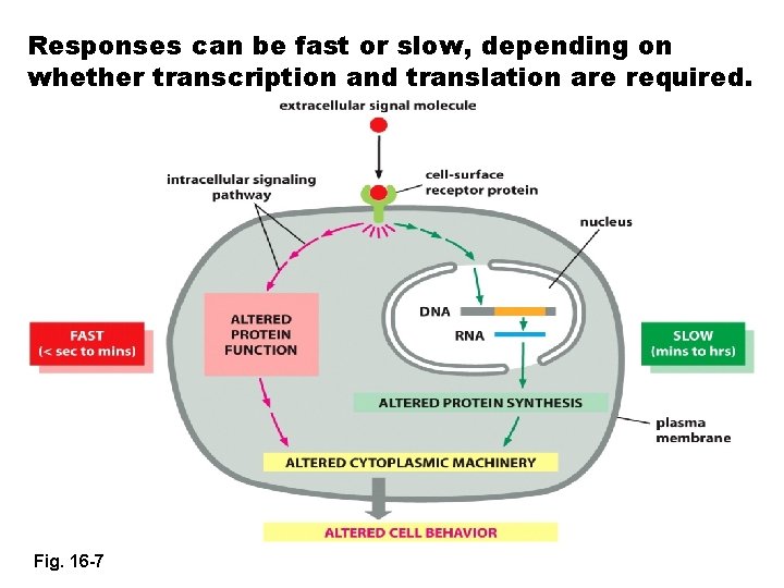
Responses can be fast or slow, depending on whether transcription and translation are required. Fig. 16 -7
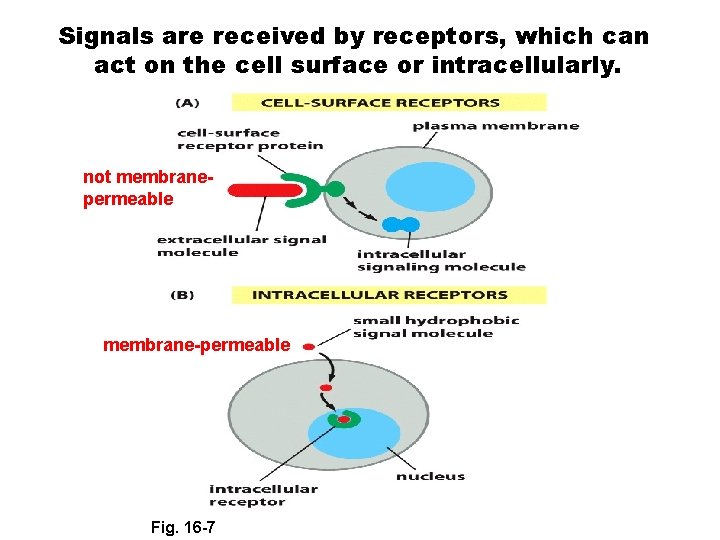
Signals are received by receptors, which can act on the cell surface or intracellularly. not membranepermeable membrane-permeable Fig. 16 -7
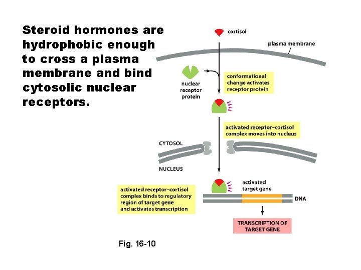
Steroid hormones are hydrophobic enough to cross a plasma membrane and bind cytosolic nuclear receptors. Fig. 16 -10
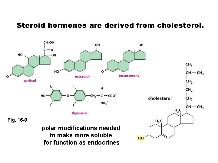
Steroid hormones are derived from cholesterol. Fig. 16 -9 polar modifications needed to make more soluble for function as endocrines
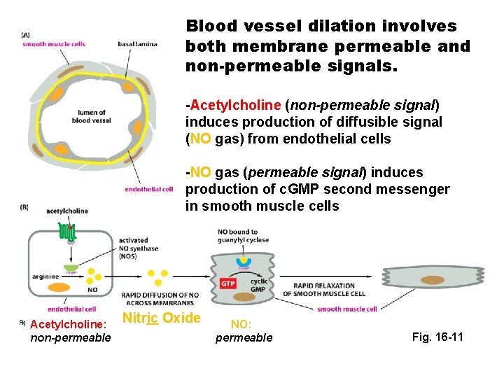
Blood vessel dilation involves both membrane permeable and non-permeable signals. -Acetylcholine (non-permeable signal) induces production of diffusible signal (NO gas) from endothelial cells -NO gas (permeable signal) induces production of c. GMP second messenger in smooth muscle cells Acetylcholine: non-permeable Nitric Oxide NO: permeable Fig. 16 -11
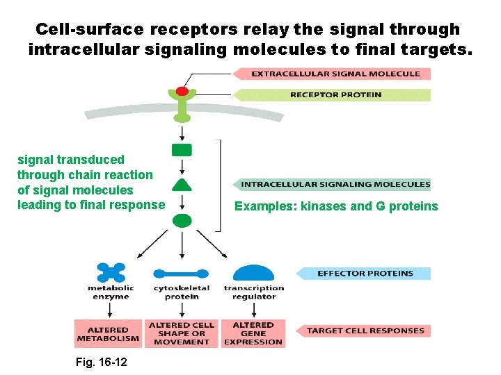
Cell-surface receptors relay the signal through intracellular signaling molecules to final targets. signal transduced through chain reaction of signal molecules leading to final response Fig. 16 -12 Examples: kinases and G proteins
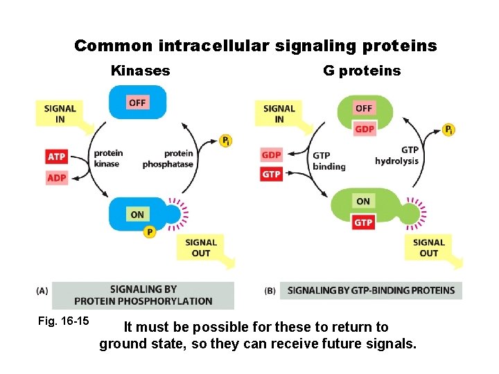
Common intracellular signaling proteins Kinases Fig. 16 -15 G proteins It must be possible for these to return to ground state, so they can receive future signals.
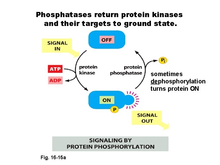
Phosphatases return protein kinases and their targets to ground state. sometimes dephosphorylation turns protein ON Fig. 16 -15 a
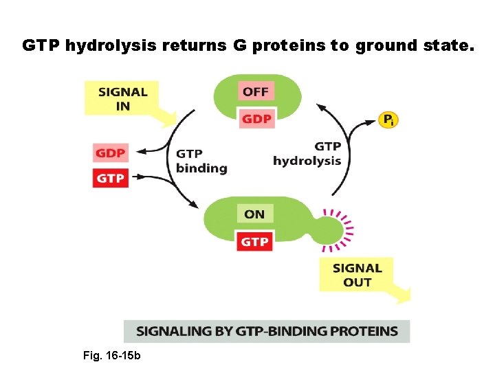
GTP hydrolysis returns G proteins to ground state. Fig. 16 -15 b 16 -15 a Fig.
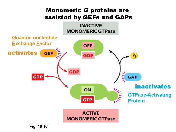
Monomeric G proteins are assisted by GEFs and GAPs Guanine nucleotide Exchange Factor activates inactivates GTPase-Activating Protein Fig. 16 -16
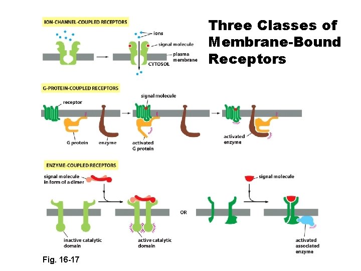
Three Classes of Membrane-Bound Receptors Fig. 16 -17
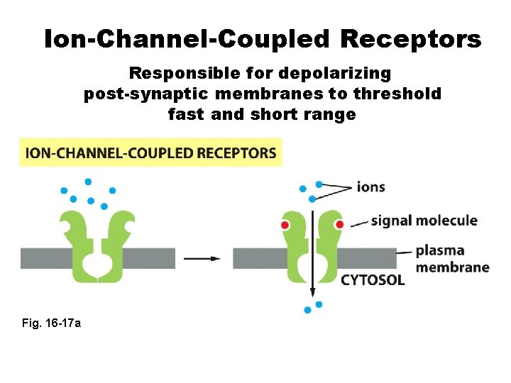
Ion-Channel-Coupled Receptors Responsible for depolarizing post-synaptic membranes to threshold fast and short range Fig. 16 -17 a
Example: Acetylcholine Receptor in skeletal muscle (Ch 4 and 17)
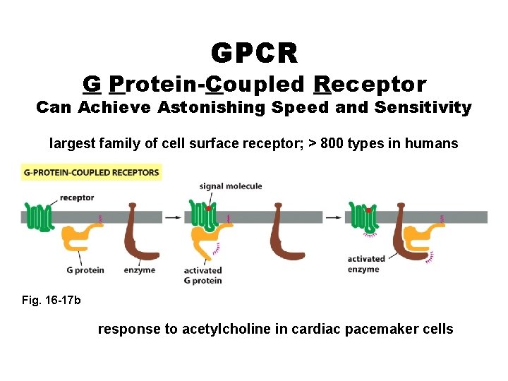
GPCR G Protein-Coupled Receptor Can Achieve Astonishing Speed and Sensitivity largest family of cell surface receptor; > 800 types in humans Fig. 16 -17 b response to acetylcholine in cardiac pacemaker cells
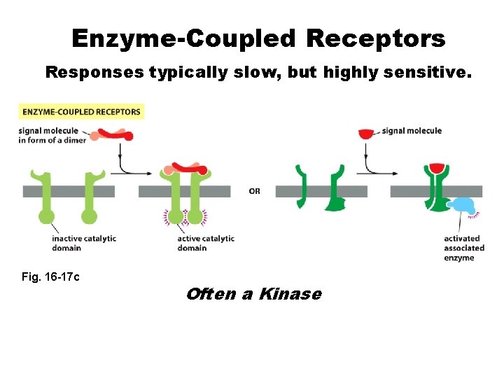
Enzyme-Coupled Receptors Responses typically slow, but highly sensitive. Fig. 16 -17 c Often a Kinase
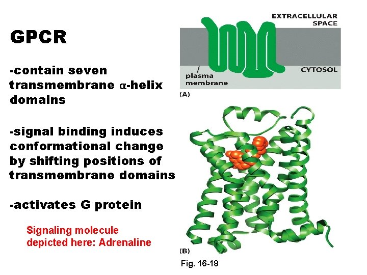
GPCR -contain seven transmembrane α-helix domains -signal binding induces conformational change by shifting positions of transmembrane domains -activates G protein Signaling molecule depicted here: Adrenaline Fig. 16 -18
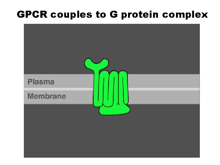
GPCR couples to G protein complex
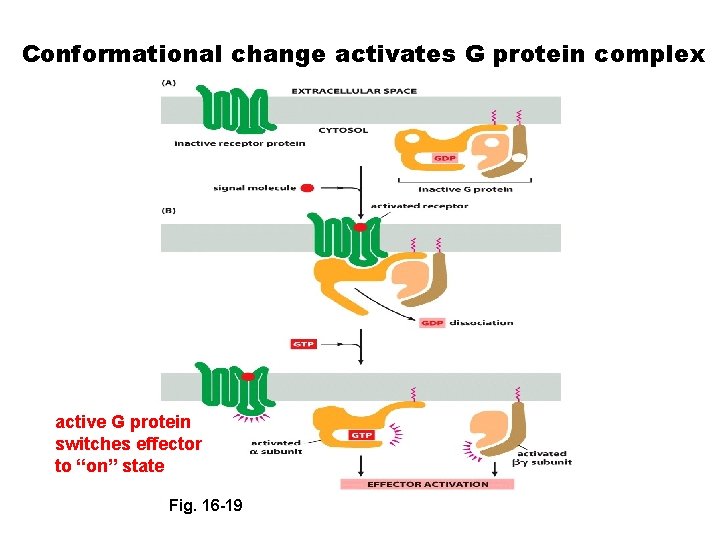
Conformational change activates G protein complex active G protein switches effector to “on” state Fig. 16 -19
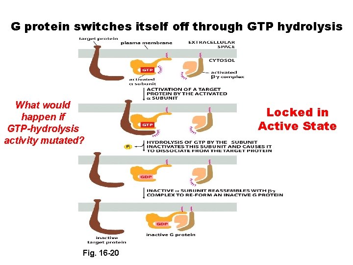
G protein switches itself off through GTP hydrolysis What would happen if GTP-hydrolysis activity mutated? Fig. 16 -20 Locked in Active State
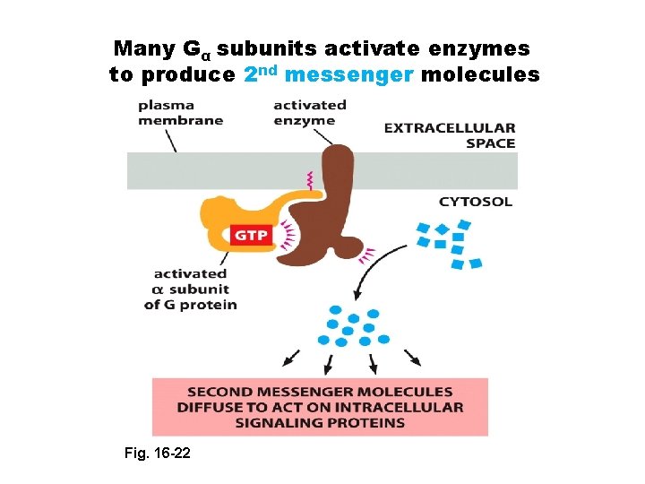
Many Gα subunits activate enzymes to produce 2 nd messenger molecules Fig. 16 -22
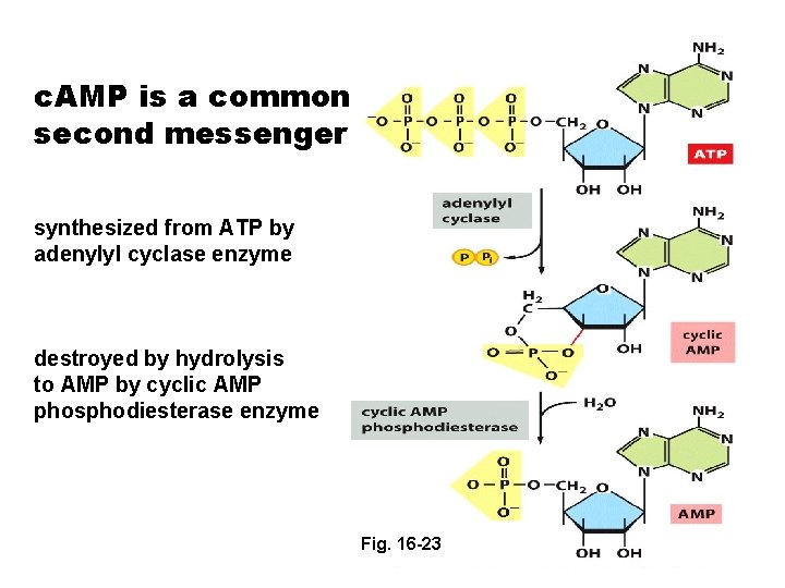
c. AMP is a common second messenger synthesized from ATP by adenylyl cyclase enzyme destroyed by hydrolysis to AMP by cyclic AMP phosphodiesterase enzyme Fig. 16 -23
G protein complexes can activate c. AMP production by Adenylyl Cyclase
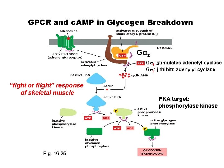
GPCR and c. AMP in Glycogen Breakdown Gαs: stimulates adenelyl cyclase Gαi: inhibits adenylyl cyclase “fight or flight” response of skeletal muscle Fig. 16 -25 PKA target: phosphorylase kinase
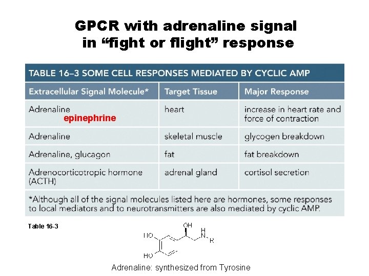
GPCR with adrenaline signal in “fight or flight” response epinephrine Table 16 -3 Adrenaline: synthesized from Tyrosine
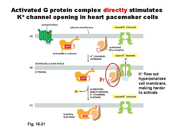
Activated G protein complex directly stimulates K+ channel opening in heart pacemaker cells βγ Fig. 16 -21 K+ flow out hyperpolarizes cell membrane, making harder to activate
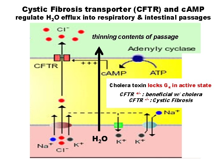
Cystic Fibrosis transporter (CFTR) and c. AMP regulate H 2 O efflux into respiratory & intestinal passages thinning contents of passage Cholera toxin locks Gα in active state CFTR +/- : beneficial w/ cholera CFTR -/- : Cystic Fibrosis H 2 O
Special Seminar Ronald Breaker, Ph. D. , Henry Ford II Professor of Molecular, Cellular, and Developmental Biology and Professor of Molecular Biophysics and Biochemistry, HHMI, Yale University http: //www. hhmi. org/scientists/ronald-r-breaker Prospects for Bacterial Noncoding RNA Discovery Date: Time: Place: MONDAY, April 25, 2016 4: 00 PM (refreshments at 3: 45 PM) 116 TH Morgan Biology Building Dr. Breaker discovered riboswitches and continues mechanistic and gene discovery studies on RNA function in gene regulation and catalysis. Dr. Breaker is an HHMI investigator and member of the National Academy of Sciences
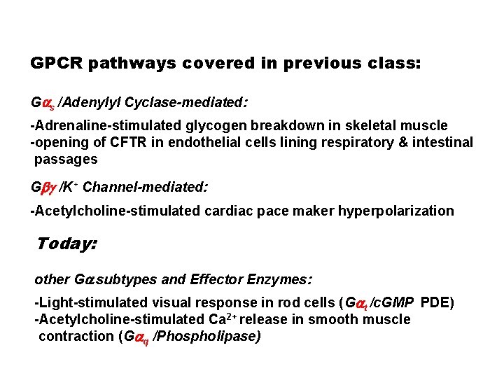
GPCR pathways covered in previous class: Gas /Adenylyl Cyclase-mediated: -Adrenaline-stimulated glycogen breakdown in skeletal muscle -opening of CFTR in endothelial cells lining respiratory & intestinal passages Gbg /K+ Channel-mediated: -Acetylcholine-stimulated cardiac pace maker hyperpolarization Today: other Ga subtypes and Effector Enzymes: -Light-stimulated visual response in rod cells (Gat /c. GMP PDE) -Acetylcholine-stimulated Ca 2+ release in smooth muscle contraction (Gaq /Phospholipase)
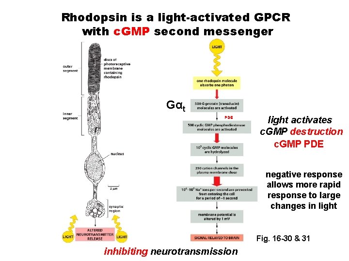
Rhodopsin is a light-activated GPCR with c. GMP second messenger Gαt PDE light activates c. GMP destruction c. GMP PDE negative response allows more rapid response to large changes in light Fig. 16 -30 & 31 inhibiting neurotransmission
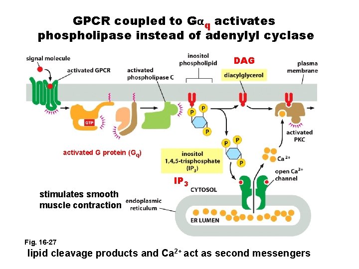
GPCR coupled to Gαq activates phospholipase instead of adenylyl cyclase DAG activated G protein (Gq) IP 3 stimulates smooth muscle contraction Fig. 16 -27 lipid cleavage products and Ca 2+ act as second messengers
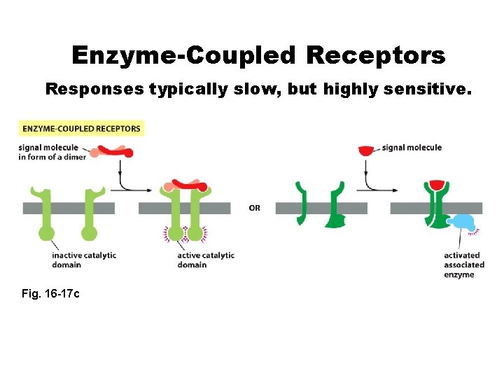
Enzyme-Coupled Receptors Responses typically slow, but highly sensitive. Fig. 16 -17 c
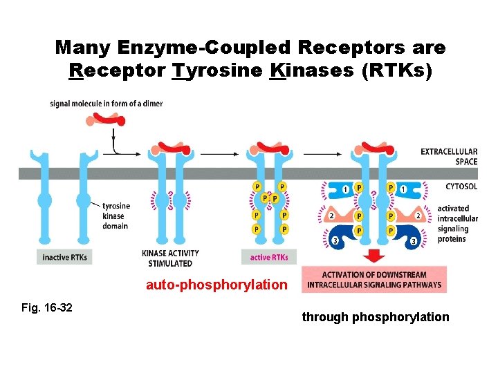
Many Enzyme-Coupled Receptors are Receptor Tyrosine Kinases (RTKs) auto-phosphorylation Fig. 16 -32 through phosphorylation
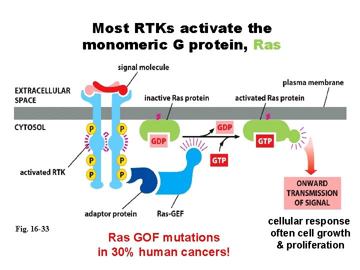
Most RTKs activate the monomeric G protein, Ras Fig. 16 -33 Ras GOF mutations in 30% human cancers! cellular response often cell growth & proliferation
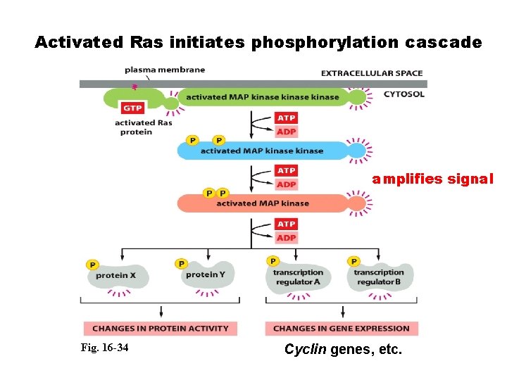
Activated Ras initiates phosphorylation cascade amplifies signal Fig. 16 -34 Cyclin genes, etc.
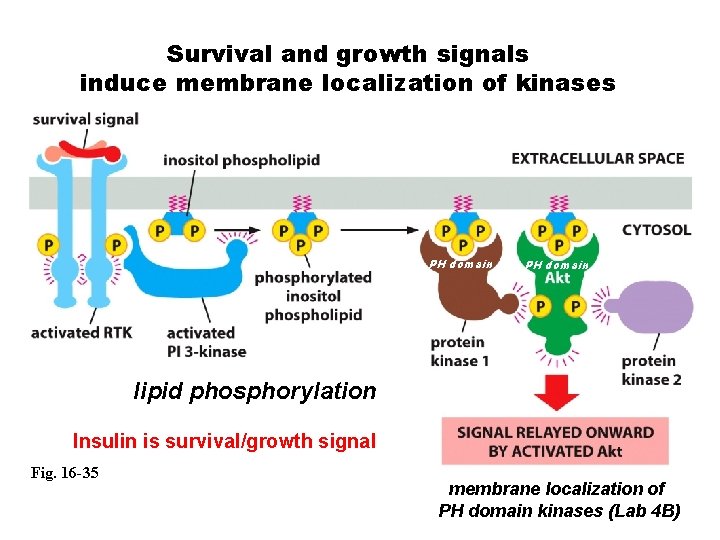
Survival and growth signals induce membrane localization of kinases PH domain lipid phosphorylation Insulin is survival/growth signal Fig. 16 -35 membrane localization of PH domain kinases (Lab 4 B)
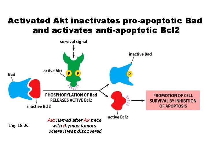
Activated Akt inactivates pro-apoptotic Bad and activates anti-apoptotic Bcl 2 Fig. 16 -36 Akt named after Ak mice with thymus tumors where it was discovered
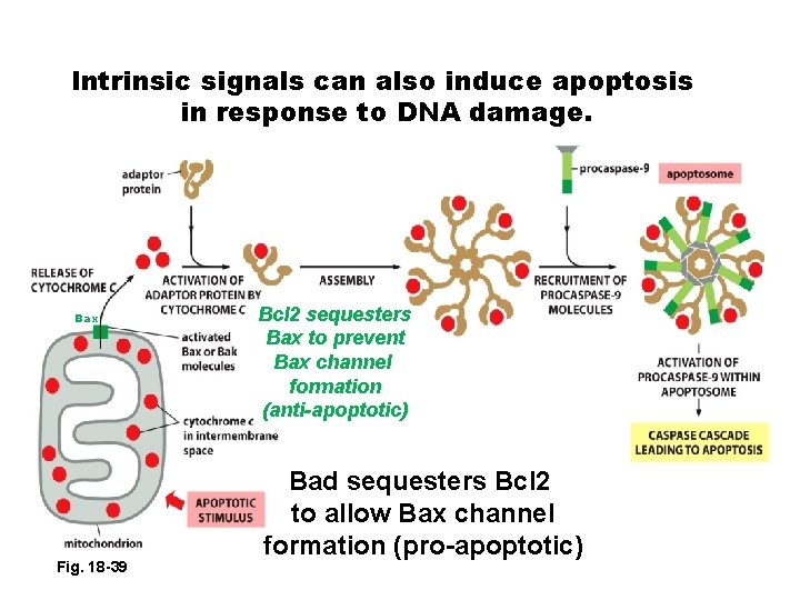
Intrinsic signals can also induce apoptosis in response to DNA damage. Bax Fig. 18 -39 Bcl 2 sequesters Bax to prevent Bax channel formation (anti-apoptotic) Bad sequesters Bcl 2 to allow Bax channel formation (pro-apoptotic)
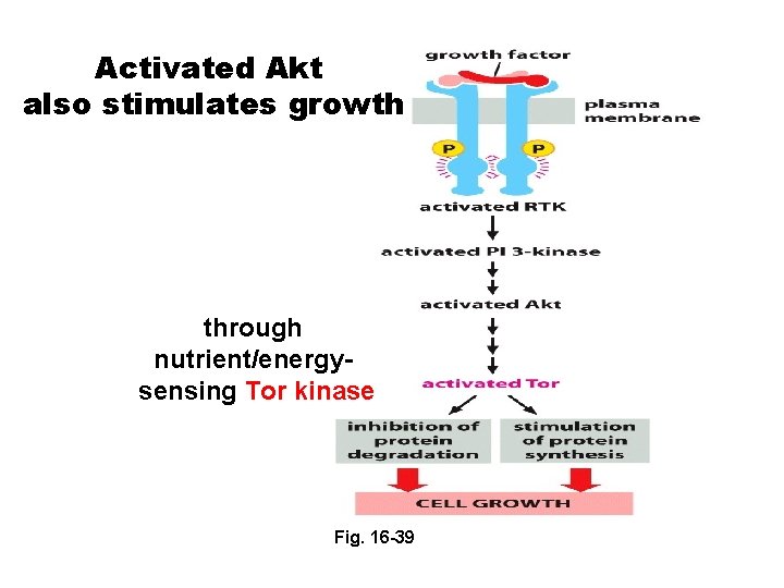
Activated Akt also stimulates growth through nutrient/energysensing Tor kinase Fig. 16 -39
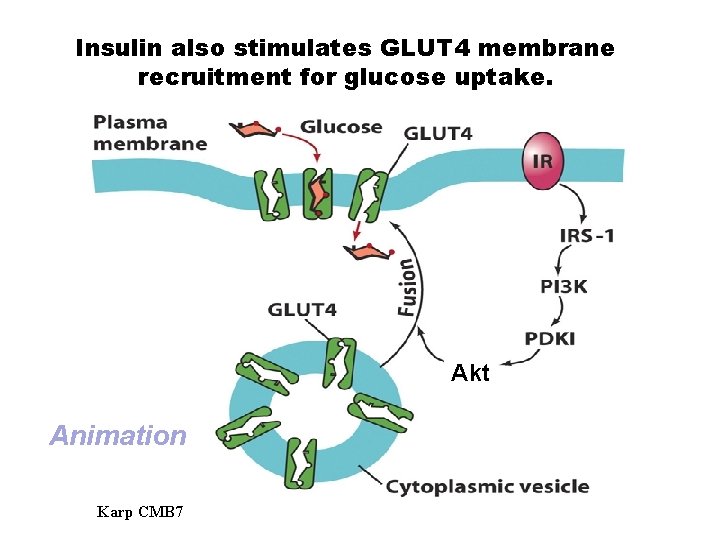
Insulin also stimulates GLUT 4 membrane recruitment for glucose uptake. Akt Animation Karp CMB 7
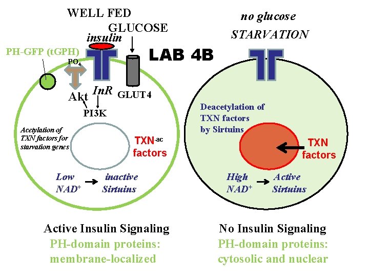
WELL FED GLUCOSE insulin LAB 4 B PH-GFP (t. GPH) PO 4 Akt In. R GLUT 4 PI 3 K Acetylation of TXN factors for starvation genes Low NAD+ no glucose STARVATION Deacetylation of TXN factors by Sirtuins TXN-ac factors inactive Sirtuins Active Insulin Signaling PH-domain proteins: membrane-localized TXN factors High NAD+ Active Sirtuins No Insulin Signaling PH-domain proteins: cytosolic and nuclear
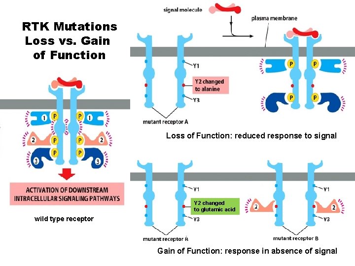
RTK Mutations Loss vs. Gain of Function Loss of Function: reduced response to signal Y 2 changed to glutamic acid wild type receptor Gain of Function: response in absence of signal
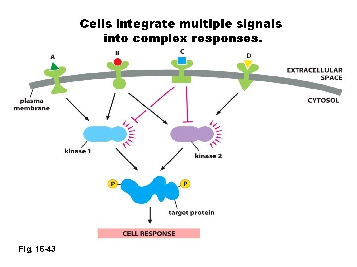
Cells integrate multiple signals into complex responses. Fig. 16 -43
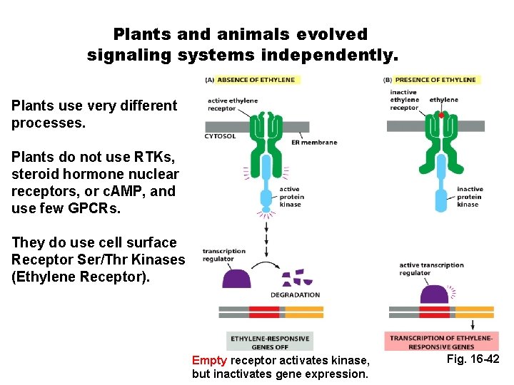
Plants and animals evolved signaling systems independently. Plants use very different processes. Plants do not use RTKs, steroid hormone nuclear receptors, or c. AMP, and use few GPCRs. They do use cell surface Receptor Ser/Thr Kinases (Ethylene Receptor). Empty receptor activates kinase, but inactivates gene expression. Fig. 16 -42
- Slides: 50