
Aktin cytoskeleton Seminar PCDU WS 1516 Vera Krieger
- Slides: 37
Aktin cytoskeleton Seminar PCDU WS 15/16 Vera Krieger - 28. 10. 15
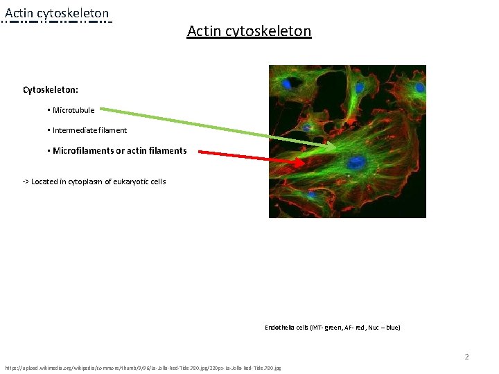
Actin cytoskeleton Cytoskeleton: • Microtubule • Intermediate filament • Microfilaments or actin filaments -> Located in cytoplasm of eukaryotic cells Endothelia cells (MT- green, AF- red, Nuc – blue) 2 https: //upload. wikimedia. org/wikipedia/commons/thumb/9/96/La-Jolla-Red-Tide. 780. jpg/220 px-La-Jolla-Red-Tide. 780. jpg
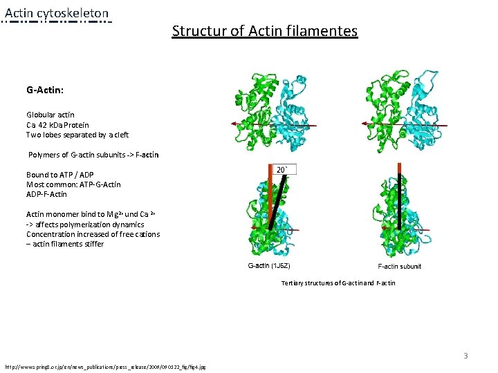
Actin cytoskeleton Structur of Actin filamentes G-Actin: Globular actin Ca. 42 k. Da Protein Two lobes separated by a cleft Polymers of G-actin subunits -> F-actin Bound to ATP / ADP Most common: ATP-G-Actin ADP-F-Actin monomer bind to Mg 2+ und Ca 2+ -> affects polymerization dynamics Concentration increased of free cations – actin filaments stiffer Tertiary structures of G-actin and F-actin 3 http: //www. spring 8. or. jp/en/news_publications/press_release/2009/090122_fig/fig 4. jpg
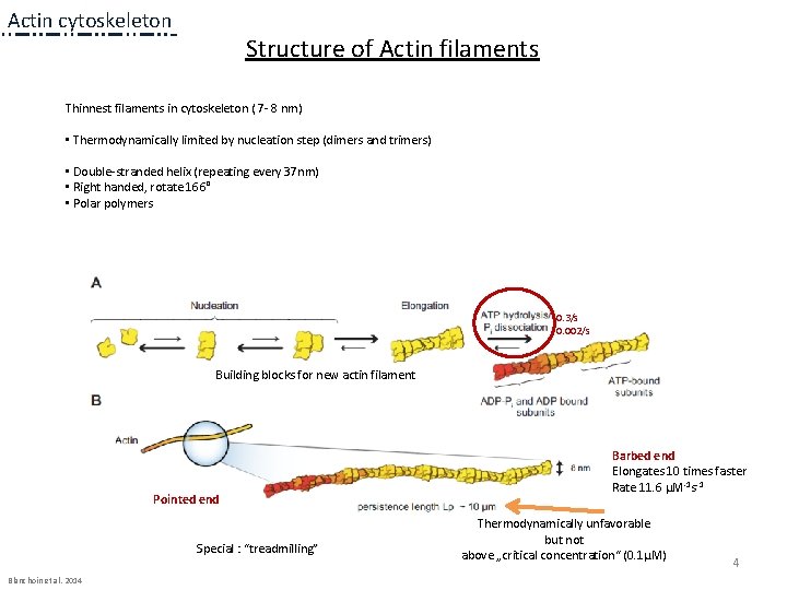
Actin cytoskeleton Structure of Actin filaments Thinnest filaments in cytoskeleton ( 7 - 8 nm) • Thermodynamically limited by nucleation step (dimers and trimers) • Double-stranded helix (repeating every 37 nm) • Right handed, rotate 166° • Polar polymers 0. 3/s 0. 002/s Building blocks for new actin filament Pointed end Special : “treadmilling” Blanchoin et al. 2014 Barbed end Elongates 10 times faster Rate 11. 6 µM-1 s-1 Thermodynamically unfavorable but not above „critical concentration“ (0. 1µM) 4
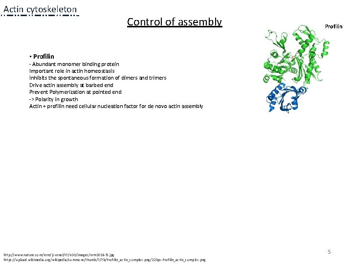
Actin cytoskeleton Control of assembly Profilin • Profilin - Abundant monomer binding protein Important role in actin homeostasis Inhibits the spontaneous formation of dimers and trimers Drive actin assembly at barbed end Prevent Polymerization at pointed end -> Polarity in growth Actin + profilin need cellular nucleation factor for de novo actin assembly http: //www. nature. com/nrm/journal/v 7/n 10/images/nrm 2026 -f 1. jpg https: //upload. wikimedia. org/wikipedia/commons/thumb/7/7 b/Profilin_actin_complex. png/223 px-Profilin_actin_complex. png 5
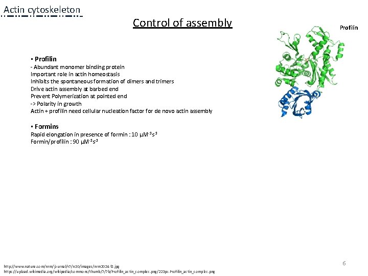
Actin cytoskeleton Control of assembly Profilin • Profilin - Abundant monomer binding protein Important role in actin homeostasis Inhibits the spontaneous formation of dimers and trimers Drive actin assembly at barbed end Prevent Polymerization at pointed end -> Polarity in growth Actin + profilin need cellular nucleation factor for de novo actin assembly • Formins Rapid elongation in presence of formin : 10 µM-1 s-1 Formin/profilin : 90 µM-1 s-1 http: //www. nature. com/nrm/journal/v 7/n 10/images/nrm 2026 -f 1. jpg https: //upload. wikimedia. org/wikipedia/commons/thumb/7/7 b/Profilin_actin_complex. png/223 px-Profilin_actin_complex. png 6
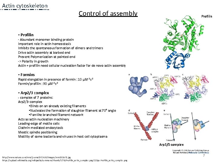
Actin cytoskeleton Control of assembly Profilin • Profilin - Abundant monomer binding protein Important role in actin homeostasis Inhibits the spontaneous formation of dimers and trimers Drive actin assembly at barbed end Prevent Polymerization at pointed end -> Polarity in growth Actin + profilin need cellular nucleation factor for de novo actin assembly • Formins Rapid elongation in presence of formin : 10 µM-1 s-1 Formin/profilin : 90 µM-1 s-1 • Arp 2/3 complex - complex of 7 proteins: Arp 2/3 -complex • Binds on an already existing filaments • Nucleates the formation of daughter filament at 70° angle • Fan-like branched filament network Acts as actin nucleation machinery Leading-edge of motile cells Clathrin mediated endocytosis Meotic spindle positioning Motility of some bacteria and viruses in host cell cytoplasma Arp 2/3 complex http: //www. nature. com/nrm/journal/v 7/n 10/images/nrm 2026 -f 1. jpg https: //upload. wikimedia. org/wikipedia/commons/thumb/7/7 b/Profilin_actin_complex. png/223 px-Profilin_actin_complex. png 7
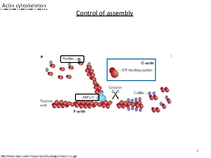
Actin cytoskeleton Control of assembly 8
Actin cytoskeleton Polymerization and association with actin regulatory proteins Variety of architectures 9
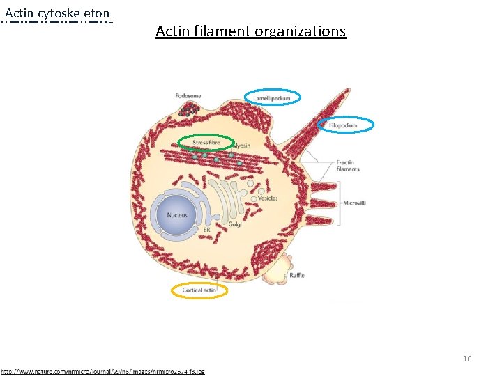
Actin cytoskeleton Actin filament organizations 10
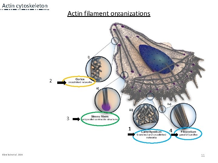
Actin cytoskeleton Actin filament organizations 2 3 1 1 a Blanchoin et al. 2014 4 11
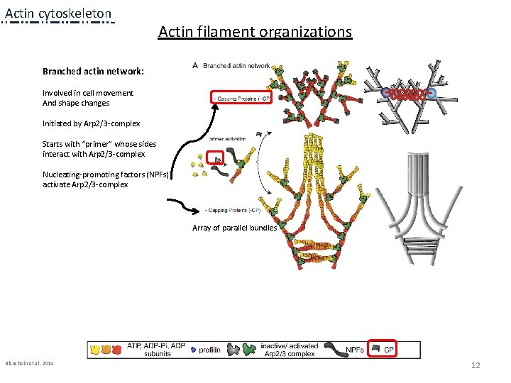
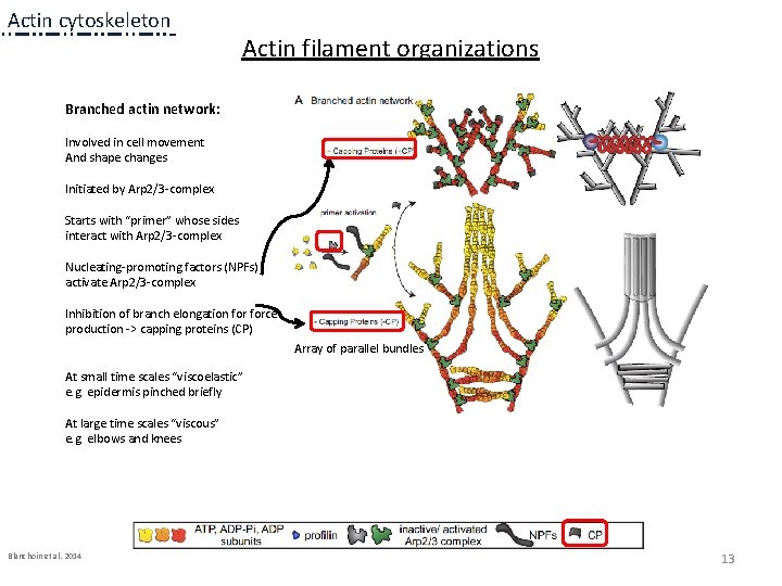
Actin cytoskeleton Actin filament organizations Branched actin network: Involved in cell movement And shape changes Initiated by Arp 2/3 -complex Starts with “primer” whose sides interact with Arp 2/3 -complex Nucleating-promoting factors (NPFs) activate Arp 2/3 -complex Array of parallel bundles 1 a Blanchoin et al. 2014 1 a 12
Actin cytoskeleton Actin filament organizations Branched actin network: Involved in cell movement And shape changes Initiated by Arp 2/3 -complex Starts with “primer” whose sides interact with Arp 2/3 -complex Nucleating-promoting factors (NPFs) activate Arp 2/3 -complex Inhibition of branch elongation force production -> capping proteins (CP) Array of parallel bundles At small time scales “viscoelastic” e. g. epidermis pinched briefly At large time scales “viscous” e. g. elbows and knees Blanchoin et al. 2014 1 a 1 a 13
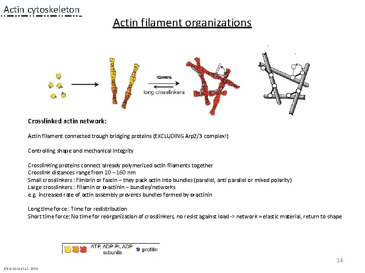
Actin cytoskeleton Actin filament organizations 1 a 1 a 1 a Crosslinked actin network: Actin filament connected trough bridging proteins (EXCLUDING Arp 2/3 complex!) Controlling shape and mechanical integrity Crosslinking proteins connect already polymerized actin filaments together Crosslink distances range from 10 – 160 nm Small crosslinkers : Fimbrin or fascin – they pack actin into bundles (parallel, anti parallel or mixed polarity) Large crosslinkers. : Filamin or α-actinin – bundles/networks e. g. increased rate of actin assembly prevents bundles formed by α-actinin Long time force : Time for redistribution Short time force: No time for reorganization of crosslinkers, no resist against load -> network = elastic material, return to shape 14 Blanchoin et al. 2014
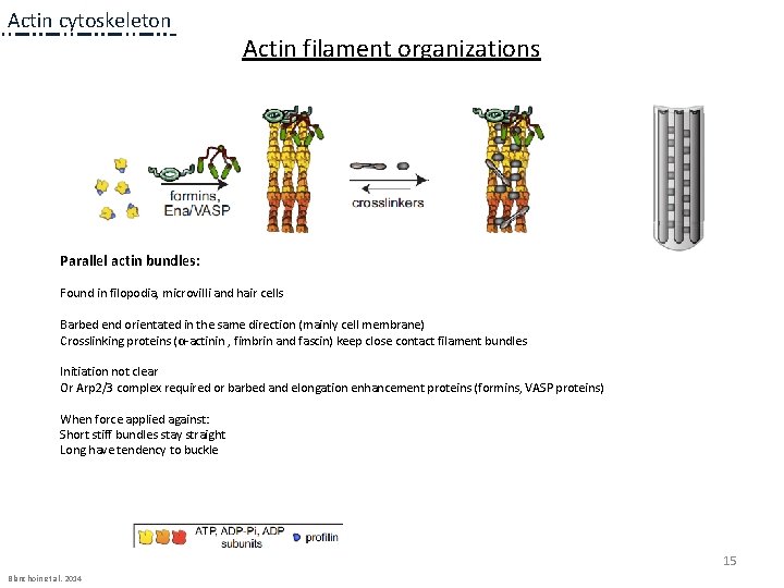
Actin cytoskeleton Actin filament organizations 1 a 1 a 1 a Parallel actin bundles: Found in filopodia, microvilli and hair cells Barbed end orientated in the same direction (mainly cell membrane) Crosslinking proteins (α-actinin , fimbrin and fascin) keep close contact filament bundles Initiation not clear Or Arp 2/3 complex required or barbed and elongation enhancement proteins (formins, VASP proteins) When force applied against: Short stiff bundles stay straight Long have tendency to buckle 15 Blanchoin et al. 2014
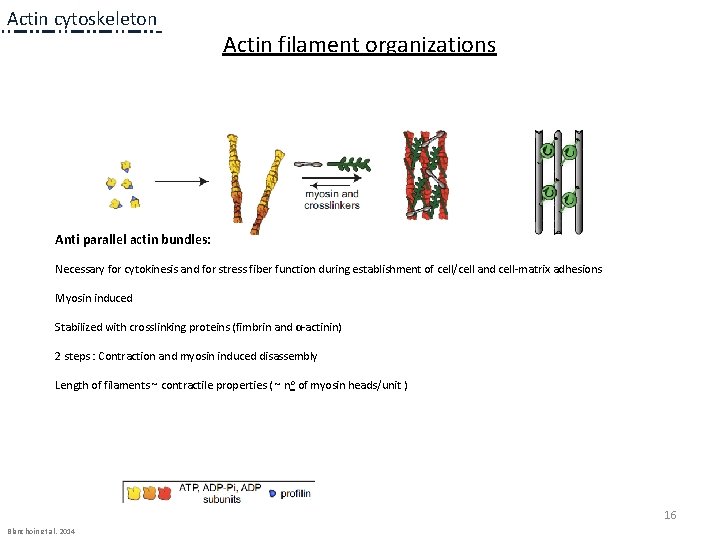
Actin cytoskeleton Actin filament organizations 1 a 1 a 1 a Anti parallel actin bundles: Necessary for cytokinesis and for stress fiber function during establishment of cell/cell and cell-matrix adhesions Myosin induced Stabilized with crosslinking proteins (fimbrin and α-actinin) 2 steps : Contraction and myosin induced disassembly Length of filaments ~ contractile properties ( ~ no of myosin heads/unit ) 16 Blanchoin et al. 2014
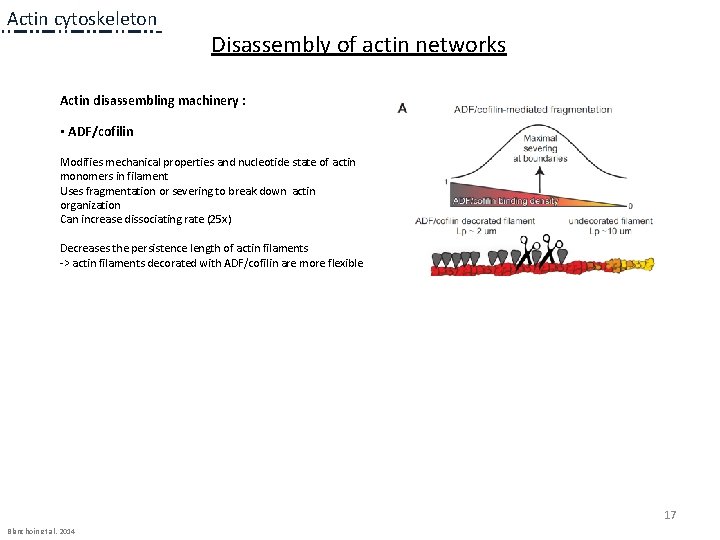
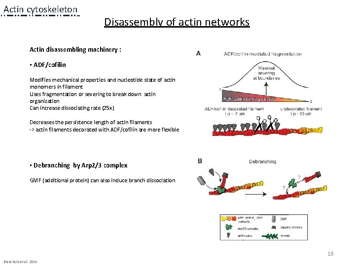
Actin cytoskeleton Disassembly of actin networks Actin disassembling machinery : • ADF/cofilin Modifies mechanical properties and nucleotide state of actin monomers in filament Uses fragmentation or severing to break down actin organization Can increase dissociating rate (25 x) Decreases the persistence length of actin filaments -> actin filaments decorated with ADF/cofilin are more flexible 1 a 17 Blanchoin et al. 2014
Actin cytoskeleton Disassembly of actin networks Actin disassembling machinery : • ADF/cofilin Modifies mechanical properties and nucleotide state of actin monomers in filament Uses fragmentation or severing to break down actin organization Can increase dissociating rate (25 x) Decreases the persistence length of actin filaments -> actin filaments decorated with ADF/cofilin are more flexible • Debranching by Arp 2/3 complex GMF (additional protein) can also induce branch dissociation 1 a 18 Blanchoin et al. 2014
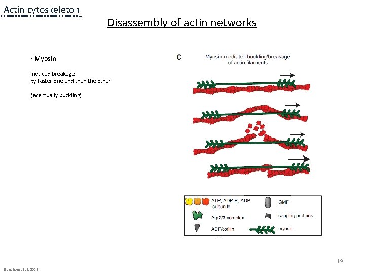
Actin cytoskeleton Disassembly of actin networks • Myosin Induced breakage by faster one end than the other (eventually buckling) 1 a 1 a 19 Blanchoin et al. 2014
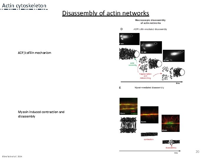
Actin cytoskeleton Disassembly of actin networks ADF/cofilin mechanism Myosin induced contraction and disassembly 1 a 1 a 20 Blanchoin et al. 2014
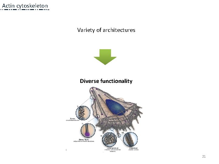
Actin cytoskeleton Variety of architectures 1 a Diverse functionality 1 a 21
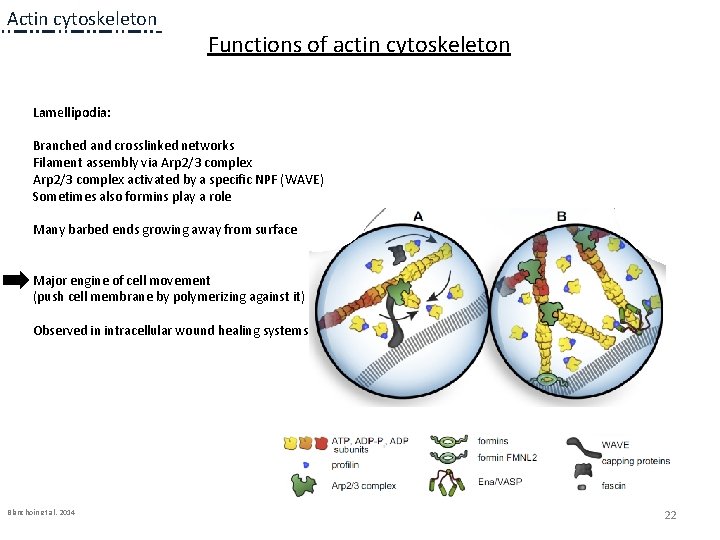
Actin cytoskeleton Functions of actin cytoskeleton Lamellipodia: Branched and crosslinked networks Filament assembly via Arp 2/3 complex activated by a specific NPF (WAVE) Sometimes also formins play a role Many barbed ends growing away from surface Major engine of cell movement (push cell membrane by polymerizing against it) Observed in intracellular wound healing systems Blanchoin et al. 2014 22
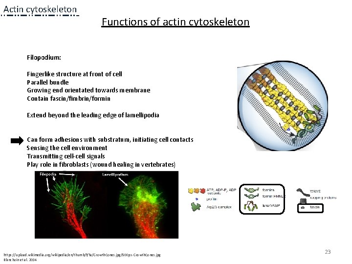
Actin cytoskeleton Functions of actin cytoskeleton Filopodium: Fingerlike structure at front of cell Parallel bundle Growing end orientated towards membrane Contain fascin/fimbrin/formin Extend beyond the leading edge of lamellipodia Can form adhesions with substratum, initiating cell contacts Sensing the cell environment Transmitting cell-cell signals Play role in fibroblasts (wound healing in vertebrates) https: //upload. wikimedia. org/wikipedia/en/thumb/f/fa/Growth. Cones. jpg/500 px-Growth. Cones. jpg Blanchoin et al. 2014 23
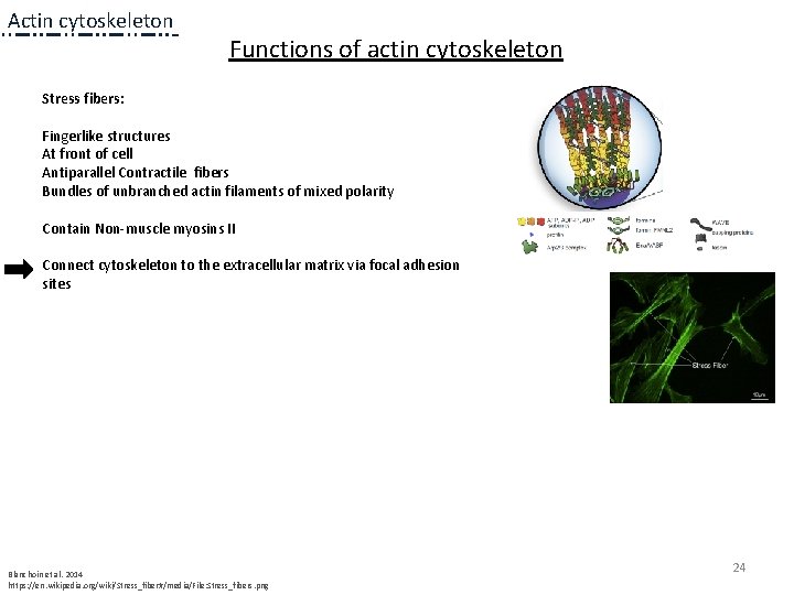
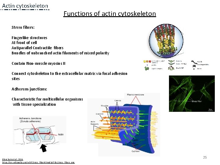
Actin cytoskeleton Functions of actin cytoskeleton Stress fibers: Fingerlike structures At front of cell Antiparallel Contractile fibers Bundles of unbranched actin filaments of mixed polarity Contain Non-muscle myosins II Connect cytoskeleton to the extracellular matrix via focal adhesion sites Blanchoin et al. 2014 https: //en. wikipedia. org/wiki/Stress_fiber#/media/File: Stress_fibers. png 24
Actin cytoskeleton Functions of actin cytoskeleton Stress fibers: Fingerlike structures At front of cell Antiparallel Contractile fibers Bundles of unbranched actin filaments of mixed polarity Contain Non-muscle myosins II Connect cytoskeleton to the extracellular matrix via focal adhesion sites Adherens junctions: Characteristic for multicellular organisms with tissue specialization Blanchoin et al. 2014 https: //en. wikipedia. org/wiki/Stress_fiber#/media/File: Stress_fibers. png 25
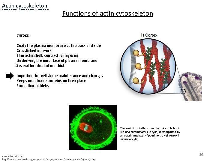
Actin cytoskeleton Functions of actin cytoskeleton Cortex: Coats the plasma membrane at the back and side Crosslinked network Thin actin shell, contractile (myosin) Underlying the inner face of plasma membrane Several hundred of nm thick Important for cell shape maintenance and changes Keeps membrane proteins on their place Formation of blebs The meiotic spindle (shown by microtubules in red and chromosomes in cyan) is transported by an F-actin meshwork (green) to the cell cortex in mouse oocytes. Blanchoin et al. 2014 http: //www. actindynamics. org/cms/uploads/images/members/Ellenberg-Lenart/Figure 2_1. jpg 26
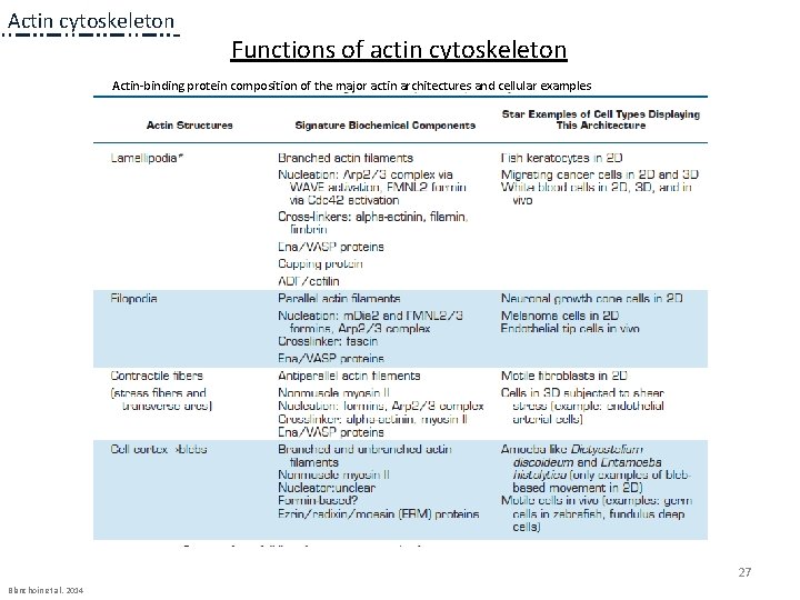
Actin cytoskeleton Functions of actin cytoskeleton Actin-binding protein composition of the major actin architectures and cellular examples 27 Blanchoin et al. 2014
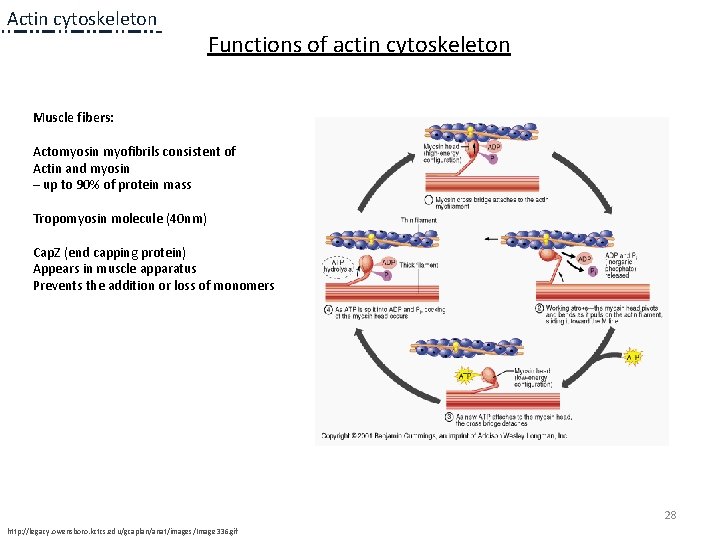
Actin cytoskeleton Functions of actin cytoskeleton Muscle fibers: Actomyosin myofibrils consistent of Actin and myosin – up to 90% of protein mass Tropomyosin molecule (40 nm) Cap. Z (end capping protein) Appears in muscle apparatus Prevents the addition or loss of monomers 28 http: //legacy. owensboro. kctcs. edu/gcaplan/anat/images/Image 336. gif
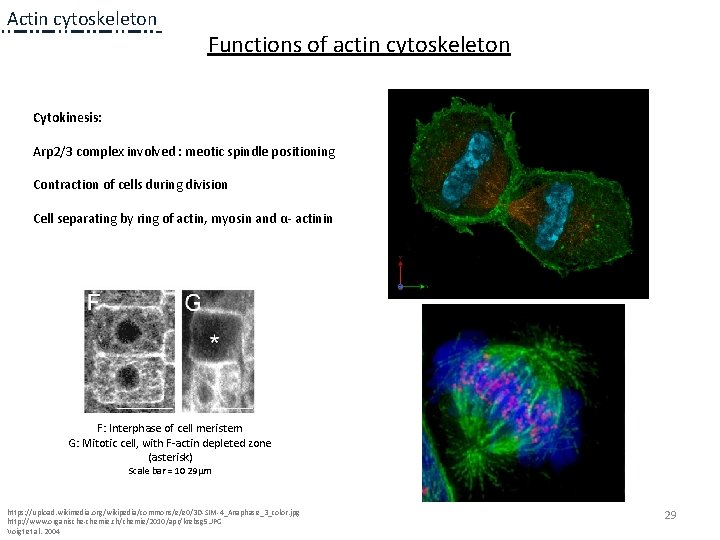
Actin cytoskeleton Functions of actin cytoskeleton Cytokinesis: Arp 2/3 complex involved : meotic spindle positioning Contraction of cells during division Cell separating by ring of actin, myosin and α- actinin F: Interphase of cell meristem G: Mitotic cell, with F-actin depleted zone (asterisk) Scale bar = 10 29µm https: //upload. wikimedia. org/wikipedia/commons/e/e 0/3 D-SIM-4_Anaphase_3_color. jpg http: //www. organische-chemie. ch/chemie/2010/apr/krebsg 5. JPG Voigt et al. 2004 29
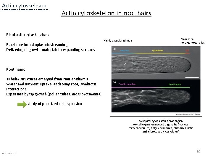
Actin cytoskeleton in root hairs Plant actin cytoskeleton: Backbone for cytoplasmic streaming Delivering of growth materials to expanding surfaces Highly vacuolated tube Clear zone no larger organelles Root hairs: Tubular structures emerged from root epidermis Water and nutrient uptake, anchoring root, symbiotic interactions Expansion by tip growth (pollen tubes, moss protonema) study of polarized cell expansion Subapical cytoplasmic dense region For cell expansion needed organelles (Nucleus, mitochondria, ER, Golgi, endosomes, ribosomes, actin and microtubule cytoskeleton) Ketalaar 2013 30
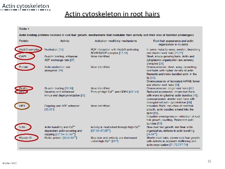
Actin cytoskeleton Ketalaar 2013 Actin cytoskeleton in root hairs 31
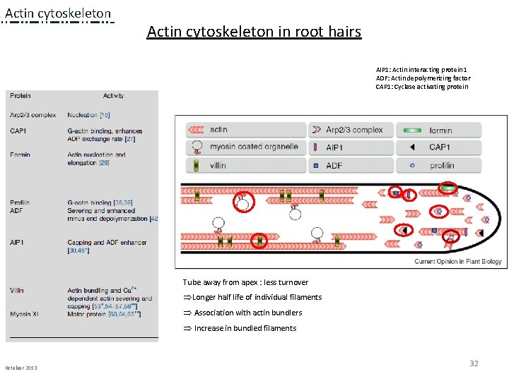
Actin cytoskeleton in root hairs AIP 1: Actin interacting protein 1 ADF: Actin depolymerizing factor CAP 1: Cyclase activating protein Tube away from apex : less turnover ÞLonger half life of individual filaments Þ Association with actin bundlers Þ Increase in bundled filaments Ketalaar 2013 32
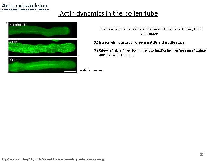
Actin cytoskeleton Actin dynamics in the pollen tube Based on the functional characterization of ABPs derived mainly from Arabidopsis. (A) Intracellular localization of several ABPs in the pollen tube (B) Schematic describing the intracellular localization and function of various ABPs in the pollen tube Scale bar = 10 μm. 33 http: //www. frontiersin. org/files/Articles/124351/fpls-05 -00786 -HTML/image_m/fpls-05 -00786 -g 003. jpg
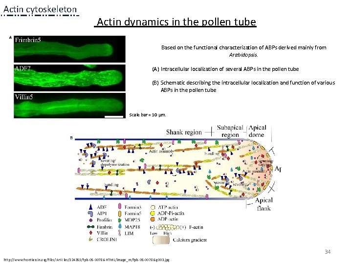
Actin cytoskeleton Actin dynamics in the pollen tube Based on the functional characterization of ABPs derived mainly from Arabidopsis. (A) Intracellular localization of several ABPs in the pollen tube (B) Schematic describing the intracellular localization and function of various ABPs in the pollen tube Scale bar = 10 μm. 34 http: //www. frontiersin. org/files/Articles/124351/fpls-05 -00786 -HTML/image_m/fpls-05 -00786 -g 003. jpg
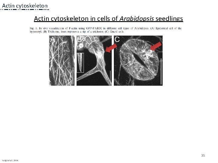
Actin cytoskeleton in cells of Arabidopsis seedlings 35 Voigt et al. 2004
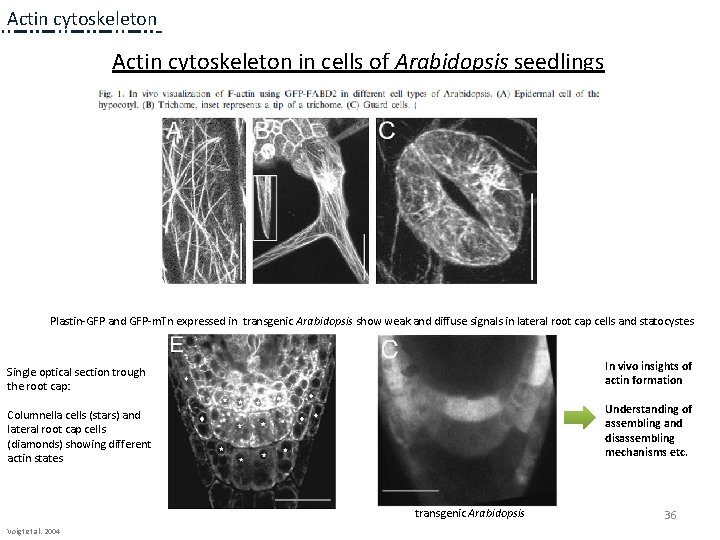
Actin cytoskeleton in cells of Arabidopsis seedlings Plastin-GFP and GFP-m. Tn expressed in transgenic Arabidopsis show weak and diffuse signals in lateral root cap cells and statocystes Single optical section trough the root cap: In vivo insights of actin formation Columnella cells (stars) and lateral root cap cells (diamonds) showing different actin states Understanding of assembling and disassembling mechanisms etc. transgenic Arabidopsis Voigt et al. 2004 36
Thank you for listening ! 37