Airway management Foreign body obstruction Maria P Ntalouka

Airway management Foreign body obstruction Maria P. Ntalouka

Airway Anatomy • Upper airway – Nasal passage – Turbinates – Oral cavity – Epiglottis – Vocal cord – Esophagus

Anatomy of the Glottis • Posterior tongue • Epiglottis • Vocal cords – True – False • Esophagus • Prehospital care providers who perform endotracheal intubation must know this anatomy Courtesy of James P. Thomas, M. D. , www. voicedoctor. net

Pediatric Airway Considerations • Larger head and tongue – Greater potential for airway obstruction – Special attention to proper positioning • Epiglottis – Proportionally larger – Floppier than adult • Trachea – Shorter and conical shape – Greater potential for main bronchus intubation
 • If the patient is talking normally, the airway")
Airway Assessment (1 of 5) • If the patient is talking normally, the airway is open – Further assessment is still required • Assessment of the airway requires the provider to: – Look – Listen – Feel
 • Look for findings that may indicate airway obstruction")
Airway Assessment (2 of 5) • Look for findings that may indicate airway obstruction or injury or may lead to pulmonary aspiration • Examples may include: – Blood and secretions – Fractured teeth – Foreign bodies
 • Examples may include (cont’d): – Vomitus – Hematomas/contusions")
Airway Assessment (3 of 5) • Examples may include (cont’d): – Vomitus – Hematomas/contusions (e. g. , tongue, neck) – Gross subcutaneous emphysema Photograph provided courtesy of J. C. Pitteloud M. D. , Switzerland
 • Listen for abnormal sounds indicating airway compromise •")
Airway Assessment (4 of 5) • Listen for abnormal sounds indicating airway compromise • Examples include: – Snoring – Stridor (inspiratory) – Gurgling (expiratory) – Hoarseness
 • Feel for abnormal masses and signs of airway")
Airway Assessment (5 of 5) • Feel for abnormal masses and signs of airway injury • Examples include: – Hematomas – Subcutaneous emphysema in the neck • Additional consideration – Measure oxygen saturation
 • Causes of airway obstruction – Tongue • Most")
Airway Obstruction (1 of 2) • Causes of airway obstruction – Tongue • Most common cause • Falls back, obstructing the airway with decreased mental status • Snoring — clinical finding
 • Causes of airway obstruction (cont’d) – Foreign body")
Airway Obstruction (2 of 2) • Causes of airway obstruction (cont’d) – Foreign body – Blood – Vomit – Teeth
 • Blunt injuries – Examples of findings may include:")
Airway Trauma (1 of 2) • Blunt injuries – Examples of findings may include: • • Swelling and edema Fractured larynx Subcutaneous emphysema Hematoma
 • Penetrating injuries – Examples of findings may include")
Airway Trauma (2 of 2) • Penetrating injuries – Examples of findings may include (cont’d): • Bleeding into the airway • Subcutaneous emphysema • Hematoma

Inhalation Injuries of the Airway • Examples of causes – Dry – Steam – Chemical • Signs and symptoms of airway burns – Swelling/edema – Stridor

Aligning the Axes (creates a more direct approach for ventilation during BVM and improved visualization of vocal cords during intubation) Oral axis Pharyngeal axis Laryngeal axis Head on bed, neutral position

Aligning the Axes Oral axis Pharyngeal axis Laryngeal axis Head Elevated, neutral position

Aligning the Axes Oral axis Pharyngeal axis Laryngeal axis Head elevated and extended

FBAO - Choking: Algorithm

FBAO - Choking: Algorithm

FBAO - Choking: Algorithm

FBAO - Choking: severe obstruction Back Blows Abdominal thrusts: position of first hand position of second hand

FBAO - Choking: Algorithm

Indications for Intubation • • Failure to maintain or protect the airway Failure of ventilation Failure of oxygenation Therapy / Evaluation is required that mandates intubation

Failure to protect or maintain airway • • • Altered mental status Excessive Secretions Bleeding Hematoma Angioedema Others

Failure to Ventilate • • • Inability to remove p. CO 2 COPD Narcotic OD Myasthenia Gravis Stroke Other

Failure to Oxygenate • • • Inability to maintain p. O 2 > 60 CHF Pneumonia ARDS Pulmonary Embolism Other

Therapy is required that mandates intubation • Severe Head Injury – Hyperventilation • Combative – Need to evaluate/treat • Other

Evaluating the airway • Dentition – prominent upper incisors, receding chin • Distortion – edema, blood, vomitus, tumor, infection • Disproportion – short chin-to-larynx distance, bull neck, large tongue, small mouth • Dysmobility – TMJ and cervical spine immobility

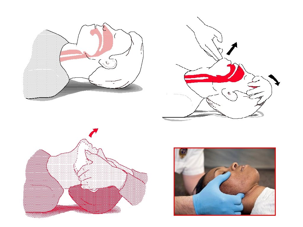
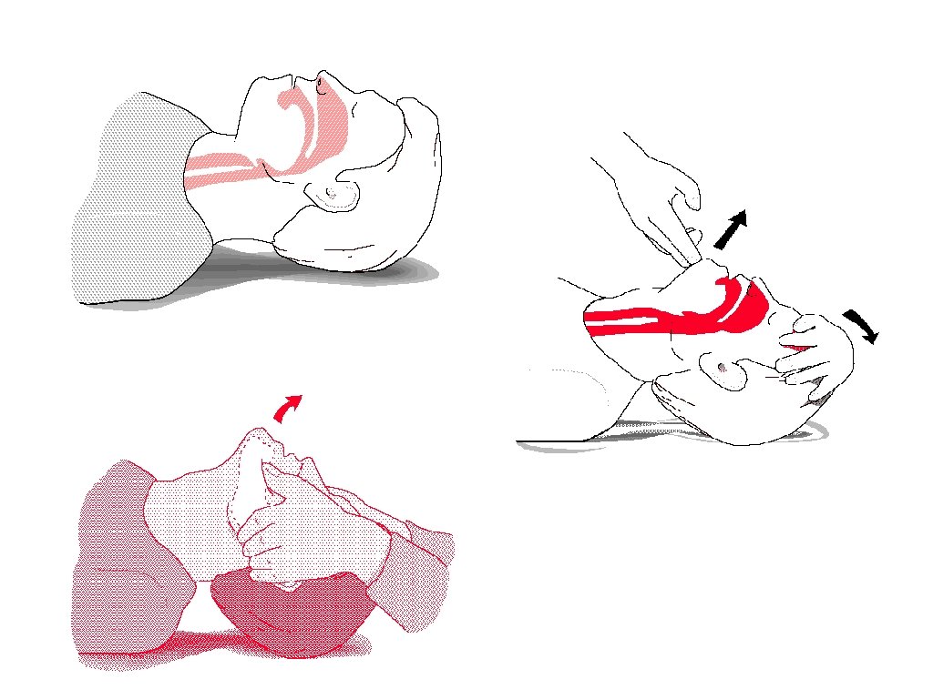
Neck Mobility • Extension of the neck at the atlanto-occipital joint brings the oral, pharyngeal, and laryngeal axes into alignment.

Bag Valve Mask • Make sure it is the right size – Adult – Child – Infant • Facial Landmarks – Bridge of nose – Malar eminences – Mandibular alveolar ridge

Bag Valve Mask • What type of bag – Duckbill inspiratory valves – One way expiratory valves • Delivers 90 to 97% oxygen – Spontaneously breathing – Ventilated
 •")
Bag valve mask • Use both hands to make a seal (two operators) • Lift the jaw • Align the axes

Single-operator BVM • Use one hand to make a seal • Use other hand to deliver breaths • Lift the jaw • Align the axes Note: always observe universal precautions, including gloves, when delivering patient care

Bag Valve Mask • Continuous Firm Pressure – Want to minimize barotrauma • Allow time to exhale – Watch Chest – Listen for exhalation • Cadence – Time with breaths

BVM Assessment – Simple tools for airway assessment in the emergency setting, including BVM assessment – Example: MOANS* *The Difficult Airway Course. TM *Walls RM, Murphy MF (2008). Manual of Emergency Airway Management; 3 rd edition. Philadelphia: Lippincott Williams and Wilkins.

MOANS* • BVM difficulty is anticipated if these factors are present: – M: Mask seal difficulty • (Beards and facial injuries) – O: Obesity, obstruction – A: Age (>55) – N: No teeth – S: Stiff (also sleep apnea and snoring) • Decreased lung compliance *Walls RM, Murphy MF (2008). Manual of Emergency Airway Management; 3 rd edition. Philadelphia: Lippincott Williams and Wilkins. *The Difficult Airway Course. TM

Bag Valve Mask • Use adjuncts – Nasopharyngeal Airway – Oropharyngeal Airway

Adjuncts • Nasopharyngeal Airway – Advance until airflow heard – Can be used in semi-conscious patient Note: always observe universal precautions, including gloves, when delivering patient care

BVM adjuncts • Oropharyngeal Airway – Unconscious / No gag – Insert with tongue blade or rotate Note: always observe universal precautions, including gloves, when delivering patient care

Difficult to Bag Valve Mask • • • Facial Hair Obesity Edentulous (leave dentures in) Upper airway obstruction Small Chin Large tongue

Lemon Law* • • • Look Externally Evaluate the 3 -3 -2 Rule Mallampati Obstruction Neck Mobility *Walls RM, Murphy MF (2008). Manual of Emergency Airway Management; 3 rd edition. Philadelphia: Lippincott Williams and Wilkins.

Look Externally • • • abnormal face shape sunken cheeks edentulous "buck teeth" receding mandible "bull-neck" narrow mouth obesity face or neck pathology

Dentition • Overbite can impose a variable degree of limitation on alignment of axes during intubation • Remove teeth that might dislodge during laryngoscopy • Remove dentures
")
Evaluate the 3 -3 -2 Rule Mouth opening > 3 F (F=fingerbreadths)
")
Evaluate the 3 -3 -2 Rule • Hyoid-chin distance > 3 F (F=fingerbreadths)

Evaluate the 3 -3 -2 Rule Thyroid cartilage - mouth floor distance > 2 F (F=fingerbreadths)

3 -3 -2 rule not met, anticipate challenging anatomy < 3 F
- Slides: 49