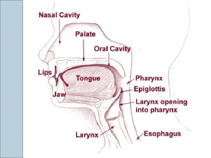
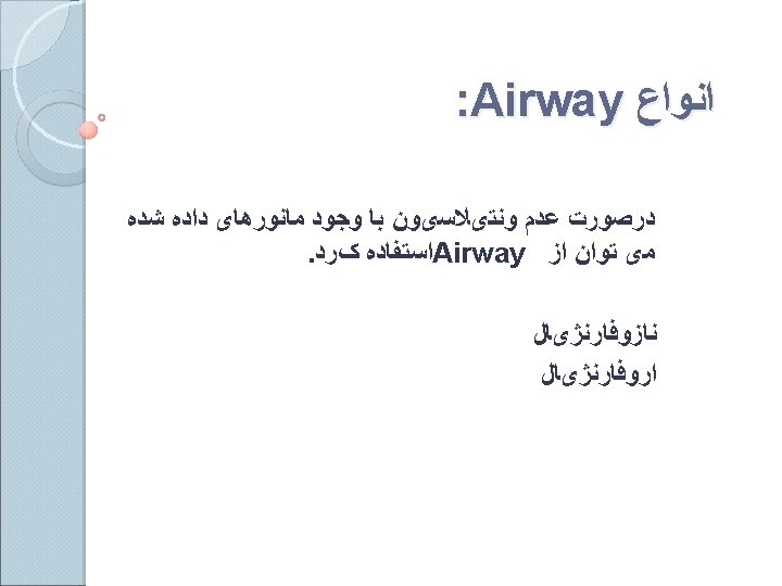
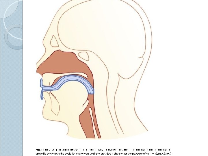
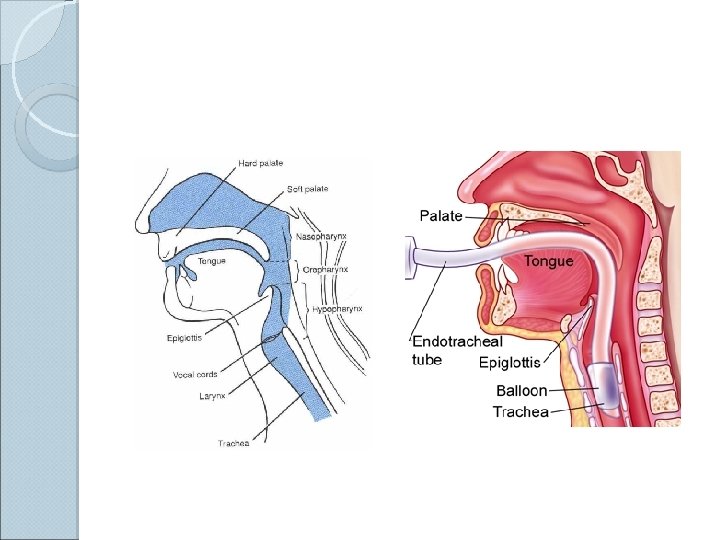
Airway management Airway Anatomy Upper Airway Pharynx Epiglottis


 can be oxygenated and ventilated")


 Condition intubation: that associated with difficult Congenital anomalies Pierre Robin syndrome")


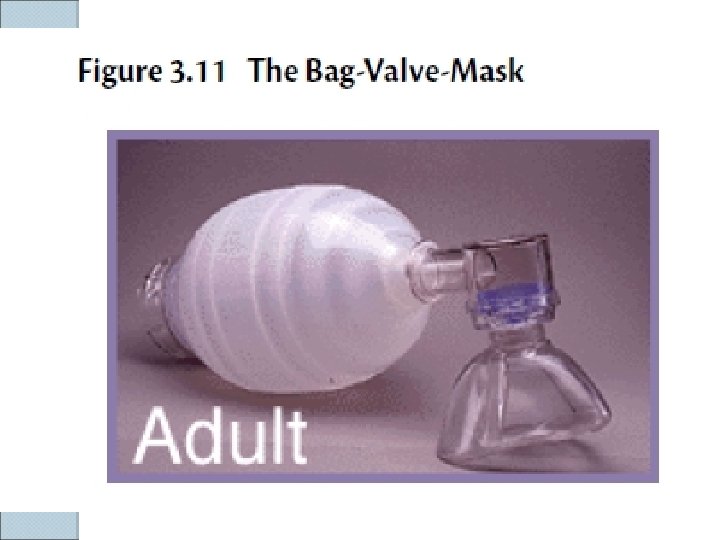
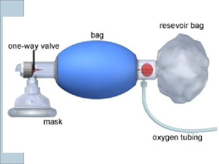
 or bag-valve unit")
 Laryngoscope : handle and blade")
 and Miller (straight) blade F Adult : Macintosh blade,")
 Endotracheal tube")
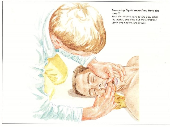
 Open the patient's mouth with the right hand, and remove")


 During intubation 2) During remained intubation 3) During extubation")

 During remained intubation Obstruction from klinking , secretion or overinflation of cuff Accidental")
 After Extubation Sore throat Hoarseness Tracheal stenosis (Prolong intubation) Laryngeal granuloma")

- Slides: 69
Airway management
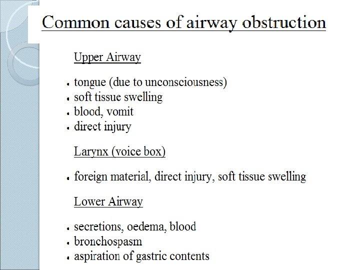
Airway Anatomy Upper ◦ ◦ ◦ Airway Pharynx Epiglottis Glottis Vocal cords Larynx Lower ◦ ◦ Airway Trachea Bronchi Alveoli Lung tissue, consisting of lobes and lobules (3 on the right and 2 on the left) ◦ Pleura
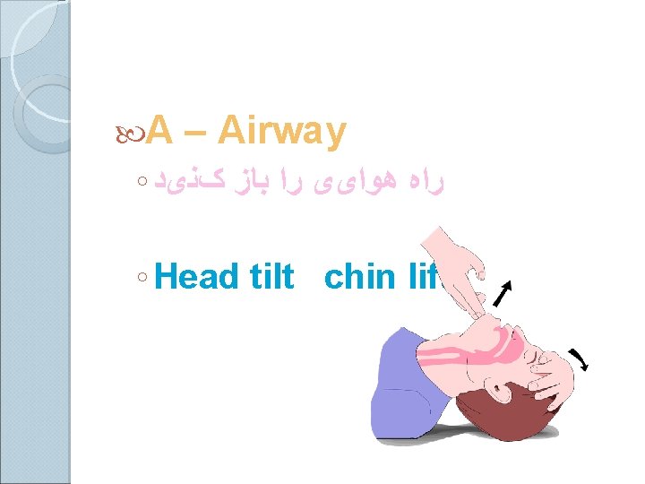
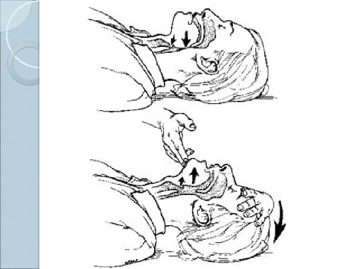
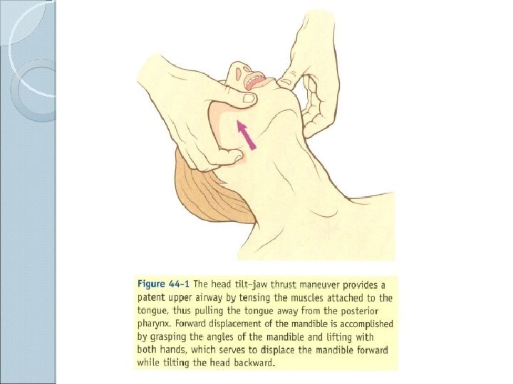
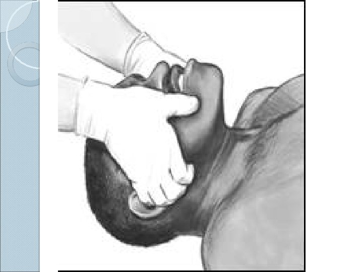
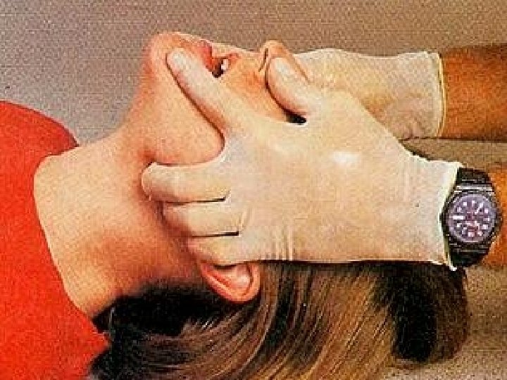
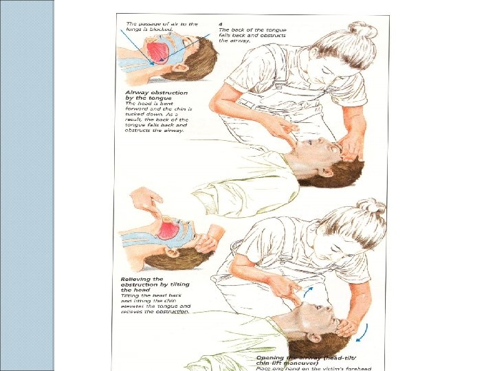
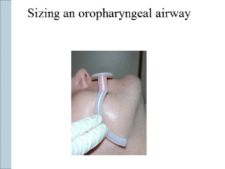
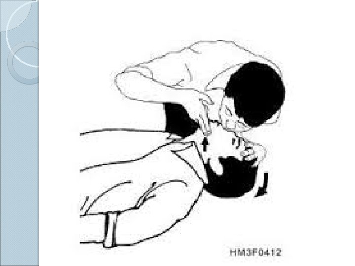
Basic Airway Maneuvers ALWAYS REMEMBER THE BASICS These skills should be used prior to initiating any advanced airway technique ◦ Head-tilt/chin lift ◦ Jaw thrust ◦ Sellick’s maneuver
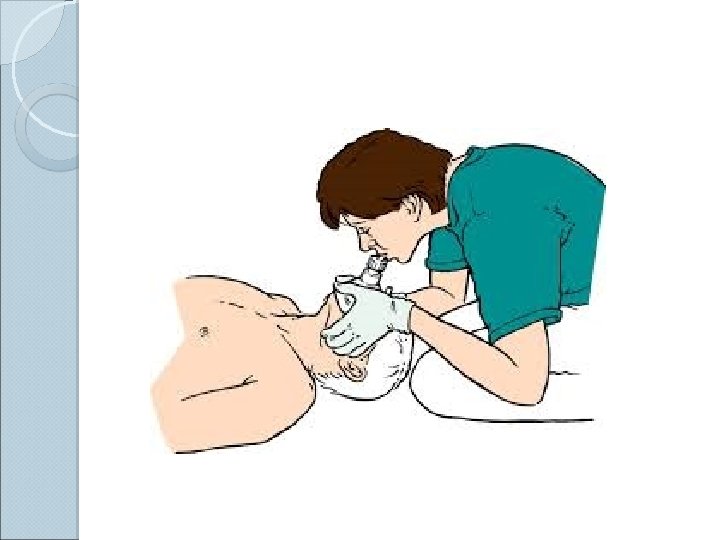
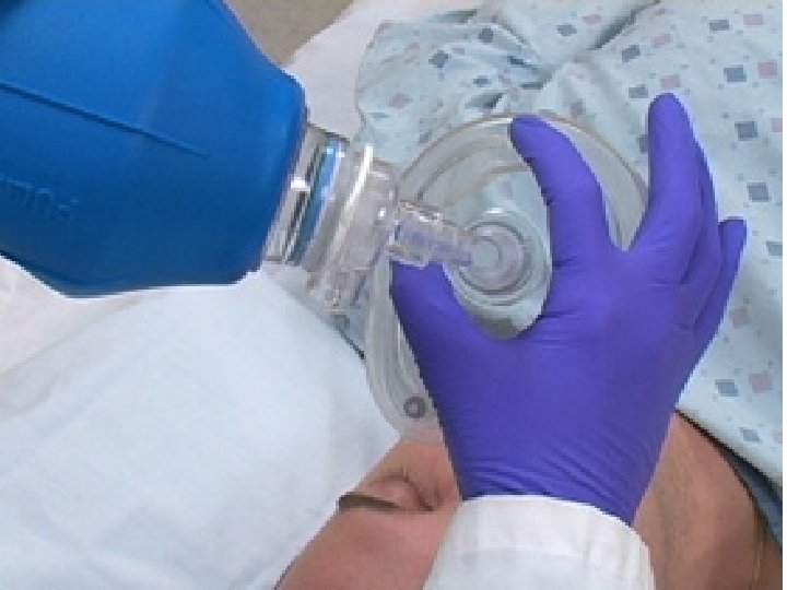
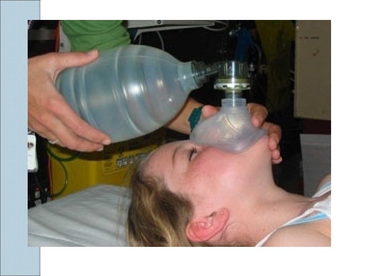
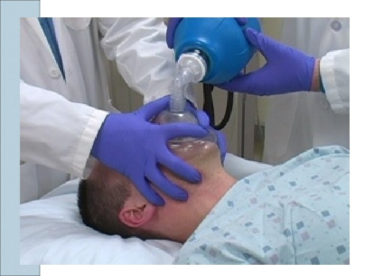
BVM Ventilation The most important airway skill Always the first response to inadequate oxygenation and ventilation The first “bail-out” maneuver to a failed intubation attempt Attenuates the urgency to intubate
Golden Rules of Bagging “ Anybody ( almost ) can be oxygenated and ventilated with a bag and a mask “ The art of bagging should be mastered before the art of intubation Manual ventilation skill with proper equipment is a fundamental premise of advanced airway
BVM Ventilation: Assessment of Efficacy Observe the chest rise and fall Good bilateral air entry Lack of air entering the stomach Feeling the bag Pulse oximetry
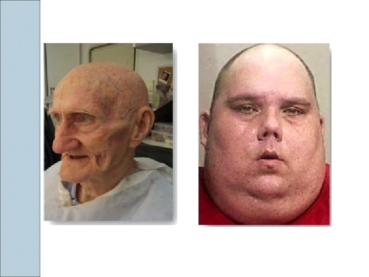
Predictors of difficult face mask ventilation 1 - age>55 years 2 -body mass index>26 Kg/m 2 3 -a beard 4 -lack of teeth 4 -a history of snoring
Anatomy
Methods: Endotracheal ◦ Orotracheal ◦ Nasotracheal intubation
Endotracheal Intubation Placement of a flexible plastic tube into the trachea to: ◦ maintain an open airway, ◦ serve as a conduit through which to administer certain drugs. Is performed in critically injured, ill or anesthetized patients: ◦ to facilitate ventilation of the lungs, including mechanical ventilation, ◦ to prevent the possibility of asphyxiation or airway obstruction.
Indications: For supporting ventilation in patient with pathologic disease: ◦ Upper airway obstruction, ◦ Respiratory failure, ◦ Loss of consciousness For supporting ventilation during general anaesthesia: ◦ Type of surgery: Operative site near the airway, Thoracic or abdominal surgery, Prone or lateral surgery, Long period of surgery Patient has risk of pulmonary aspiration Difficult mask ventilation
BASIC AIRWAY EVALUATION Previous anaesthetic problems and general appearance of the patient. 2. Neck, face, maxilla and mandible with jaw movements. 3. Head extension and movements, teeth, oropharanx and soft tissue of the neck. 1.
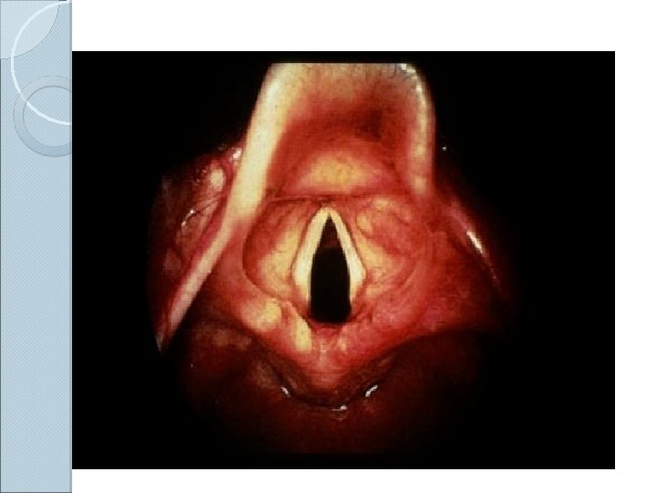
ASSESSMENT OF AIRWAY Mallampati classification larangoscopic view.
Laryngoscopic view Grade 3, 4 risk for difficult intubation!
Airway Assessment Interincisor gap : normal more than 3 cms
Movement of temperomandibular joint (TMJ ( Grinding
Airway Assessment 1) Condition intubation: that associated with difficult Congenital anomalies Pierre Robin syndrome , Down’s syndrome Infection in airway Retropharyngeal abscess, Epiglottitis Tumor in oral cavity or larynx Enlarge thyroid gland trachea shift to lateral or compressed tracheal lumen
Continuation. . . Maxillofacial , cervical or laryngeal trauma Temperomandibular joint dysfunction Burn scar at face and neck Morbidly obese or pregnancy
Anatomical factors affecting Larangoscopy 1. 2. 3. 4. 5. Short Neck. Protruding incisor teeth. Long high arched palate. Poor mobility of neck. Increase in either anterior depth or Posterior depth of the mandible decrease in Atlanto Occipital distance that's why role of Radiology has increased in our specialty
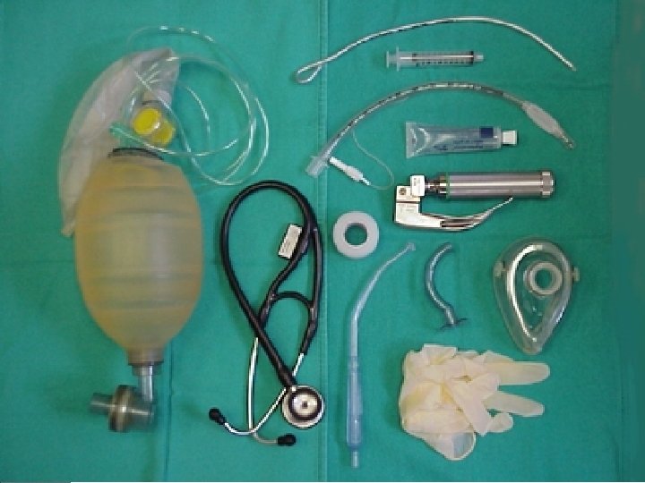
Instruments used. . . 1. Self-refilling bag-valve combination (eg, Ambu bag) or bag-valve unit (Ayres bag), connector, tubing, and oxygen source. Assemble all items before attempting intubation. 2. Tincture of benzoin and precut tape. 3. Introducer (stylets or Magill forceps). 4. Suction apparatus (tonsil tip and catheter suction). 5. Syringe, 10 -m. L, to inflate the cuff. 6. Mucosal anesthetics (eg, 2% lidocaine) 7. Water-soluble sterile lubricant. 8. Gloves.
1) Laryngoscope : handle and blade
LARYNGOSCOPIC BLADE F Macintosh (curved) and Miller (straight) blade F Adult : Macintosh blade, small children : Miller blade Macintosh blade
2) Endotracheal tube
Curved blade technique a) Open the patient's mouth with the right hand, and remove any dentures. b) Grasp the laryngoscope in the left hand c) Spread the patient's lips, and insert the blade between the teeth, being careful not to break a tooth. d) Pass the blade to the right of the tongue, and advance the blade into the hypopharynx, pushing the tongue to the left. e) Lift the laryngoscope upward and forward, without changing the angle of the blade, to expose the vocal cords.
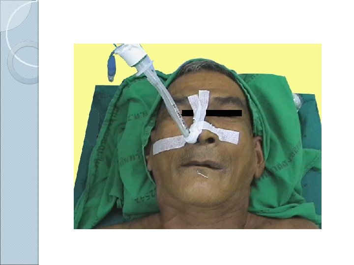
Nasoendotracheal intubation F Advantages: (1 Comfortable for prolong intubation in postoperative period (2 Suitable for oral surgery : tonsillectomy , mandible surgery (3 For blind nasal intubation (4 Can take oral feeding (5 Resist for kinking and difficult to accidental extubation Disadvantages: (1 Trauma to nasal mucosa (2 Risk for sinusitis in prolong intubation (3 Risk for bacteremia (4 Smaller diameter than oral route difficult for suction
Contraindication for nasoendotracheal intubation (1 Fracture base of skull (2 Coagulopathy (3 Nasal cavity obstruction (4 Retropharyngeal abscess
Complication of endotracheal intubation 1) During intubation 2) During remained intubation 3) During extubation 4) After extubation
(1 During intubation Trauma to lip, tongue or teeth Hypertension and tachycardia or arrhythmia Pulmonary aspiration Laryngospasm Bronchospasm Laryngeal edema Arytenoid dislocation hoarseness Increased intracranial pressure Spinal cord trauma in cervical spine injury Esophageal intubation
2) During remained intubation Obstruction from klinking , secretion or overinflation of cuff Accidental extubation or endobronchial intubation Disconnection from breathing circuit Pulmonary aspiration Lib or nasal ulcer in case with prolong period of intubation Sinusitis or otitis in case with prolong nasoendotracheal intubation 3) During Extubation • Laryngospasm • Pulmonary aspiration • Edema of upper airway
4) After Extubation Sore throat Hoarseness Tracheal stenosis (Prolong intubation) Laryngeal granuloma
Thanks for your attention!