
Airway evaluation and Management Dr Adel Elshimy College


- Slides: 54
Airway evaluation and Management Dr. Adel Elshimy College of Medicine Anesthesia Department
Lecture Objectives Students at the end of the lecture will be able to : § Describe the applied anatomy of the airway. § Conduct a preoperative airway assessment. § Identify a potentially difficult airway. § Learn about management of airway. § Become familiar with airway equipment. § Understand issues around aspiration prophylaxis. § Become familiar with controlled ventilation.
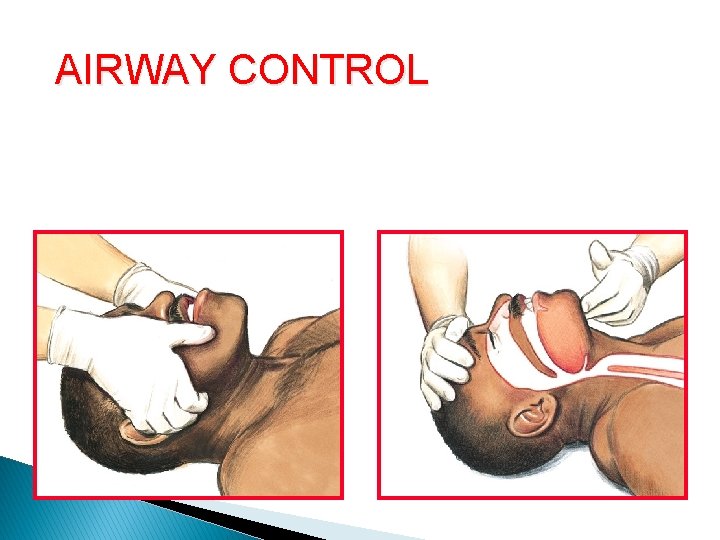
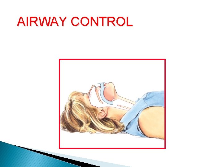
AIRWAY CONTROL Opening the Airway Jaw thrust Head tilt–chin lift
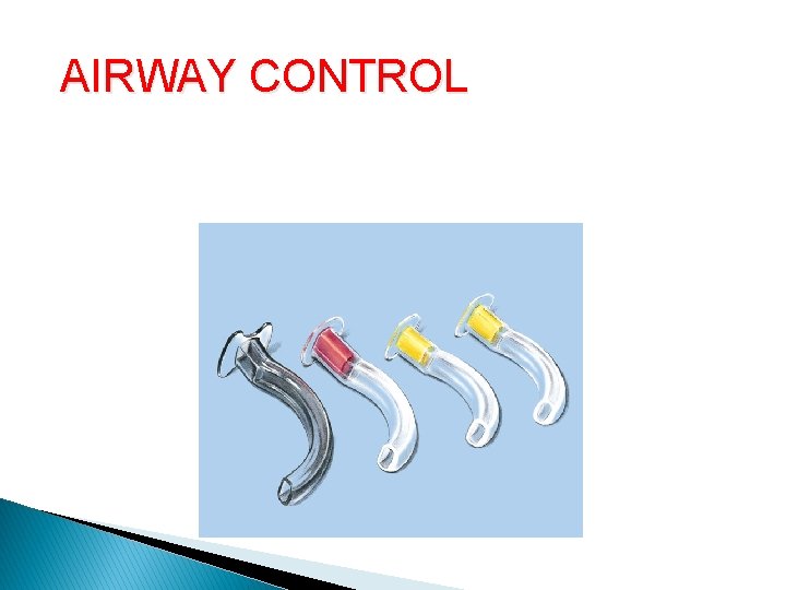
AIRWAY CONTROL Oropharyngeal Airway
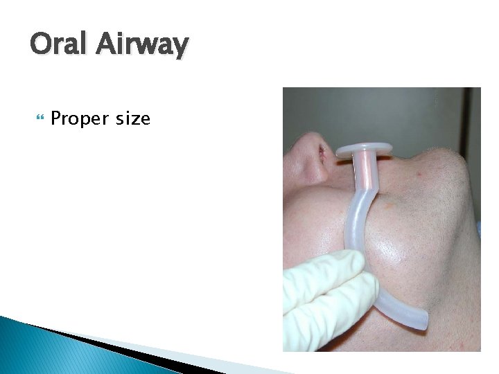
Oral Airway Proper size
AIRWAY CONTROL Oropharyngeal Airway (cont. )
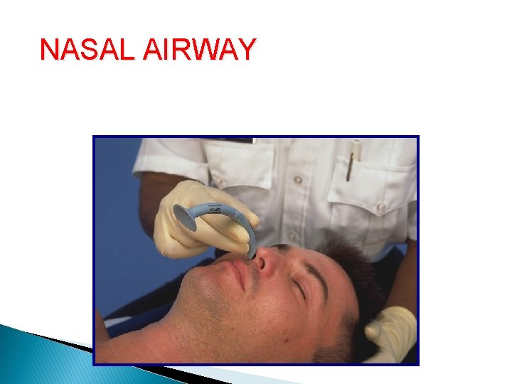
NASAL AIRWAY Nasopharyngeal Airway (cont. )
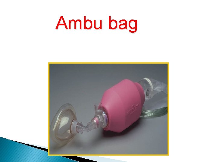
Ambu bag Bag-Valve-Mask (cont. ) With oxygen reservoir
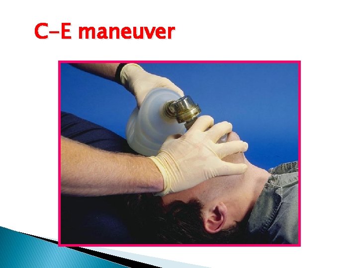
C-E maneuver
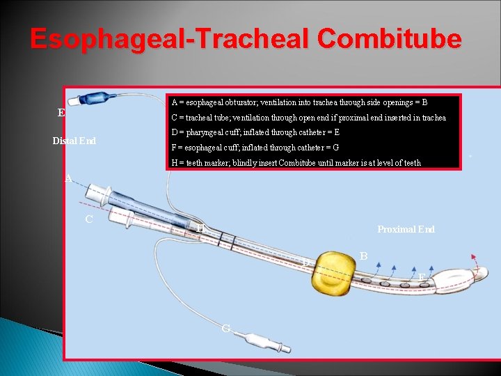
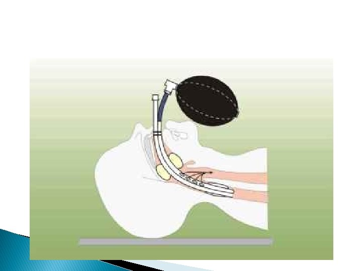
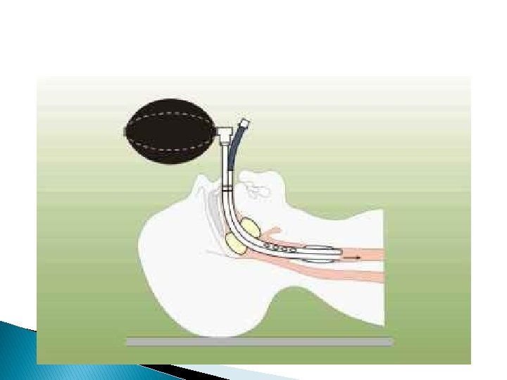
Esophageal-Tracheal Combitube A = esophageal obturator; ventilation into trachea through side openings = B E C = tracheal tube; ventilation through open end if proximal end inserted in trachea Distal End D = pharyngeal cuff; inflated through catheter = E F = esophageal cuff; inflated through catheter = G H = teeth marker; blindly insert Combitube until marker is at level of teeth A C H Proximal End D B F G
Indications of endotracheal intubation Resuscitation (CPR) Prevention of lung soiling Positive pressure ventilation (GA) Pulmonary toilet Patent airway (coma or near coma) Respiratory failure(CO 2 retention )
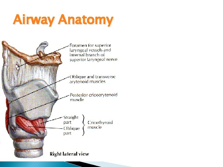
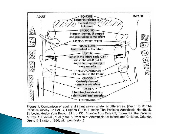
Airway Anatomy Innervation Vagus n. ◦ Superior laryngeal n. External branch – motor to cricothyroid m. Internal branch – sensory larynx above TVC’s ◦ Recurrent laryngeal n. Right – subclavian Left – Aortic arch (board question) Motor to all other muscles, Sensory to TVC’s and trachea
Management I-History: previous history of difficulty is the best predictor Inquire about: -Nature of difficulty -No of trials -Ability to ventilate bet trials -Maneuver used -Complications II-Snoring and sleep apnea. III-Predictors of DMV (obese ).
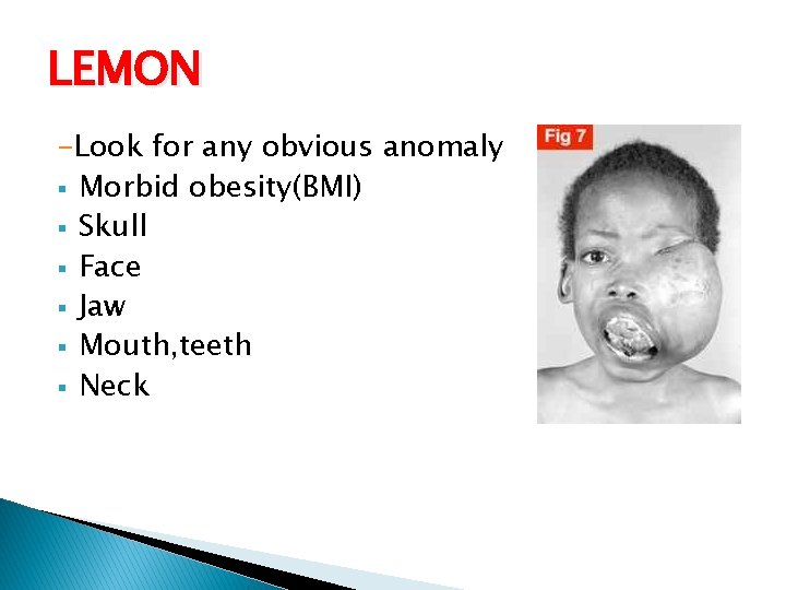
LEMON -Look for any obvious anomaly § Morbid obesity(BMI) § Skull § Face § Jaw § Mouth, teeth § Neck
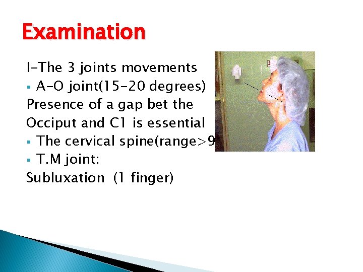
Examination I-The 3 joints movements § A-O joint(15 -20 degrees) Presence of a gap bet the Occiput and C 1 is essential § The cervical spine(range>90) § T. M joint: Subluxation (1 finger)
Examination Mouth opening: 3 fingers Thyromental distance: >6. 5 cm Sternomental distance >12. 5 cm.
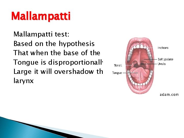
Mallampatti test: Based on the hypothesis That when the base of the Tongue is disproportionally Large it will overshadow the larynx
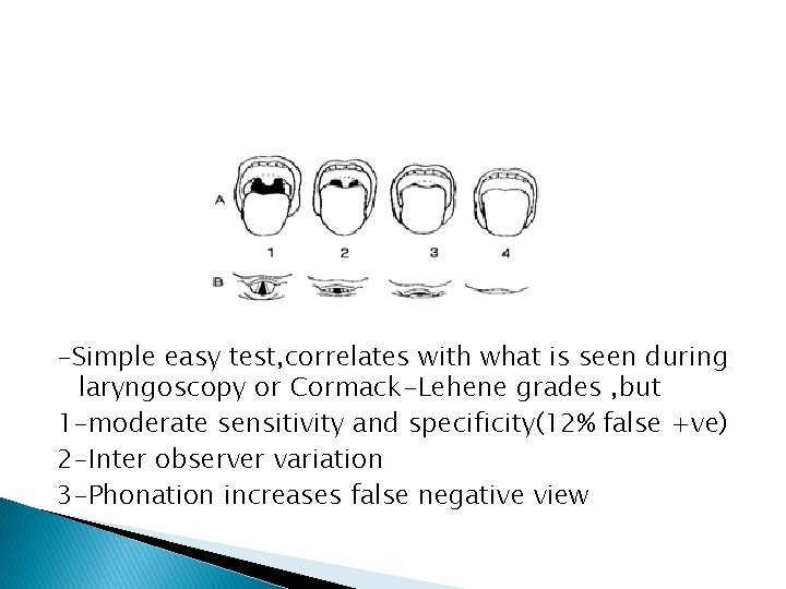
-Simple easy test, correlates with what is seen during laryngoscopy or Cormack-Lehene grades , but 1 -moderate sensitivity and specificity(12% false +ve) 2 -Inter observer variation 3 -Phonation increases false negative view
Obstruction Apparent cause e. g. goitre OSA Noisy breathing or stridor Signs of upper airway obstruction Other causes
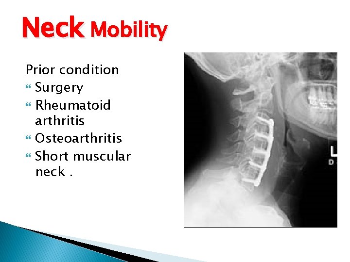
Neck Mobility Prior condition Surgery Rheumatoid arthritis Osteoarthritis Short muscular neck.
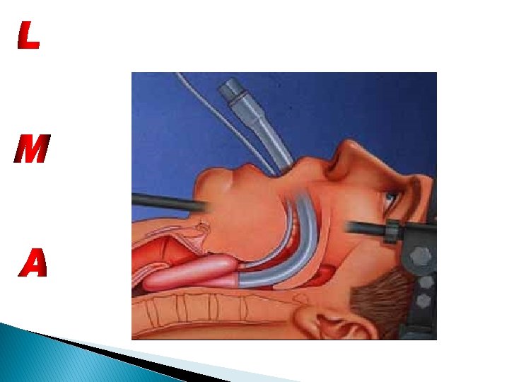
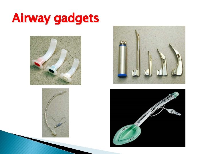
Proper Equipment -Bag and mask, oxygen source -Airways oro , nasopharyngeal and LMA -Laryngosopes different blades -ETT different sizes -suction on
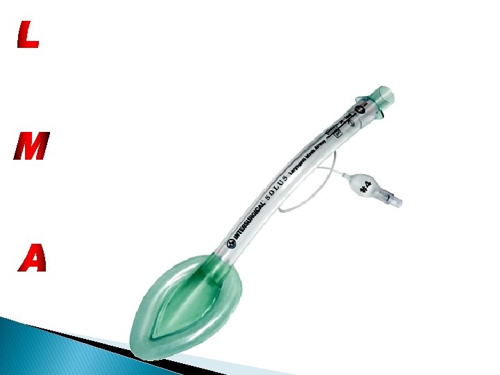
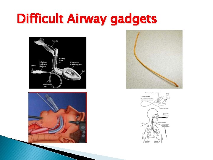
Airway gadgets
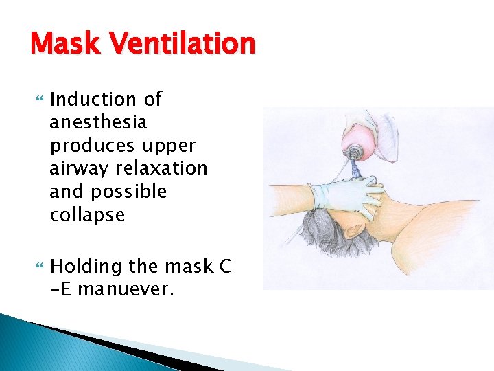
Mask Ventilation Induction of anesthesia produces upper airway relaxation and possible collapse Holding the mask C -E manuever.
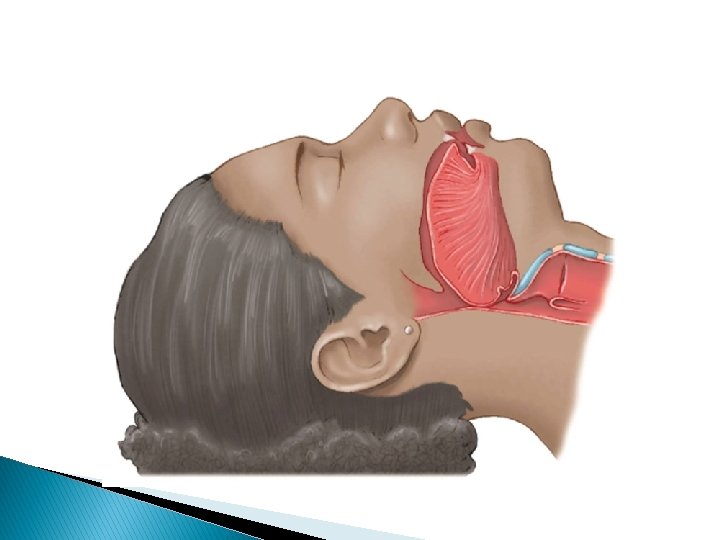
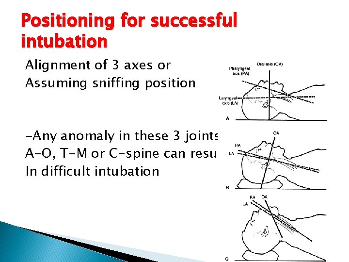
Positioning for successful intubation Alignment of 3 axes or Assuming sniffing position -Any anomaly in these 3 joints A-O, T-M or C-spine can result In difficult intubation
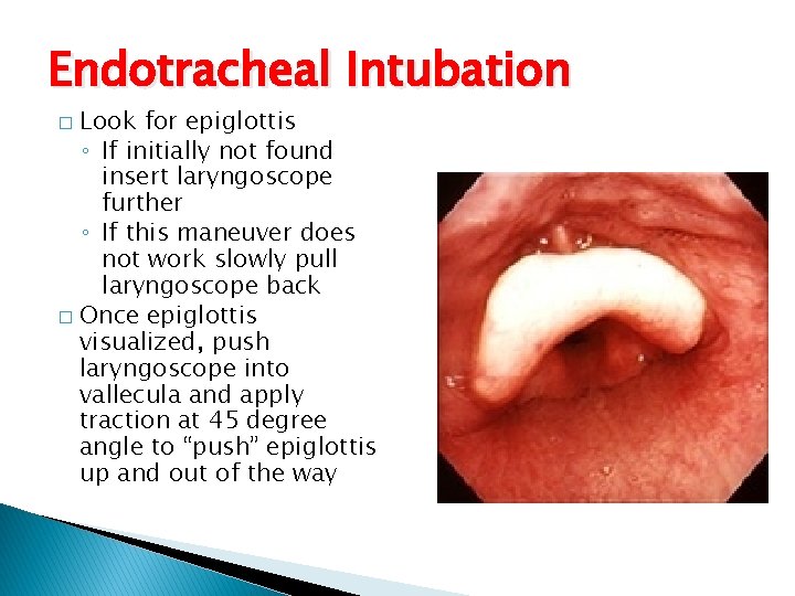
Endotracheal Intubation Look for epiglottis ◦ If initially not found insert laryngoscope further ◦ If this maneuver does not work slowly pull laryngoscope back � Once epiglottis visualized, push laryngoscope into vallecula and apply traction at 45 degree angle to “push” epiglottis up and out of the way �
Confirm tube position Direct visualization of ETT between cords Continuous trace of capnography 3 point auscultation Bronchoscopy ; carina seen Esophageal detector device Other as bilateral chest movement, mist in the tube, CXR
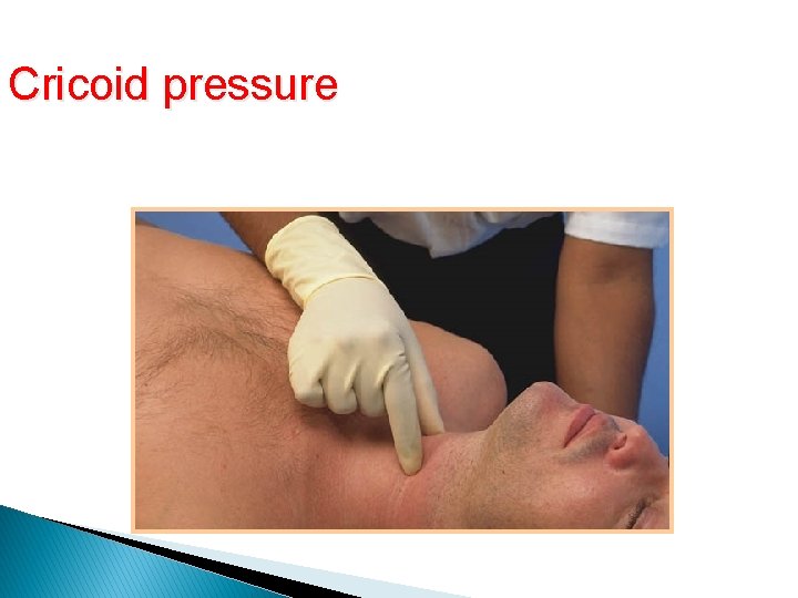
Rapid sequence induction Indications Technique: -Preoxygenation -IV induction with sux -Cricoid pressure -Intubate, inflate the cuff , confirm position -Release cricoid and fix the tube
Cricoid pressure Cricoid Pressure
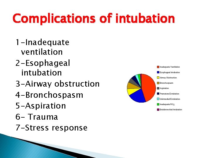
Complications of intubation 1 -Inadequate ventilation 2 -Esophageal intubation 3 -Airway obstruction 4 -Bronchospasm 5 -Aspiration 6 - Trauma 7 -Stress response
Problems with ETT and Cuff ❏ Too long - endobronchial intubation ❏ Too short - accidental extubation ❏ Too large - trauma to surrounding tissues ❏ Too narrow - increased airway resistance ❏Too soft - kinks ❏ Too hard - tissue damage ❏ Prolonged placement - vocal cord granulomas, tracheal stenosis ❏ Poor curvature - difficult to intubate ❏ Cuff insufficiently inflated - allows leaking and aspiration ❏ Cuff excessively inflated - pressure necrosis
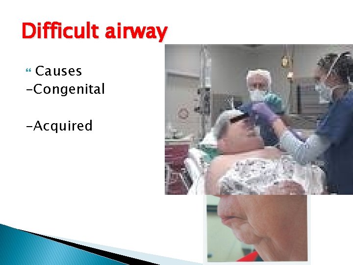
Difficult airway Causes -Congenital -Acquired
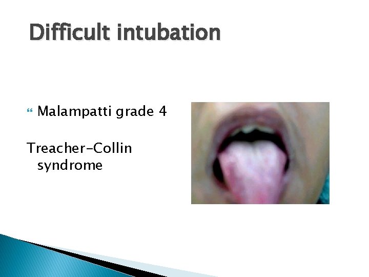
Difficult intubation Malampatti grade 4 Treacher-Collin syndrome
Difficult Airway gadgets
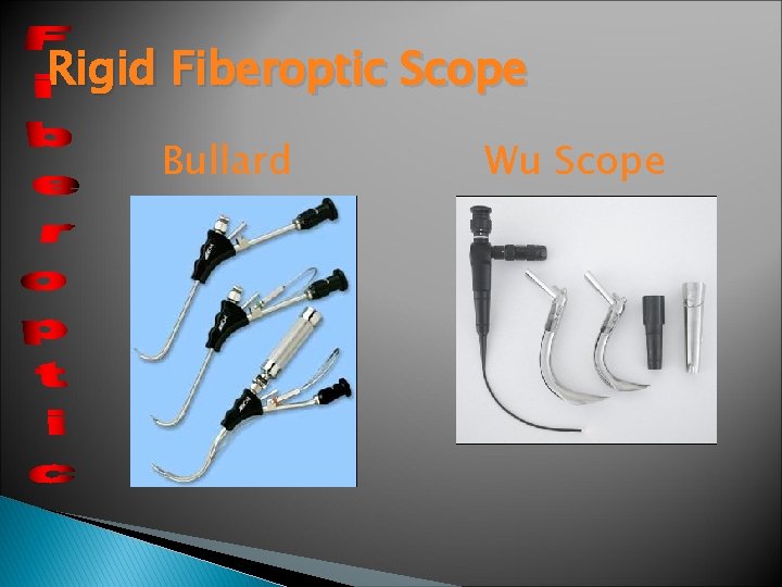
Rigid Fiberoptic Scope Bullard Wu Scope
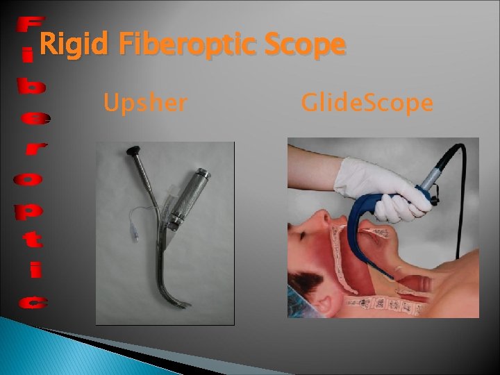
Rigid Fiberoptic Scope Upsher Glide. Scope
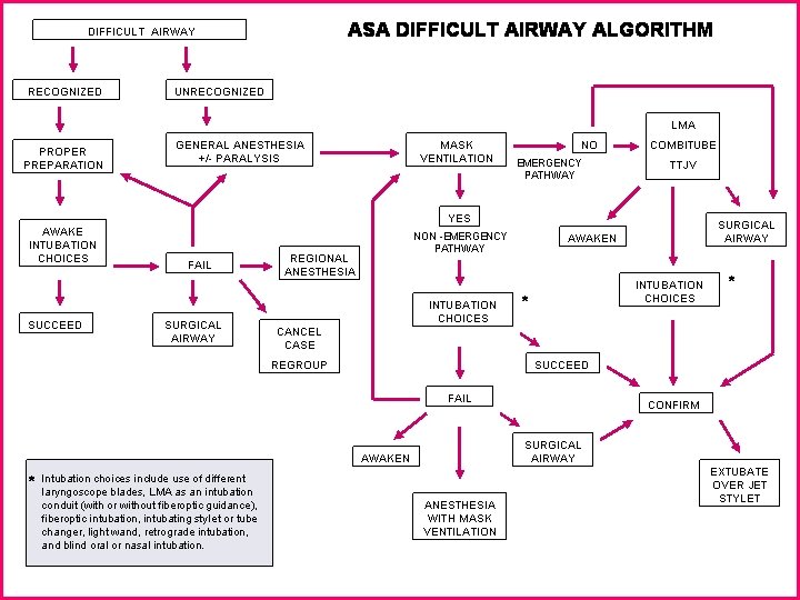
Management of Difficult airway Expected from history, examination Secure airway while awake under LA q Unexpected different options Priority for maintenance of patent airway and oxygenation
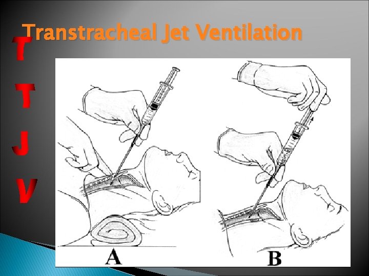
Transtracheal Jet Ventilation
Ventilation Spontaneous ventilation Controlled ventilation Pressure cycled and volume cycled ventilator -Tidal volume 10 mls/kg -Respiratory rate to maintain normocarbia -I: E ratio -PEEP
Recommendations Adequate airway assessment to pick up expected D. A to be secured awake Difficult intubation cart always ready Pre oxygenation as a routine Maintenance of oxygenation not the intubation should be your aim Use the technique you are familiar with Always have plan B, C, D in unexpected D. A
EXTUBATION General guidelines: check that neuromuscular function and hemodynamic status is normal Ø check that patient is breathing spontaneously with adequate rate and tidal volume Ø allow patient to breathe 100% O 2 for 3 -5 minutes Ø suction secretions from pharynx Ø deflate cuff, remove ETT on inspiration (vocal cords abducted) Ø ensure patient breathing adequately after extubation Ø ensure face mask for O 2 delivery available Ø proper positioning of patient during transfer to recovery room, e. g. sniffing position, side lying. Ø
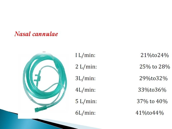
Oxygen delivery systems: Nasal cannulae ◦ inspired oxygen concentration is dependent on the oxygen flow rate, the nasopharyngeal volume and the patient’s inspiratory flow rate. ◦ Increases inspired oxygen concentration by 3 -4%. ◦ Oxygen flow rates greater than 3 liters are poorly tolerated by patients due to drying and crusting of the nasal mucosa.
Nasal cannulae
Face masks : ◦ Three types of facemask are available; open, Venturi, non-rebreathing. Open facemasks : ◦ Are the most simple of the designs available. ◦ They do not provide good control over the oxygen concentration being delivered to the patient causing variability in oxygen treatment. ◦ A 6 l/min flow rate is the minimum necessary to prevent the possibility of rebreathing. ◦ Maximum inspired oxygen concentration ~ 50 -60%.
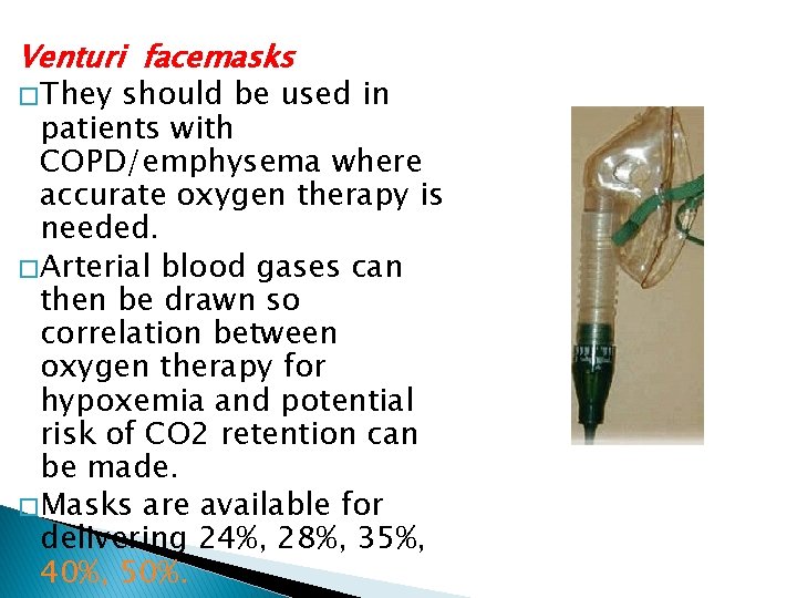
Venturi facemasks � They should be used in patients with COPD/emphysema where accurate oxygen therapy is needed. � Arterial blood gases can then be drawn so correlation between oxygen therapy for hypoxemia and potential risk of CO 2 retention can be made. � Masks are available for delivering 24%, 28%, 35%, 40%, 50%.
Non-rebreathing facemasks ◦ have an attached reservoir bag and one-way valves on the sides of the facemask. ◦ With flow rates of 10 liters an oxygen concentration of 95% can be achieved. ◦ These masks provide the highest inspired oxygen concentration for nonintubated patients.
Reference book and the relevant page numbers. . American Society of Anesthesiologists (http: //www. asahq. org/publications. Services. htm), accessed January 30, 2006. Anesthesia Patient Safety Foundation (http: //www. apsf. org) accessed January 30, 2006. Cooper JB, Gaba DM. A strategy for preventing anesthesia accidents. Int Anesthesiol Clin 1989; 27: 148– 152. Cooper JB, Newbower RS, Kitz RJ. An analysis of major errors and equipment failures in anesthesia management: considerations for prevention and detection. Anesthesiology 1984; 60: 34– 42. Gaba DM. Anaesthesiology as a model for patient safety in health care. BMJ 2000; 320: 785 -“ 788. Available at: http: //www. bmj. com/cgi/content/full/320/7237/785.
Thank You Dr. Adel Elshimy Date: 20/1/2017