Airway anatomy Supervised by Dr ashraf dmour Done

Airway anatomy Supervised by : Dr ashraf dmour Done by : Rawan zidanen

CONTENT * Review of airway anatomy *Airway Assessment & evaluation * Symptoms of airway obstruction

ANATOMY consisting of: Upper ● Nasal and oral cavities Middle ● Pharynx ● Larynx Lower ● Trachea ● main bronchi
is 4 to 6 cm . Lips. Teeth.")
the normal mouth opening ( ADULT )is 4 to 6 cm . Lips. Teeth. Palate. Uvula. Tonsils. tongue

Pharynx • Mascular tube • common opening of the digestive and respiratory systems. • Receives both air from the nasal cavity , food and water from the mouth.

Larynx

Vocal folds

Trachea *Cartilaginous tube *connects the larynx to the primary bronchi of the lungs * 15 -20 "C"-shaped pieces of cartilage * Posteriorly contains no cartilage and consists of a ligamentous membrane and smooth muscle

Bronchi # Right bronchus is shorter and wider and is more vertical than the left bronchus. # Foreign bodies usually enter the right bronchus

Airway Assessment DONE BY: RAZAN TARAWNEH
 Why it is necessary ? ? PURPOSE - TO DIAGNOSE")
Airway Assessment 1 ) Why it is necessary ? ? PURPOSE - TO DIAGNOSE THE POTENTIAL FOR DIFFICULT AIRWAY FOR Optimal patient preparation Proper selection of equipment Participation of personnel experienced in difficult airway management

History – Patient/notes/chart/previous anesthesia records – Surgery/burns/trauma/tumor in & around oral cavity – Concurrent disease – Reflux/recent meals General examination – Do they just look difficult? – Recognition of anatomic factors that can cause difficult airway • Specific tests/indices • Investigations.
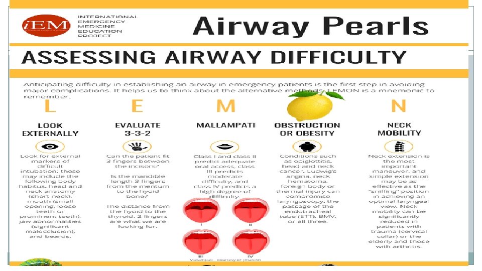

LOOK Externally Receding mandible Beards or facial hair • Morbidly obese patients • Facial or neck trauma Short immobile muscular neck Receding mandible Protruding maxillary incisors Long high-arched palate Loose or capped , Missing teeth Enlarged tonsils & Tongue Tumor that could obstruct air flow Limited temporomandibular joint mobility
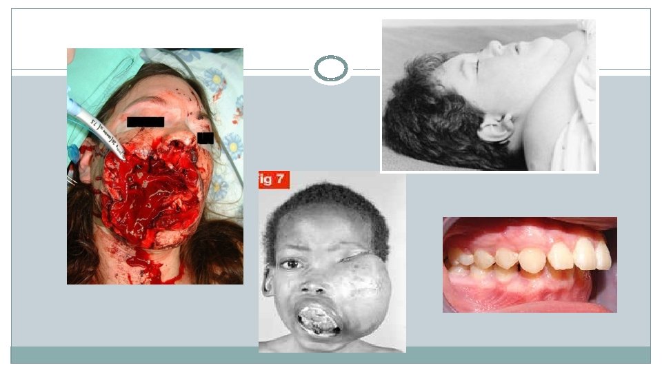

Predictors of difficulty to ventilate 3 rd Trimester Pregnancy � - The Bearded - The Elderly (older than 55 y) - The Edentulous (=BONES) Obesity � Heavy chest Abdominal contents inhibit movement of the diaphragm Increased supraglottic airway resistance huge cheeks Difficult mask seal Quicker desaturation Increased body mass Quick desaturation Increased Mallampati Score Gravid uterus inhibits movement of the diaphragm
 - Mouth Opens at least 3 finger widths (>6")
EVALUATE 3 -3 -2 (LEMON) - Mouth Opens at least 3 finger widths (>6 cm). � -Thyromental distance Three finger widths(>6 cm � -hyomental distance Two finger widths � ( Mouth opening ) Less than 3 fingers width

Measured from the mentum to the top of the hyoid bone >2 fingers -The position of the hyoid bone marks the entrance to the larynx. -A more caudal hyoid bone thus indicates a relatively caudal larynx. <2 = Less space to displace tongue tend to be more difficult to intubate. .

Thyromental Distance : Distance from the mentum to the thyroid notch. Ideally done with the neck fully extended. If the thyromental distance is short, <3 finger widths, the laryngeal axis makes a more acute angle with the pharyngeal axis and it will be difficult to achieve alignment. Less space to displace tongue

AIRWAY ASSESSMENT DONE BY : MALLAK ALJAFARI

Mallampati score -for assessing the adequacy of the oropharynx for laryngoscopy and intubation. - good predictor in pregnancy , obesity and acromegaly - relates to tongue size to pharyngeal size. - Pt should be sitting, head in neutral position, mouth wide open, and tongue extended out as far as possible. -The classification is based on the structures that are visible
 Severe Difficult intubation")
Mallampati score : Class IV: <1% prevalence (hard palate only visible) Severe Difficult intubation Class III: <13% prevalance (soft palate, base of uvula visible) Moderate Difficult intubation Class II: 40% prevalence (soft palate, uvula, fauces visible) No Difficult intubation Class I: 46% prevalence (soft palate, uvula, fauces, pillars visible) No Difficult intubation A Class I view is a Grade I Intubation 99% of the time A Class IV view is a Grade III or IV intubation 99% of the time

Grade 1: Full aperture visible Grade 2: Lower part of cords visible Grade 3: Only epiglottis visible t. Grade 1: Full aperture visible Grade 2: Lower part of cords visible Grade 3: Only epiglottis visible Grade 4: Epiglottis not visible
 - note any thing might interfere with visualization or endotracheal tube placement.")
Obstruction (LEMON) - note any thing might interfere with visualization or endotracheal tube placement. -patients are evaluated for stridor , foreign bodies and other forms of sub- and supraglottic obstructions including tumors , abcesses , inflammed glottis or expanding hematoma. ** other Potential causes of airway obstruction : -Tongue -Dentures -Food stuffs -Vomit -Blood -Secretions

Airway obstruction can be either complete or partial : Symptoms of Partial Airway Obstruction 1. noisy breathing , some air escaping from mouth. 2. coughing. 3. high-pitched sound upon inspiration (stridor) 4. Hypoxemia 5. Hypercapnia Symptoms of Complete Airway Obstruction 1. Lack of any air movement perceived by feeling with the hand over the mouth or placing the ear over the mouth 2. Lack of breath sounds with stethoscope. 3. violent coughing. 4. Retraction of the sternum and rib cage. 5. Hypoxemia 6. Hypercapnia
 neck mobility should be assessed as part of the")
Neck mobility ( LEMON ) neck mobility should be assessed as part of the patients ability to achieve the sniffing position. ** the best position for intubation is the sniffing position , it requires flexion of the neck to 35 degrees and head extension ( atlanto-occipital extension ) to 15 degrees. - airway has three axes : oral , pharyngeal , laryngeal : - at acute angle in neutral position , , , we align the PA and LA to facilitate visualization of the airway by sniffing position. - obese patients may require a ramped position.

Neck mobility : Ideally the neck should be able to extend back approximately 35° Problems: - Cervical Spine Immobilization - Ankylosing Spondylitis - Rheumatoid Arthritis - Halo fixation

When the atlanto-occipital joint cannot be extended, Attempts to do so cause the convexity of the cervical spine to bulge anteriorly, pushing the larynx more anterior And more to be difficult

- Slides: 29