Aim What are the steps in the visual


")


 induces an experience in")


 bipolar cells ganglion cells (axons = optic nerve •")


 • won Nobel Prize for")


 2. Intensity (brightness) 3. Saturation (purity) 59")
 Hue (color) is the dimension of color determined by the wavelength of")
? Violet Indigo 400 nm Short wavelengths Blue Green Yellow")
 Intensity Amount of energy in a wave determined by the amplitude. 62")
 63")
 Saturated 64")
- Slides: 64
Aim: What are the steps in the visual process? DO NOW: • What are the 5 senses? How are they alike? Different? HW: Have a Happy Halloween
How our Senses are Alike • Vision, hearing, smell, taste, touch, pain and body position are all similar for three reasons. – First, they all transduce stimulus energy into neural impulses. – Second, they are all more sensitive to change than to constant stimulation (habituate). habituate – Third, they all provide us with information about the environment we are in.
Sensory Receptors Name the category for each (photoreception, mechanoreception, or chemoreception)
Categories of Sense Organs and Sensory Receptors sensory receptors – specialized cells that detect stimulus information and transmit it to sensory (afferent) nerves and the brain – TYPES • photoreception – detection of light – perceived as sight • mechanoreception –detection of pressure vibration, and movement – perceived as touch, hearing and equilibrium • chemoreception – detection of chemical stimuli – perceived and smell and taste
What is Transduction? • Conversion of one form of energy into another – i. e. change stimulus energies, such as sights, sounds, and smells, into neural impulses our brains can interpret • • Vision (Retina) Hearing (Cochlea) Touch (Skin) Taste (Tongue) Smell (Olfactory Bulb) Vestibular (Balance) Kinesthetic (body position and movement) • • Light Sound Waves Pressure, heat, cold Chemical Receptors in inner ear Receptors in muscles
How Our Senses are Different • Each sense taps a different form of stimulus, and sends the information it gathers to a diff. part of the brain. • Each senses extracts different information and sends it to its own specialized processing are in brain.
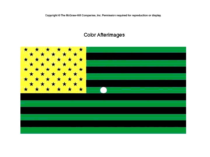
Synesthesia • an experience in which one sense (say, sight) induces an experience in another sense (say, hearing) – Possible Outcomes: see music, taste color
Structure of the Eye
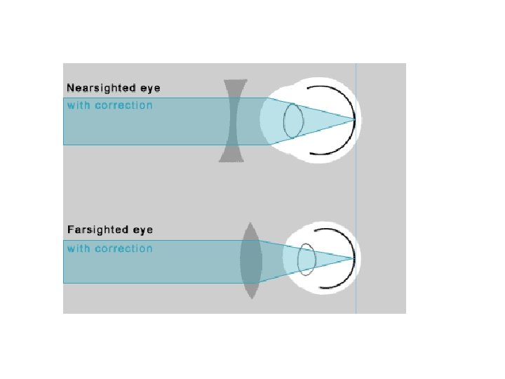
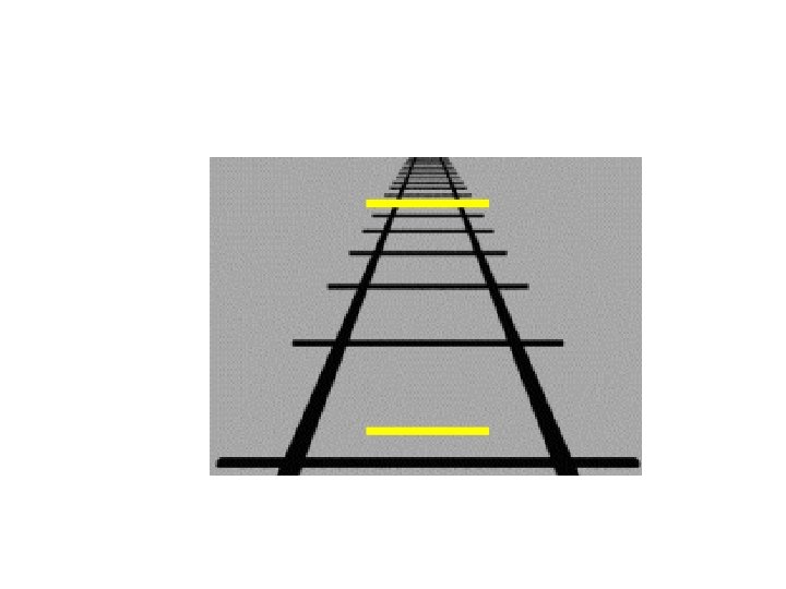
Acuity- the sharpness of vision Light rays from distant objects focus in front…when image reaches the back, the rays spread out creating a blur. Light rays from near objects focus behind the retina creating a blur.
Retina’s Reaction to Light- Receptors **Rods are more sensitive to light than the cones which is why the world looks colorless at night. Cones – near center of retina (fovea) – fine detail and color vision – daylight or well-lit conditions Rods – peripheral retina – detect black, white and gray – twilight or low light **Nocturnal animals such as mice, toads, rats and bats have retinas made up almost entirely of rods.
Vision- Receptors in the Human Eye Cones Rods Number 6 million 120 million Location in retina Center Periphery Sensitivity in dim light Low High Color sensitive? Yes No
Aim: What are the steps in the visual process? DO NOW: • What are the differences between the two visual receptors (rods and cones)? • What is the optic disk? HW: Read “ Watch out for Visual Cliff” packet pps. To W/ Questions
Photoreceptors & Light Control Where are these? • blind spot – the place on the retina where the optic nerve leaves the eye on its way to the brain – NO photoreceptors – cannot see anything that reaches only this part of the retina – brain uses top-down processing to to “fill in the gaps” • fovea – a tiny area in the center of the retina at which vision is at its best • contains only cones • explains difficulty of reading out of corner of eye
• • • Steps in Transduction Concept Map Watch Video & Add to Concept Map Bipolar cells • • Cones • Cornea Ganglion cells • • Iris Lens • Rods Light wave • retina Pupil Occipital lobe Optic nerve https: //www. youtube. com/watch? v=g. Bdy. U 1 b 0 ADQ
Step 1 - Gathering Light • Gather Light – -light reflected off object, and travels though space in waves • Waves determines hue or color • color we see depends upon light intensity & hue Wavelength – distance from one wave to next • The length of the wave gives us it’s hue (color) ROY G BIV • The longer the wave the more red, The shorter the wavelength the more violet. Amplitude – waves height determines it’s intensity (brightness).
Path of the Light Wave Through the Eye How is the eye similar to a camera? A camera must gather light and focus it, just like an eye! • cornea – transparent membrane covering front of eye, Bends light waves inward (brings image into focus) • pupil – the opening in the center of the iris (muscle controls size of pupil), through which light passes • lens –brings image into focus – Curvature – allows eye to focus on things up close (more curved) or far away (flatter) – Ability to curve decreases with age (thus stylish reading glasses)
Path of the Light Wave Through the Eye – Cont’d • Image of object is reflected upside down on the retina (light -sensitive layer of cells in the back of the eye, converts light waves to neural impulses for processing in the brain ) It contains two types of photoreceptors …. • rods –sensitive to light, not only very useful for color vision • Found almost everywhere on the retina except the fovea • Gives ability to detect fainter spots of light on the peripheral • cones –allow for color perception https: //www. youtube. com/watch? v=Wy. Lfm 5 Hs. Hc&feature=youtu. be&app=desktop
Transduction cont’d • Retina (rods/cones) bipolar cells ganglion cells (axons = optic nerve • optic nerve – the structure at the back of the eye, made up of axons of the ganglion cells, that carries visual information for further processing
Transduction Continued • Optic nerve sends info to thalamus- area called lateral geniculate nucleus (LGN). • Then sent to cerebral cortexes. • Where the optic nerves cross is called the optic chiasm.
Visual Processing • visual information originating in the right halves of the two retinas (from the left visual field) is transmitted to the right side of the occipital lobe in cerebral cortex; left half of retinas (right visual field) to left occipital lobe
Phase Four: In the Brain • Goes to the Visual Cortex located in the Occipital Lobe of the Cerebral Cortex. • Feature Detectors. • Parallel Processing We have specific cells that see the lines, motion, curves and other features of this turkey. These cells are called feature detectors.
Feature Detectors • feature detectors – neurons in the brain’s visual system that respond to particular features of a stimulus • Specialized nerve cells in brain respond to specific features of visual stimuli (lines, edge, movement, angle). Discovered by Hubel & Weisel http: //www. youtube. com/watch? v=IOHayh 06 LJ 4&safety_mode=t rue&persist_safety_mode=1&safe=active
Visual Information Processing Feature Detectors – neurons in the visual cortex respond to specific features – shape – angle – movement Cell’s responses Stimulus Illusory Contours So what if these are damaged? Subjective Contours http: //www. youtube. com/watch? v=I 4 cj. MV 98 e 10
The Visual Cortex: David Hubel and Torsten Wiesel (1963) • won Nobel Prize for research on feature detectors (in cats) – recorded activity of a single neuron in a cat while it looked at patterns that varied in size, shape, color , and movement – found that the visual cortex has neurons that are individually sensitive to different types of lines and angles – Noted that when deprived of certain types of visual stimulation early one, kittens lost the ability to perceive these patterns • Suggests there might be a critical period in visual development and that the brain requires stimulation it its efforts to delegate its resources to different perceptual tasks brain “learns” to perceive through experience. So what if these are damaged? http: //www. youtube. com/watch? v=I 4 cj. MV 98 e 10
Feature Detectors • feature detectors – neurons in the brain’s visual system that respond to particular features of a stimulus • Specialized nerve cells in brain respond to specific features of visual stimuli (lines, edge, movement, angle). Discovered by Hubel & Weisel http: //www. youtube. com/watch? v=IOHayh 06 LJ 4&safety_mode=t rue&persist_safety_mode=1&safe=active
Aim: How do we see color/ depth? DO NOW: • Take out your HW. • Read 20 Amazing Facts about your eye http: //discoveryeye. org/20 -facts-about-theamazing-eye/ HW:
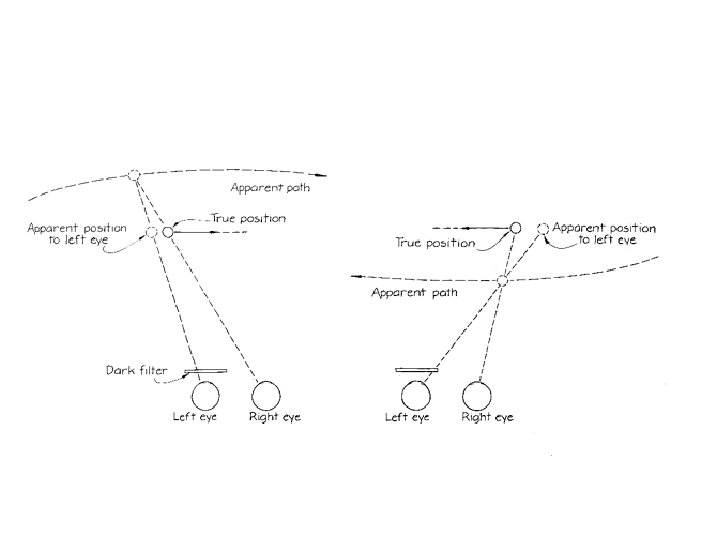
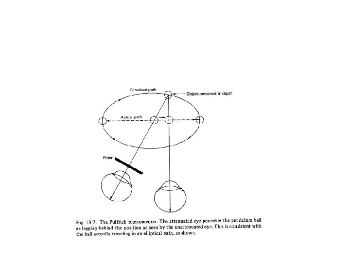
Pulfrich Illusion • When a dark lens placed in front the right eye the pendulum seems to take on an elliptical orbit, and appears to be revolving counterclockwise. • If the left eye is covered, the pendulum would appear to be revolving clockwisefrom-top, appearing closer as it swings toward the left and farther as it swings toward the right.
Pulfrich Illusion • Eye behind the dark lens perceives the image later than the eye behind the clear lens. • Pulfrich illusion occurs because of the time delay between your two eyes. The delay/ difference is perceives as depth. • Retinal Disparity
Depth Cues • Eleanor Gibson and her Visual Cliff Experiment. • If you are old enough to crawl, you are old enough to see depth perception. • We see depth by using two cues that researchers have put in two categories: • Monocular Cues • Binocular Cues
Binocular Cues https: //docs. google. com/a/napls. us/view er? a=v&pid=sites&srcid=bm. Fwb. HMud. X • We need both of our eyes to use these cues. • Convergence (as an object comes closer our eyes have to come together to keep focused on the object). • Retinal Disparity (as an object comes closer to us, the differences in images between our eyes becomes greater.
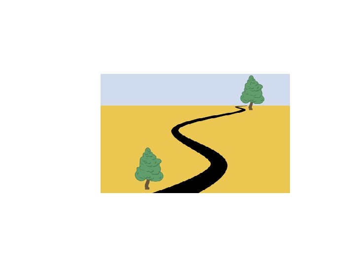
Monocular Cues • You really only need one eye to use these (used in art classes to show depth). • Linear Perspective • Interposition • Relative size • Texture gradient • Shadowing • Height in Field
IDENTIFY THE MONOCULAR CUE USED….
Linear Perspective
Texture Gradient
Interposition
Relative Size Cue
Vision • Our most dominating sense. • Visual Capture
Color Vision Two Major Theories
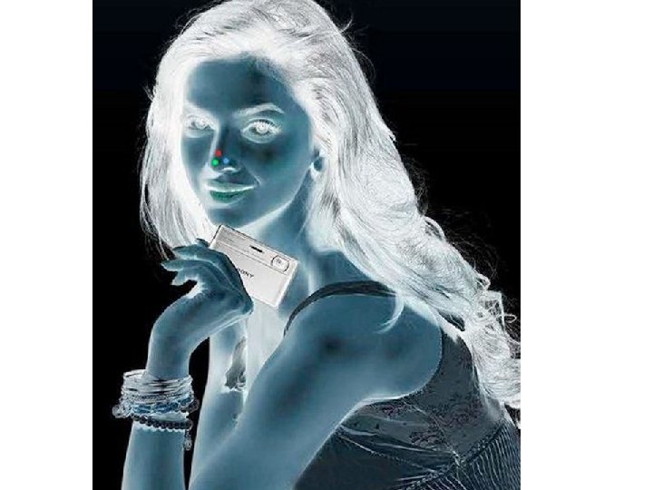
Trichromatic Theory Three types of cones: • Red • Blue • Green • These three types of cones can make millions of combinations of colors. • Does not explain afterimages, but DOES explain color blindness.
Visual Information Processing Parallel Processing – simultaneous processing of several dimensions through multiple pathways (color, motion, form, depth) Trichromatic (three color) Theory – Young (1802) and Helmholtz (1850) – three different retinal color receptors • red You see colors according to their • green response to the wavelengths of light • blue striking the retina---short-preferring (blue), middle-preferring (green), and long-preferring (red).
Visual Information Processing Opponent-Process Theory- opposing retinal processes enable color vision “OFF” green red yellow white black “ON” red green blue yellow blue black white
Visual Information Processing Color Constancy Perceiving familiar objects as having consistent color, even if changing illumination alters the wavelengths reflected by the object
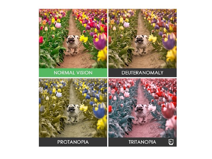
Are You Colorblind? Color Blindness – inability to perceive colors – lacks cones or malfunction cones Color Weakness – inability to distinguish some colors Red-green most common in men, recessive trait on X chromosome
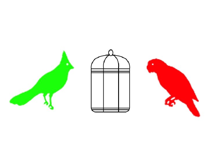
Opponent-Process theory The sensory receptors come in pairs. • Red/Green • Yellow/Blue • Black/White • If one color is stimulated, the other is inhibited.
Both Photos: Thomas Eisner The Stimulus Input: Light Energy Visible Spectrum 57
What is changing in each panel?
Light Characteristics 1. Wavelength (hue/color) 2. Intensity (brightness) 3. Saturation (purity) 59
Wavelength (Hue) Hue (color) is the dimension of color determined by the wavelength of the light. 60
What determines the wavelength (Hue)? Violet Indigo 400 nm Short wavelengths Blue Green Yellow Orange Red 700 nm Long wavelengths 61
Intensity (Brightness) Intensity Amount of energy in a wave determined by the amplitude. 62
Intensity (Brightness) 63
Purity (Saturation) Saturated 64