Agarose Polysaccharide extracted from sea weed Gel casted


- Slides: 17
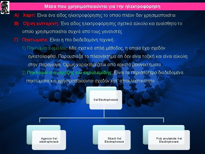
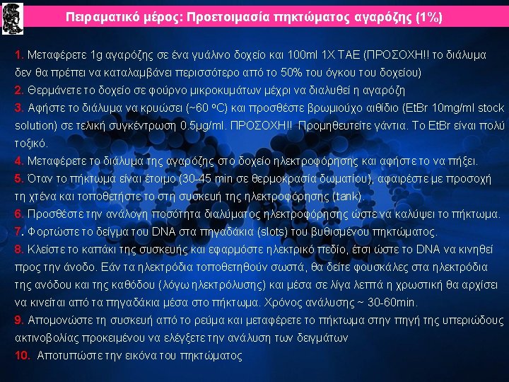
Είδη πηκτωμάτων Agarose • • • Polysaccharide extracted from sea weed. Gel casted horizontally Non-toxic. Separate large molecules Commonly used for DNA separations. Staining can be done before or pouring the gel. Polyacrylamide Gel • • • Cross-linked polymer of acrylamide. Gel casted vertically. Potent neuro-toxic. Separate small molecules. Used for DNA or protein separations. Staining can be done after pouring the gel.
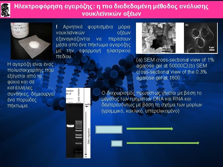
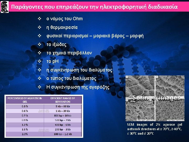
TYPES OF AGAROSE • Standard Agarose - LE Ø Gels at 35 -38 o. C; Melts at 90 -95 o. C Ø Becomes opaque at high concentrations • Low Melting Agarose (Nu. Sieve) Ø Gels at 35 o. C; Melts at 65 o. C Ø Often used to isolate DNA fragments from gel v Intermediate forms or combinations of LE and Nu. Sieve can provide sturdy, translucent gels at high agarose concentrations. % Agarose (w/v) Size Range (kb pairs)for Optimal Separation • 0. 5 2 -30 • 0. 75 0. 7 -20 • 1. 0 0. 5 -10 • 1. 5 0. 2 -3 • 2. 0 0. 1 -2 • 3. 0 (Nu-Sieve) 0. 07 -1. 5 • 4. 0 (N-S) 0. 04 -0. 9 • 5. 0 (N-S) 0. 03 -0. 6 • 6. 0 (N-S) 0. 01 -0. 4
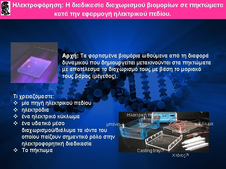
Applied voltage • • voltage, rate of migration The higher the voltage, the more quickly the gel runs But if voltage is too high, gel melts The best separation will apply voltage at no more than 5 V/cm of gel length.
Buffers • • During electrophoresis water undergoes hydrolysis : H 2 O H + OH- • Most common buffer used is called TRIS • • • Buffers prevent the p. H from changing by reacting with the H+ or OHproducts – [tris(hydroxymethyl)aminomethane] Another compound is added to make Tris an effective buffer — either boric or acetic acid Another compound is added to bind metals EDTA The buffer is either TBE or TAE Ø TBE is made with Tris/Boric Acid/EDTA Ø TAE is made with Tris/Acetic Acid/ EDTA
Staining of DNA • • • To make DNA fragments visible after electrophoresis, the DNA must be stained The favorite—ethidium bromide: a fluorescent dye that intercalates between bases of nucleic acids and allows very convenient detection of DNA fragments in gels. When bound to DNA it fluoresces under ultraviolet light (reddish –orange colour) Convenient because it can be added directly to the gel Sensitive—detects 0. 1 ug of DNA • The standard concentration used in staining DNA in gels is 0. 5 -1 ug/m. L • Detection limit of bound DNA is 0. 5 -5 ng/band. • Ethidium bromide is mutagenic so care must be taken while handling the dye Other alternatives for ethidium bromide : Ø Methylene blue Ø Syber safe Ø xylene cyanol Ø bromphenol blue
Results A 1% agarose 'slab' gel prior to UV illumination, behind a perspex UV shield. Only the marker dyes can be seen The gel with UV illumination, the ethidium bromide stained DNA glows orange Digital photo of the gel. Lane 1. Commercial DNA Markers (1 kbplus), Lane 2. empty, Lane 3. a PCR product of just over 500 bases, Lane 4. Restriction digest showing the a similar fragment cut from a 4. 5 kb plasmid vector
DNA ladder • • • It is a solution of DNA molecules of different length DNA Ladder consists of known DNA sizes used to determine the size of an unknown DNA sample. The DNA ladder usually contains regularly spaced sized samples which when run on an agarose gel looks like a "ladder".