AEROBIC PHASE OF RESPIRATION 1 2 3 4

AEROBIC PHASE OF RESPIRATION 1. 2. 3. 4. Questions for consideration: Aerobic phase of respiration Generation of ATP Glyoxylate cycle Ways of regulation of respiration.

An overview of cellular respiration

Aerobic respiration phase

• • Plant mitochondria have two membranes: a smooth outer membrane that completely surrounds a highly invaginated inner membrane. Most inorganic ions and charged organic molecules are not able to diffuse freely into the matrix space. The inner membrane is the osmotic barrier; the outer membrane is permeable to solutes that have a molecular mass of less approximately 10, 000 Da (i. e. , most cellular metabolites and ions, but not proteins). The lipid fraction of both membranes is primarily made up of phospholipids, 80% of which are either phosphatidylcholine or phosphatidylathanolamine. Pyruvate is a charged molecule, it must enter mitochondrion via active transport, with the help of a transport protein. Next, a complex of several enzymes (the pyruvate dehydrogenase complex) catalyzes the three numbered steps. The acetyl group of acetyl Co. A will enter the citric acid cycle. The CO 2 molecule will diffuse out of the cell.
 activation of pyruvate; B) Oxidation of")
Aerobic respiration phase Goes in 3 stages: A) activation of pyruvate; B) Oxidation of pyruvate in the TAC (Krebs cycle); C) transfer of H + and ē to the ETC, while O 2 of air is connected to this process only at the last stage. • During oxidative phosphorylation electron transport chains convert the chemical energy to a form used for ATP synthesis in the process chemiosmosis • •

Conversation of pyruvate to acetyl Co. A, the junction between glycolysis and the citric acid cycle pyruvate dehydrogenase (PDH)
 • Pyruvate dehydrogenase exists as a large complex of several enzymes")
Pyruvate dehydrogenase (PDH) • Pyruvate dehydrogenase exists as a large complex of several enzymes that catalyze the overall reaction in a three-step process: decarboxylation, oxidation, and conjugation to Co. A.

• Activation of pyruvate takes place with the participation of a polyenzyme complex (3 enzymes + 5 coenzymes - Co. A, lipoic acid, thiamine pyrophosphate, FAD, NAD). • It should be noted that acetyl - Co. A – • 1) one of the most important intermediates of metabolism, the nodal point of crossing many paths, • 2) the link between the metabolism of fats and carbohydrates; • 3) the point at which many biosynthetic processes start.
")
1939 А. Chibnell (for plants)
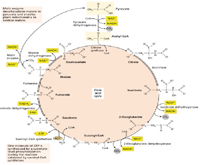

The stepwise oxidation of one molecule of pyruvate in the mitochondrion gives rise to three molecules of CO 2, and much of the free energy released during these oxidations is conserved in the form of 4 NADH and FADH 2. In addition, one molecule of ATP is produced by a substrate-level phosphorylation during the citric cycle

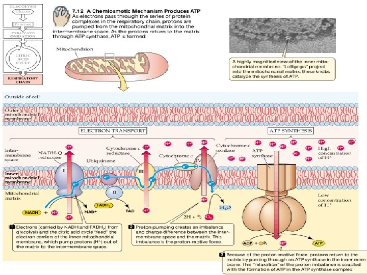
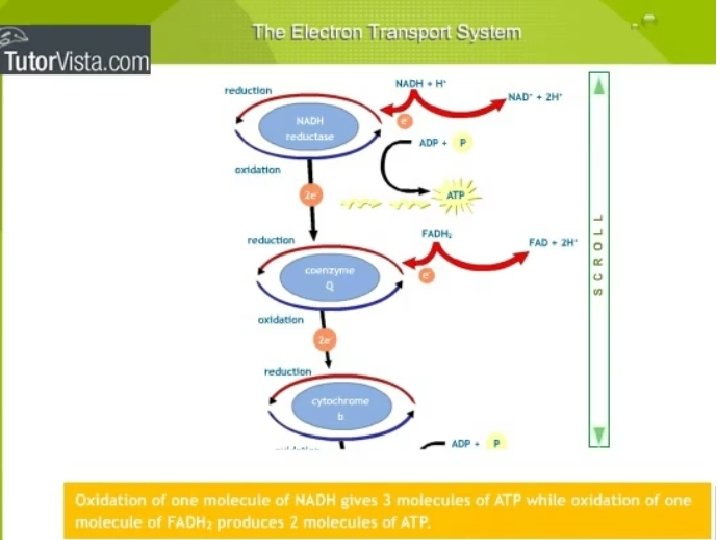
ELECTRON TRANSPORT AND ATP SYNTHESIS ATP is the energy carrier used by cells to drive living processes, and chemical energy conserved during the citric acid cycle in the form of NADH and FADH 2 (redox equivalents with high-energy electrons) must be converted to ATP to perform useful work in the cell. This O 2 -dependent process, called oxidative phosphorylation, occurs in the inner mitochondrial membrane. The electron transport chain catalyzes a flow of electrons from NADH to O 2.
 to")
The electron transport chain catalyses an electron flow from NADH (and FADH 2) to oxygen, the final electron acceptor of the respiratory process. • The electron transport chain of plants contains the same set of electron carriers found in mitochondria from other organisms. The individual electron transport proteins are organized into four multiprotein complexes (identified by Roman numerals I through IV), all of which are localized in the inner mitochondrial membrane:
. • Electrons from NADH generated in the mitochondrial matrix during")
Complex I (NADH dehydrogenase). • Electrons from NADH generated in the mitochondrial matrix during the citric acid cycle are oxidized by complex I (an NADH dehydrogenase). The electron carriers in complex I include a tightly bound cofactor (flavin mononucleotide – FMN), which is chemically similar to FAD; and several iron-sulfur centers. Complex I then transfers these electrons to ubiquinone. Four protons are pumped from the matrix to the inter membrane space for every electron pair passing through the complex.

Ubiquinone, • a small lipid-soluble electron and proton carrier, is located within the inner membrane. It is not tightly associated with any protein, and it can diffuse within the hydrophobic core of the membrane bilayer.
. • Oxidation of succinate in the citric acid cycle is")
Complex II (succinate dehydrogenase). • Oxidation of succinate in the citric acid cycle is catalyzed by this complex, and the reducing equivalents are transferred via the FADH 2 and a group of iron-sulfur (Fe. S) proteins into the ubiquinone pool. • This complex does not pump protons.
. • This complex oxidizes reduced ubiquinone (ubiquinol) and")
Complex III (cytochrome bc 1 complex). • This complex oxidizes reduced ubiquinone (ubiquinol) and transfers the electrons via an iron-sulfur center, two b-type cytochromes (b 565 and 560) and a membrane-bound cytochrome c 1 to cytochrome c. • Four protons per electron pair are pumped by complex III.

Cytochrome с • is a small protein loosely attached to the outer surface of the inner membrane and serves as a mobile carrier to transfer electrons between complexes III and IV.
. • This complex contains two copper centers (Cu. A")
Сomplex IV (cytochrome c oxidase). • This complex contains two copper centers (Cu. A and Cu. B) and cytochromes a and a 3. Complex IV is the terminal oxidase and brings about the fourelectron reduction of 02 to two molecules of H 20. Two protons are pumped per electron pair. • Both structurally and functionally, ubiquinone and the cytochrome bc 1 complex are very similar to plastoquinone and the cytochrome b 6 f complex, respectively, in the photosvnthetic electron transport chain.

• It is interesting to note that plant mitochondria contain some components not found in mammalian mitochondria. • Note that none of these additional enzymes pump protons and that energy conservation is therefore lower whenever they are used:

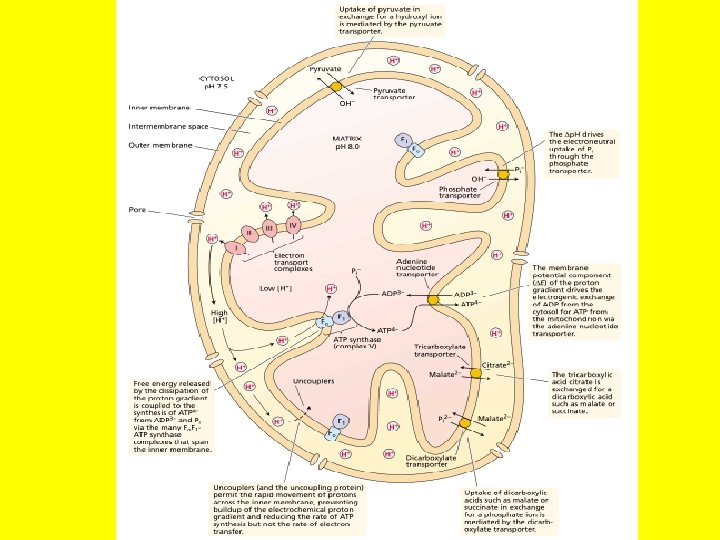
Organization of the electron transport chain and ATP synthesis in the inner membrane of plant mitochondria.

• In addition to the five standard protein complexes found in nearly all other mitochondria, the electron transport chain of plant mitochondria contains five additional enzymes in green. • None of these additional enzymes pumps protons. Specific inhibitors, rotenone for complex I, antimycin for complex III, cyanide for complex IV, and salicylhydroxamic acid (SHAM) for the electron transport chain of plant mitochondria.
H dehydrogenases, both Ca 2+dependent, attached to the outer surface of the inner")
Two NAD(P)H dehydrogenases, both Ca 2+dependent, attached to the outer surface of the inner membrane facing the intermembrane space can oxidize cytosolic NADH and. NADPH. • Plant mitochondria have two pathways for oxidizing matrix NADH. Electron flow through complex I is sensitive to inhibition by several compounds, including rotenone and piericidin. In addition, plant mitochondria have a rotenone-resistant dehydrogenase.

• Most, if not all, plants have an "alternative" respiratory pathway for the reduction of oxygen. • This pathway involves the so-called alternative oxidase that, unlike cytochrome c oxidase, is insensitive to inhibition by cyanide, azide, or carbon monoxide.

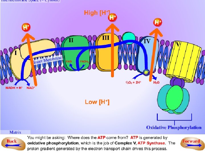
Generation of ATP • In oxidative phosphorylation, the transfer of electrons to oxygen via complexes I to IV is coupled to the synthesis of ATP from ADP and Pi via the ATP synthase (complex V). • The number of ATPs synthesized depends on the nature of the electron donor. • The mechanism of mitochondrial ATP synthesis is based on the chemiosmotic hypothesis, which was first proposed in 1961 by Nobel laureate Peter Mitchell as a general mechanism of energy conservation across biological membranes.

According to the chemiosmotic theory, the orientation of electron carriers within the mitochondrial inner membrane allows for the transfer of protons (H+) across the inner membrane during electron flow. • Numerous studies have confirmed that mitochondrial electron transport is associated with a net transfer of protons from the mitochondrial matrix to the intermembrane space. • Because the inner mitochondrial membrane is impermeable to H+, an electrochemical proton gradient can build up. The free energy associated with the formation of an electrochemical proton gradient (ΔμH+, also referred to as a proton motive force, Δp, when expressed in units of volts) is made up of an electric transmembrane potential component (ΔE) and a chemical-potential component (Δp. H).
 across")
• ΔE results from the asymmetric distribution of a charged species (H+) across the membrane, and Δp. H is due to the proton concentration difference across the membrane. Because protons are translocated from the mitochondrial matrix to the intermembrane space, the resulting ΔE across the inner mitochondrial membrane is negative.

• The free-energy input required to generate ΔμH+ comes from the free energy released during electron transport. How electron transport is coupled to proton translocation is not well understood in all cases. Because of the low permeability (conductance) of the inner membrane to protons, the proton electrochemical gradient is reasonably stable, once generated, and the free energy ΔμH+ can be utilized to carry out chemical work (ATP synthesis). • The ΔμH+ is coupled to the synthesis of ATP by an additional protein complex associated with the inner membrane, the F 0 F 1 -ATP synthase
 consists of two major")
The F 0 F 1 -ATP-synthase (also called complex V) consists of two major components, F 1 and F 0. F 1 is a peripheral membrane protein complex that is composed of at least five different subunits and contains the catalytic site for converting ADP and Pi to ATP. This complex is attached to the matrix side of the inner membrane. F 0 is an integral membrane protein complex that consists of at least three different polypeptides that form the channel through which protons cross the inner membrane. The passage of protons through the channel is coupled to the catalytic cycle of the F 1 component of the ATP synthase, allowing the ongoing synthesis of ATP and the simultaneous utilization of the ΔμH+ each ATP synthesized, 2 H+ pass through the F 0 from the intermembrane space to the matrix down the electrochemical proton gradient. The structure and function of the mitochondrial ATP synthase is similar to that of the CF 0 -CF 1 ATP synthase in photophosphorylation.

The operation of a chemiosmotic mechanism of ATP synthesis has several implications. • First, the true site of ATP formation on the mitochondrial inner membrane is the ATPsynthase, not complex I, III, or IV. These complexes serve as sites of energy conservation whereby electron transport is coupled to the generation of a ΔμH+. • Second, the chemiosmotic theory explains the action mechanism of uncouplers, a wide range of chemically unrelated compounds (including 2, 4 -dinitrophenol and FCCP [p-trifluonumethoxycarbonylcyanide phenylhydrazone]) that decreases mitochondrial ATP synthesis but often stimulates the rate of electron transport. • All of these compounds make the inner membrane leaky to protons, which prevents the buildup of a sufficiently large ΔμH+ to drive ATP synthesis.

• ADP provides a substrate that stimulates dissipation of the ΔμH+ through the F 0 F 1 ATPsynthase during ATP synthesis. Once all the ADP has been converted to ATP, the ΔμH+ builds up again and reduces the rate of electron flow (state 4), The ratio of the rates with and without ADP (state 3: . state 4) is referred to as the respiratory control ratio.

Glyoxylate cycle

• After germinating, oil-containing seeds metabolize stored triacylglycerols by converting lipids to sucrose. • This process involves several steps that are located in different cellular compartments: oleosomes, glyoxysomes, mitochondria, and cytosol.

Ways of regulation of respiration • The substrates of ATP synthesis—ADP and Pi—appear to be key regulators of the rates of glycolysis in the cytosol, as well as the citric acid cycle and oxidative phosphorylation in the mitochondria. Control points exist at all three stages of respiration. • The best-characterized site of regulation of the citric add cycle is at the pyruvate dehydrogenase complex, which is reversibly phosphorylated by a regulatory kinase and a phosphatase. • Pyruvate dehydrogenase is inactive in the phosphorylated state, and the regulatory kinase is inhibited by pyruvate, allowing the enzyme to be active when substrate is available. • In addition, several citric acid cycle enzymes, including pyruvate dehydrogenase and 2 -oxoglutarate dehydrogenase, are directly inhibited by NADH.

• The conversion of lipids to sucrose in oilseeds is triggered by germination and begins with the hydrolysis of triacylglycerols stored in the oil bodies to free fatty acids, followed by oxidation of the fatty acids to produce acetyl. Co. A. • The fatty acids are oxidized in a type of peroxisome called a glyoxysome, an organelle enclosed by a single bilayer membrane that is found in the oil-rich storage tissues of seeds. Acetyl-Co. A is metabolized in the glyoxysome to produce succinate, which is transported from the glyoxysome to the mitochondrion, where it is converted first to oxaloacetate and then to malate. • The process ends in the cytosol with the conversion of malate to glucose via gluconeogenesis, and then to sucrose.
 activity by reversible phosphorylation and by other metabolites")
Regulation of pyruvate dehydrogenase (PDH) activity by reversible phosphorylation and by other metabolites

Concept of bottom-up regulation of plant respiration
- Slides: 43