ADVANCED ASSESSMENT ECG INTERPRETATION Version 2014 OBHG Education

ADVANCED ASSESSMENT ECG INTERPRETATION Version 2014 OBHG Education Subcommittee

ADVANCED ASSESSMENT ECG INTERPRETATION AUTHOR REVIEWERS/CONTRIBUTORS Tim Dodd, AEMCA, ACP Al Santos A-EMCA, ACP Hamilton Base Hospital Donna L. Smith AEMCA, ACP Hamilton Base Hospital References – Emergency Medicine 2014 Version OBHG Education Subcommittee

Lecture Overview 2014 Version Anatomy review Electrical impulse conduction ECG paper constants Limb placements Heart rate determination Steps to rhythm strip analyzes ECG interpretations! OBHG Education Subcommittee

Objectives To gain a basic knowledge of cardiac electrophysiology To understand the representations of the cardiac cycle on the ECG To master techniques used for learning the characteristics of the different dysrhythmias To learn measurement and count techniques essential for dysrhythmia interpretation 2014 Version OBHG Education Subcommittee

Lead Placement and ECG “View” Our cardiac monitors have the ability to display ECG rhythms In manual mode it is possible to capture various leads or “views” of the electrical activity No matter which view is being displayed, the underlying rhythm does NOT change 2014 Version OBHG Education Subcommittee

Bipolar Leads or Limb Leads (I, III) Version 2014 OBHG Education Subcommittee

Limb Leads Einthoven’s triangle is composed of the standard (bipolar) limb leads: Lead I: negative pole is right arm & positive is left arm Lead II: negative pole is right arm & positive left leg Lead III: negative pole is left arm & positive left leg 2014 Version OBHG Education Subcommittee

Lead Placement and Views Negative Lead I Positive Ground 2014 Version OBHG Education Subcommittee

Lead Placement and Views Negative Le ad Ground II Positive 2014 Version OBHG Education Subcommittee

Lead Placement and Views Negative Ground Lead III Positive 2014 Version OBHG Education Subcommittee

Bi. Polar Leads I, II & III Lead I Le ad 2014 Version Lead III II OBHG Education Subcommittee

Limb Leads Lead I - Right Arm + - - Lead II Left Arm Lead III + + Left Leg 2014 Version OBHG Education Subcommittee

Cardiac Monitoring and Lead Placement Version 2014 OBHG Education Subcommittee

3 Lead Electrode Placement RA (White): Place near right mid-clavicular line, directly below the clavicle. The original design in monitoring. Seldom used as an added ground wire reduces interference 2014 Version LA (Black): Place near left mid-clavicular line, directly below the clavicle LL (Red): Place between 6 th & 7 th intercostal Space on left midclavicular line OBHG Education Subcommittee

4 Lead Electrode Placement RA (White): Place near right mid-clavicular line, directly below the clavicle. RL (Green): Place between 6 th and 7 th intercostal Space on right mid-clavicular line 2014 Version LA (Black): Place near left mid-clavicular line, directly below the clavicle LL (Red): Place between 6 th & 7 th intercostal Space on left midclavicular line OBHG Education Subcommittee

5 Lead Electrode Placement RA (White): Place near right mid-clavicular line, directly below the clavicle. LA (Black): Place near left mid-clavicular line, directly below the clavicle V (Brown): Place to Right of sternum at the 4 th intercostal Space RL (Green): Place between 6 th and 7 th intercostal Space on right mid-clavicular line 2014 Version LL (Red): Place between 6 th & 7 th intercostal Space on left midclavicular line OBHG Education Subcommittee

12 Lead Electrode Placement Cardiac monitoring: limb leads may go on the torso 12 lead acquisition: limb leads must go on the extremities 2014 Version OBHG Education Subcommittee

Lead Groups I a. VR V 1 V 4 II a. VL V 2 V 5 III a. VF V 3 V 6 Limb Leads 2014 Version Chest Leads OBHG Education Subcommittee

Inferior Wall II, III and a. VF Left Leg 2014 Version I a. VR V 1 V 4 II a. VL V 2 V 5 III a. VF V 3 V 6 OBHG Education Subcommittee

Inferior Wall 2014 Version I a. VR V 1 V 4 II a. VL V 2 V 5 III a. VF V 3 V 6 Inferior Wall OBHG Education Subcommittee

Septal Wall V 1, V 2 Along sternal borders 2014 Version I a. VR V 1 V 4 II a. VL V 2 V 5 III a. VF V 3 V 6 OBHG Education Subcommittee

Septal Wall • V 1, V 2 2014 Version I a. VR V 1 V 4 II a. VL V 2 V 5 III a. VF V 3 V 6 OBHG Education Subcommittee

Anterior Wall V 3 and V 4 Left anterior chest 2014 Version I a. VR V 1 V 4 II a. VL V 2 V 5 III a. VF V 3 V 6 OBHG Education Subcommittee

Anterior Wall • V 3 and V 4 2014 Version I a. VR V 1 V 4 II a. VL V 2 V 5 III a. VF V 3 V 6 OBHG Education Subcommittee

Lateral Wall - High I and a. VL Left Arm 2014 Version I a. VR V 1 V 4 II a. VL V 2 V 5 III a. VF V 3 V 6 OBHG Education Subcommittee

Lateral Wall - Low V 5 and V 6 Left lateral chest 2014 Version I a. VR V 1 V 4 II a. VL V 2 V 5 III a. VF V 3 V 6 OBHG Education Subcommittee

Lateral Wall I, a. VL, V 5 and V 6 2014 Version I a. VR V 1 V 4 II a. VL V 2 V 5 III a. VF V 3 V 6 OBHG Education Subcommittee

Cardiac Conduction Version 2014 OBHG Education Subcommittee

Cardiac Conduction Essentially 2 pumps Atria Ventricles Must operate in unison and in order Three Primary Pacemakers Sinoatrial Node Atrio-Ventricular Junction Purkinje System 2014 Version OBHG Education Subcommittee

Electromechanics Resting cells: negative interior, positive exterior Changes in this resting state cause depolarization and repolarization 2014 Version OBHG Education Subcommittee

Electrical Flow Towards negative electrode: downward deflection on paper Towards positive electrode: upward deflection on paper As energy travels away from this axis amplitude on the ECG decreases 2014 Version OBHG Education Subcommittee

ECG “View” and Electrical Flow Lead II offers the best “view” The normal electrical axis travels the 11 -5 o’clock vector which is 59 degrees Lead II is at 60 degrees 2014 Version OBHG Education Subcommittee

Sino. Atrial Node Contraction of the Atria Normally depolarizes 60 -80 times/min. Can depolarize up to 300 times/min. Follows special pathways – - Intra. Atrial: Anterior, Middle, Posterior Internodal Tracts 2014 Version OBHG Education Subcommittee

Atrio. Ventricular Node Pauses depolarization to allow ventricular filling Capable of depolarizing 40 -60 times/min. (in the absence of the SA Node input) 2014 Version OBHG Education Subcommittee

Ventricular Pathways Bundle of His Left and Right Bundle Branches Left bundle gives rise to anterior and posterior fascicles Ends at the Purkinje fibers Inherent depolarization rate < 40/min. 2014 Version OBHG Education Subcommittee

2014 Version OBHG Education Subcommittee

Conduction Pathways 2014 Version OBHG Education Subcommittee

Pathway – structure relationship 2014 Version OBHG Education Subcommittee

ECG Paper Horizontal lines: distance in millimeters and time in seconds Vertical lines: voltage (amplitude) in millimeters Uses the Metric system Is very good for accuracy 2014 Version OBHG Education Subcommittee

ECG Paper Light vertical lines are 0. 04 second (1 mm) apart Dark vertical lines are 0. 20 second (5 mm) apart 5 dark squares is 1 second 2014 Version OBHG Education Subcommittee

Cardiac Conduction 2014 Version OBHG Education Subcommittee

Cardiac Conduction: P Wave: Atrial impulse 0. 04 - 0. 08 seconds 2014 Version OBHG Education Subcommittee

Cardiac Conduction: PR Interval P Wave 0. 04 - 0. 08 sec. PR Interval 0. 12 - 0. 20 Sec. 2014 Version OBHG Education Subcommittee

Cardiac Conduction: QRS Complex P Wave 0. 04 - 0. 08 sec. PRI 0. 12 - 0. 20 sec. QRS 0. 06 - 0. 12 sec. 2014 Version OBHG Education Subcommittee

Analyzing a Rhythm Strip Version 2014 OBHG Education Subcommittee

Dysrhythmia Interpretation: 5 Step Approach Step 1: What is the rate? Step 2: Is the rhythm regular or irregular? Step 3: Is the P wave normal? Step 4: P-R Interval/relationship? Step 5: Normal QRS complex? 2014 Version OBHG Education Subcommittee

Step 1 - Rate Method 1 Count the number of R waves for a six second interval and multiply by ten. 6 sec 3 sec (can be used for regular & irregular) 2014 Version OBHG Education Subcommittee

Step 1 - Rate Method 2: Count the number of 5 mm squares and divide into 300 (or memorize) 300 150 100 75 60 50 2014 Version 43 37 33 30 … slow OBHG Education Subcommittee

Step 1 - Rate RATE: Tachycardia exists if the rate is greater than 100 beats/min. Bradycardia exists if the rate is less than 60 beats/min. 2014 Version OBHG Education Subcommittee

Step 2 - Rhythm RHYTHM: Determine if the ventricular rhythm is regular or irregular (pattern to irreg. ? ) R-R intervals should measure the same P-P intervals should also measure the same 2014 Version OBHG Education Subcommittee

REGULAR Step 2 - Rhythm IRREGULAR 2014 Version OBHG Education Subcommittee

STEP 2 - Rhythm Example Irregularly Irregular 2014 Version OBHG Education Subcommittee

STEP 3 – Is the P Wave Normal Identify and examine P waves: Present? Appearance? Consistency? Relation to QRS? 2014 Version OBHG Education Subcommittee

STEP 3 - Is the P Wave Normal Same Shape P wave with no QRS complex Associated with a QRS Complex? 2014 Version OBHG Education Subcommittee

STEP 4 – PR Interval/Relationship Consistent PRI of < 0. 20 secs is normal, lengthened or variant PRI’s indicate an AV block 2014 Version OBHG Education Subcommittee

STEP FIVE –QRS DURATION • A narrow QRS complex (< 0. 12), indicates the impulse has followed the normal conduction pathway • A widened QRS complex (> 0. 12), may indicate the impulse was generated somewhere in the ventricles 2014 Version OBHG Education Subcommittee

REMEMBER!!! Use a systematic approach Go through all the steps Take your time! Compare with your characteristics list Interpret (name) the dysrhythmia 2014 Version OBHG Education Subcommittee

Sinus Rhythms Version 2014 OBHG Education Subcommittee

Sinus Node Normal Sinus Rhythm (NSR) Sinus Bradycardia Sinus Tachycardia Sinus Arrhythmia Sinus Arrest/Pause Sinoatrial Exit Block 2014 Version OBHG Education Subcommittee

Normal Sinus Rhythm (NSR) Rate: 60 -100 beats/min Rhythm: regular P waves: uniform, + (upright) in lead II, one precedes each QRS complex PR interval: 0. 12 - 0. 20 seconds and constant QRS duration: < 0. 12 sec 2014 Version OBHG Education Subcommittee

Normal Sinus Rhythm (NSR) 2014 Version OBHG Education Subcommittee

Sinus Bradycardia Rate: less than 60 beats/min Rhythm: regular P waves: uniform in appearance, upright and one precedes each QRS complex PR interval: 0. 12 - 0. 20 sec and constant QRS duration: < 0. 12 sec 2014 Version OBHG Education Subcommittee

Sinus Bradycardia 2014 Version OBHG Education Subcommittee

Sinus Tachycardia Rate: typically between 100 -150 beats/min Rhythm: regular P waves: uniform, upright, one precedes each QRS PR interval: 0. 12 - 0. 20 sec and constant QRS duration: < 0. 12 sec 2014 Version OBHG Education Subcommittee

Sinus Tachycardia 2014 Version OBHG Education Subcommittee

Sinus Arrhythmia Rate: 60 -100 beats/min Rhythm: irregular P waves: uniform in appearance, upright, one precedes each QRS PR interval: 0. 12 - 0. 20 sec and constant QRS duration: < 0. 12 sec 2014 Version OBHG Education Subcommittee

Sinus Arrhythmia 2014 Version OBHG Education Subcommittee

Sinus Arrest/Pause Rate: usually normal but varies due to the arrest Rhythm: irregular; the arrest is of undetermined length P waves: uniform in appearance, upright, one precedes each QRS PR interval: 0. 12 - 0. 20 sec and constant QRS duration: < 0. 12 sec 2014 Version OBHG Education Subcommittee

Sinus Arrest/Pause 2014 Version OBHG Education Subcommittee

Sinoatrial Exit Block Rate: varies due to the pause Rhythm: irregular; each pause is the same as the distance between two other P-P intervals P waves: uniform in appearance, upright, one precedes each QRS PR interval: 0. 12 - 0. 20 sec and constant QRS duration: < 0. 12 sec 2014 Version OBHG Education Subcommittee

Sinoatrial Exit Block 2014 Version OBHG Education Subcommittee

Atrial Dysrhythmias Version 2014 OBHG Education Subcommittee

Atrial Dysrhythmias Premature Atrial Complexes (PACs) Wandering Atrial Pacemaker (WAP) Supraventricular Tachycardia (SVT) Wolff-Parkinson-White Syndrome (WPW) Atrial Flutter Atrial Fibrillation (A-Fib) 2014 Version OBHG Education Subcommittee

Premature Atrial Complexes (PACs) Rate: depends on underlying rhythm Rhythm: regular with premature beats P waves: premature (earlier than expected) and may differ in shape from sinus P waves PR interval: 0. 12 - 0. 20 sec and constant QRS duration: < 0. 12 sec Note: Ectopic complex 2014 Version OBHG Education Subcommittee

Premature Atrial Complexes (PACs) 2014 Version OBHG Education Subcommittee

Wandering Atrial Pacemaker (WAP) Rate: usually 60 -100 beats/min; if greater than 100 beats/min, it’s termed multifocal atrial tachycardia Rhythm: may be normal or irregular due to shift in ectopic atrial locations P waves: size, shape and direction change (3 or more different P waves) PR interval: may be normal or variable QRS duration: < 0. 12 sec 2014 Version OBHG Education Subcommittee

Wandering Atrial Pacemaker (WAP) 2014 Version OBHG Education Subcommittee

Supraventricular Tachycardia (SVT/PSVT) Rate: 120 -250 beats/min Rhythm: regular P waves: not present PR interval: not measurable QRS duration: < 0. 12 sec Note: PSVT is an SVT that starts/ends suddenly 2014 Version OBHG Education Subcommittee

Supraventricular Tachycardia (SVT) 2014 Version OBHG Education Subcommittee

Wolff-Parkinson-White Syndrome (WPW) Rate: usually 60 -100 beats/min (can be associated with runs/episodes of A-Fib) Rhythm: regular P waves: uniform in appearance, upright, one precedes each QRS PR interval: 0. 12 - 0. 20 sec and constant QRS duration: usually > 0. 12 sec; slurred upstroke of the QRS complex (delta wave) 2014 Version OBHG Education Subcommittee

Wolff-Parkinson-White Syndrome (WPW) 2014 Version OBHG Education Subcommittee

Atrial Flutter Rate: atrial rate 250 -450 beats/min; ventricular rate will not usually exceed 180 beats/min Rhythm: atrial regular; ventricular regular or irregular P waves: saw-toothed “flutter” waves PR interval: not measurable QRS duration: < 0. 12 sec 2014 Version OBHG Education Subcommittee

Atrial Flutter 2014 Version OBHG Education Subcommittee

Atrial Fibrillation (Afib) Rate: variable Rhythm: irregularly irregular P waves: no identifiable P waves PR interval: not measurable QRS duration: < 0. 12 sec 2014 Version OBHG Education Subcommittee

Atrial Fibrillation (A-fib) 2014 Version OBHG Education Subcommittee

Junctional Rhythms Version 2014 OBHG Education Subcommittee

Junctional Rhythms Premature Junctional Complex (PJC) Junctional Rhythm Accelerated Junctional Rhythm Junctional Tachycardia 2014 Version OBHG Education Subcommittee

Premature Junctional Complexes (PJC) Rate: usually normal but depends on the underlying rhythm Rhythm: regular with premature beats P waves: may occur before, during or after the QRS (inverted if visible) PR interval: if P visible, < or = to 0. 12 sec QRS duration: < 0. 12 sec Note: Ectopic complex 2014 Version OBHG Education Subcommittee

Premature Junctional Complexes (PJC) 2014 Version OBHG Education Subcommittee

Junctional Rhythm Rate: 40 -60 beats/min Rhythm: regular P waves: not present or may occur before, during or after the QRS (inverted if present) PR interval: not measurable, but if present usually < or = to 0. 12 sec QRS duration: < 0. 12 sec 2014 Version OBHG Education Subcommittee

Junctional Rhythm 2014 Version OBHG Education Subcommittee

Junctional Rhythm 2014 Version OBHG Education Subcommittee

Accelerated Junctional Rhythm Rate: 61 -100 beats/min Rhythm: regular P waves: not present or may occur before, during or after the QRS (inverted if present; may be notched or S waved) PR interval: not measurable, but if present usually < or = to 0. 12 sec QRS duration: < 0. 12 sec 2014 Version OBHG Education Subcommittee

Accelerated Junctional Rhythm 2014 Version OBHG Education Subcommittee

Junctional Tachycardia Rate: 101 -180 beats/min Rhythm: regular P waves: not present or may occur before, during or after the QRS (inverted if present) PR interval: not measurable, but if present usually < or = to 0. 12 sec QRS duration: < 0. 12 sec 2014 Version OBHG Education Subcommittee

Junctional Tachycardia 2014 Version OBHG Education Subcommittee

Ventricular Rhythms Version 2014 OBHG Education Subcommittee

Ventricular Rhythms Premature Ventricular Complexes (PVC's) Idioventricular Rhythm Ventricular Tachycardia (VT) Torsades de Pointes (Td. P) Ventricular Fibrillation (VF) Asystole 2014 Version OBHG Education Subcommittee

Premature Ventricular Complexes (PVC's) Rate: depends on underlying rhythm Rhythm: regular with premature beats P waves: absent PR interval: none QRS duration: > 0. 12 sec, wide and bizarre! Note: Ectopic complex 2014 Version OBHG Education Subcommittee

Premature Ventricular Complexes (PVC's) Note – Complexes not Contractions 2014 Version OBHG Education Subcommittee

PVC’s Uniformed/Multiformed Couplets/Salvos/Runs Bigeminy/Trigeminy/Quadrageminy 2014 Version OBHG Education Subcommittee

Uniformed PVC’s 2014 Version OBHG Education Subcommittee

R on T Phenomena 2014 Version OBHG Education Subcommittee

Multiformed PVC’s 2014 Version OBHG Education Subcommittee

PVC Couplets 2014 Version OBHG Education Subcommittee

PVC Salvos and Runs 2014 Version OBHG Education Subcommittee

Bigeminy PVC’s 2014 Version OBHG Education Subcommittee

Trigeminy PVC’s 2014 Version OBHG Education Subcommittee

Quadrageminy PVC’s 2014 Version OBHG Education Subcommittee

Ventricular Escape Beats Rate: depends on underlying rhythm Rhythm: regular with late beats; occurs after the next expected sinus beat P waves: absent PR interval: none QRS duration: > 0. 12 sec. wide and bizarre Note: Ectopic complex 2014 Version OBHG Education Subcommittee

Ventricular Escape Beats 2014 Version OBHG Education Subcommittee

Idioventricular Rhythm Rate: 20 -40 beats/min Rhythm: regular P waves: absent PR interval: none QRS duration: > 0. 12 sec Note: “Wide and Slow, (Oh No!) it’s Idio” 2014 Version OBHG Education Subcommittee

Idioventricular Rhythm 2014 Version OBHG Education Subcommittee

Ventricular Tachycardia (VT) Rate: 101 -250 beats/min Rhythm: regular P waves: absent PR interval: none QRS duration: > 0. 12 sec. often difficult to differentiate between QRS and T wave Note: Monomorphic means the same shape and amplitude 2014 Version OBHG Education Subcommittee

Ventricular Tachycardia (VT) 2014 Version OBHG Education Subcommittee

V Tach 2014 Version OBHG Education Subcommittee

Torsades de Pointes (Tde. P) Rate: 150 -300 beats/min Rhythm: regular or irregular P waves: none PR interval: none QRS duration: > 0. 12 sec. gradual alteration in amplitude and direction of the QRS complexes 2014 Version OBHG Education Subcommittee

Torsades de Pointes (Tde. P) 2014 Version OBHG Education Subcommittee

Ventricular Fibrillation (VF) Rate: CNO as no discernible complexes Rhythm: rapid and chaotic P waves: none PR interval: none QRS duration: none Note: Fine vs. coarse? 2014 Version OBHG Education Subcommittee

Ventricular Fibrillation (VF) 2014 Version OBHG Education Subcommittee

Ventricular Fibrillation (VF) 2014 Version OBHG Education Subcommittee

Asystole (Cardiac Standstill) Rate: none Rhythm: none P waves: none PR interval: not measurable QRS duration: absent 2014 Version OBHG Education Subcommittee

Asystole (Cardiac Standstill) 2014 Version OBHG Education Subcommittee

Asystole 2014 Version OBHG Education Subcommittee

Atrio. Ventricular (AV) Blocks Version 2014 OBHG Education Subcommittee

Atrioventricular (AV) Blocks First-degree AV block Second-degree AV block, type I (Wenckebach or Mobitz I) Second-degree AV block, type II (Mobitz II) Third-degree AV block (Complete AV Block) 2014 Version OBHG Education Subcommittee

First-degree AV block Rate: depends on underlying rhythm (usually regular) Rhythm: regular P waves: normal in size and shape PR interval: > 0. 20 sec but constant QRS duration: < 0. 12 sec 2014 Version OBHG Education Subcommittee

First-degree AV block 2014 Version OBHG Education Subcommittee

2 nd Degree AV block Type I (Wenckebach/Mobitz Type I) Rate: atrial rate > ventricular rate Rhythm: atrial reg. ; ventricular irreg. P waves: normal in size and shape PR interval: lengthens with each cycle until a P appears with no QRS complex QRS duration: < 0. 12 sec (Periodically dropped QRS complex!) 2014 Version OBHG Education Subcommittee

2 nd Degree AV block Type I (Wenckebach/Mobitz Type I) 2014 Version OBHG Education Subcommittee

2 nd Degree AV block Type II (Mobitz Type II) Rate: atrial rate > the ventricular rate Rhythm: atrial reg. ; ventricular irreg. P waves: normal in size and shape PR interval: within normal limits or slightly prolonged but constant QRS duration: < 0. 12 sec (Periodically dropped QRS complex!) 2014 Version OBHG Education Subcommittee

2 nd Degree AV block Type II (Mobitz Type II) 2014 Version OBHG Education Subcommittee

3 rd Degree AV block (Complete AV Block) Rate: atrial rate is > ventricular rate Rhythm: atrial reg. ; ventricular reg. (P waves plot through) P waves: normal in size and shape PR interval: no relationship (independent) QRS duration: narrow or wide depending on location of pacemaker 2014 Version OBHG Education Subcommittee

3 rd Degree AV block (Complete AV Block) 2014 Version OBHG Education Subcommittee
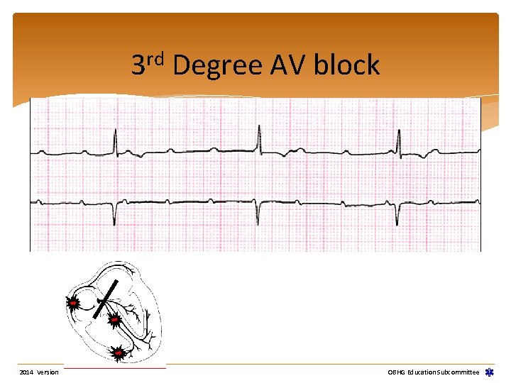
rd 3 2014 Version Degree AV block OBHG Education Subcommittee
Pacemaker Rhythms Version 2014 OBHG Education Subcommittee
Cardiac Pacemakers Definition Delivers artificial stimulus to heart Causes depolarization and contraction Uses Bradyarrhythmias Asystole Tachyarrhythmias (overdrive pacing) NOTE – some monitors require programming to enable pacer detection 2014 Version OBHG Education Subcommittee
Cardiac Pacemakers Fixed Fires at constant rate Can discharge on T-wave Very rare Demand Senses patient’s rhythm Fires only if no activity sensed after preset interval (escape interval) Most common type found 2014 Version OBHG Education Subcommittee
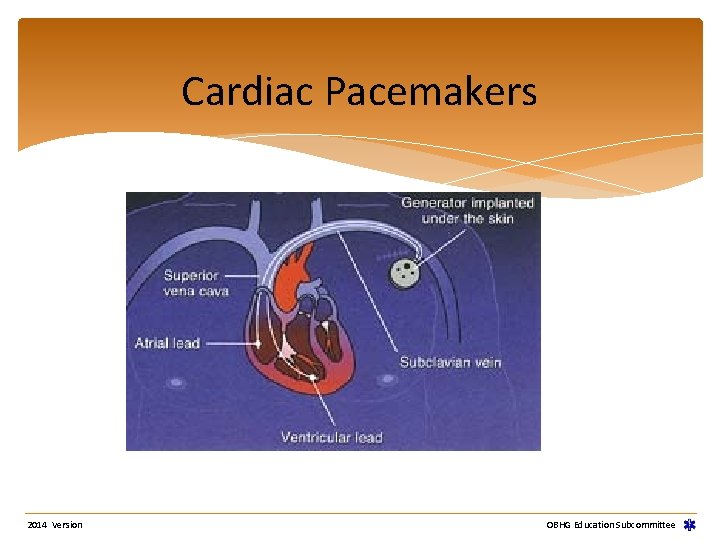
Cardiac Pacemakers 2014 Version OBHG Education Subcommittee
Cardiac Pacemakers Demand Pacemaker Types Atrial Impulse stimulates the atria to contract Atrial wave of depolarization is conducted to the ventricles causing them to contract Requires intact AV conduction Ventricular Impulse stimulates the ventricles to contract 2014 Version OBHG Education Subcommittee
Cardiac Pacemakers Demand Pacemaker Types Atrial Synchronous Senses atrial activity Impulse initiates ventricular contraction AV Sequential Two electrodes (atrial and ventricular) Impulses coordinate atrial/ventricular contractions in sequence 2014 Version OBHG Education Subcommittee
Cardiac Pacemakers Special Considerations Pacemaker does NOT affect treatment of cardiac arrest Do NOT defibrillate directly over pacemaker generator Pacemakers may keep AEDs from advising shock 2014 Version OBHG Education Subcommittee
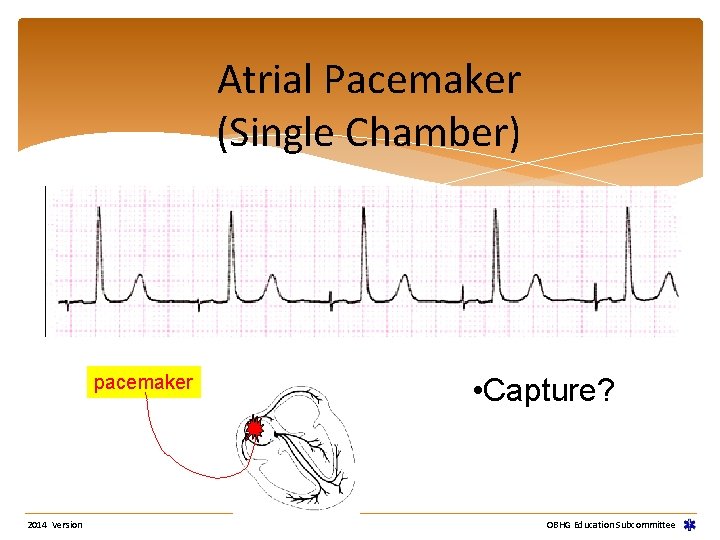
Atrial Pacemaker (Single Chamber) pacemaker 2014 Version • Capture? OBHG Education Subcommittee
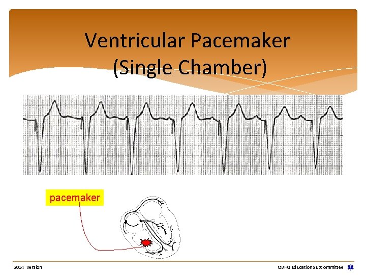
Ventricular Pacemaker (Single Chamber) pacemaker 2014 Version OBHG Education Subcommittee
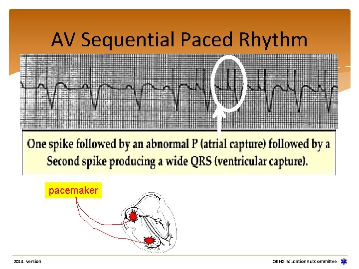
AV Sequential Paced Rhythm pacemaker 2014 Version OBHG Education Subcommittee
Special Notes Version 2014 OBHG Education Subcommittee
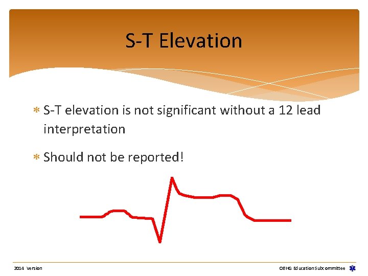
S-T Elevation S-T elevation is not significant without a 12 lead interpretation Should not be reported! 2014 Version OBHG Education Subcommittee
S-T Elevation Monitors filter rhythms and show you what they think it should ST elevation is common in patients who show no elevation on 12 Lead interpretation 2014 Version OBHG Education Subcommittee
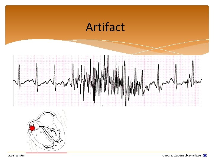
Artifact 2014 Version OBHG Education Subcommittee
60 Cycle Interference 60 cycle artifact (noise) Problem solve causes Electromagnetic emanations: i. e. TV’s, electric motors, fluorescent lights 2014 Version OBHG Education Subcommittee
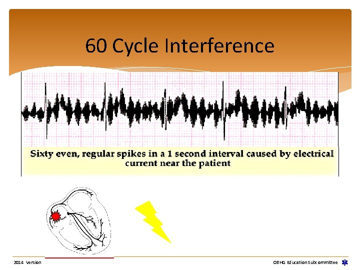
60 Cycle Interference 2014 Version OBHG Education Subcommittee
Aberrancy Bundle Branch Block 12 Lead ECG required to diagnose BBB (Left or right BBB? ) Describe widened QRS (>0. 12 sec) as “aberrant” unless using 12 lead 2014 Version OBHG Education Subcommittee
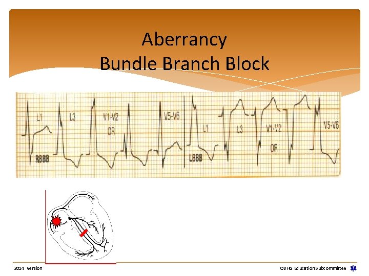
Aberrancy Bundle Branch Block 2014 Version OBHG Education Subcommittee
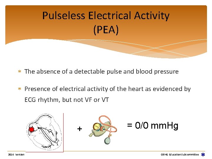
Pulseless Electrical Activity (PEA) The absence of a detectable pulse and blood pressure Presence of electrical activity of the heart as evidenced by ECG rhythm, but not VF or VT + 2014 Version = 0/0 mm. Hg OBHG Education Subcommittee
Practice USE IT OR LOSE IT!!!! Version 2014 OBHG Education Subcommittee

Well Done! Ontario Base Hospital Group Self-directed Education Program 2014 Version OBHG Education Subcommittee
- Slides: 156