Adult Nursing Second Stage Head injuries Head Injuries


 fracture is a break in the continuity of")


 may be seen")
 scan is used to")


 brain injury occurs when the head accelerates and then rapidly")


- Slides: 31
Adult Nursing Second Stage Head injuries ﻓﺎﻃﻤﺔ ﺟﺒﺮ. ﻡ. ﻡ
Head Injuries is a broad classification that includes injury to the scalp, skull, or brain. A head injury may lead to conditions ranging from mild concussion to coma and death; the most serious form is known as a traumatic brain injury (TBI). The most common causes of TBIs are falls (35. 2%), motor vehicle crashes (17. 3%), being struck by objects (16. 5%), and assaults (10%). In every age group, TBI rates are higher for males than females.
Pathophysiology Damage to the brain from traumatic injury takes two forms: primary injury and secondary injury. Primary injury is the initial damage to the brain that results from the traumatic event. This may include contusions, lacerations, and torn blood vessels due to impact; acceleration/deceleration; or foreign object penetration.
Pathophysiology Secondary injury evolves over the ensuing hours and days after the initial injury and results from inadequate delivery of nutrients and oxygen to the cells. These processes include intracranial hemorrhage, cerebral edema, increased intracranial pressure (ICP), hypoxic brain damage, and infection
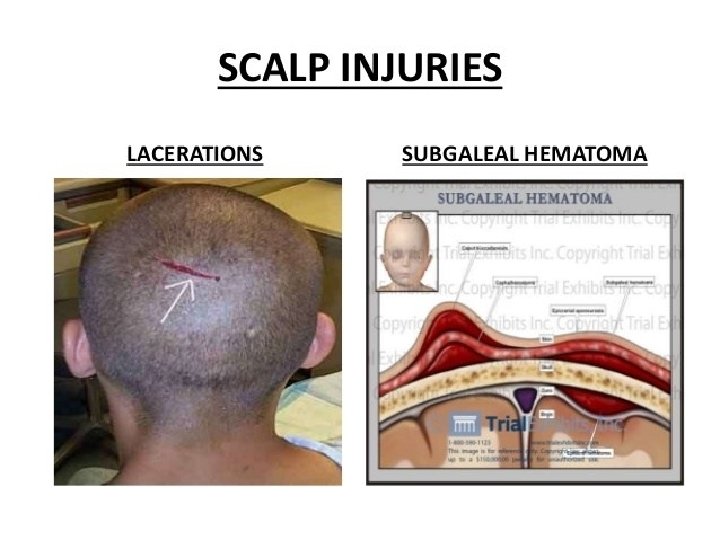
Scalp Injury Isolated scalp trauma is generally classified as a minor injury. Because its many blood vessels constrict poorly, the scalp bleeds profusely when injured. Trauma may result in an abrasion (brush wound), contusion, laceration, or hematoma beneath the layers of tissue of the scalp (subgaleal hematoma). A large avulsion (tearing away) of the scalp may be potentially life threatening and is a Diagnosis of a scalp injury is based on physical examination, inspection, and palpation. true emergency.
Management of scalp injury Scalp wounds are potential portals of entry for organisms that cause intracranial infections. Therefore, the area is irrigated before the laceration is sutured to remove foreign material and to reduce the risk for infection. Subgaleal hematomas (hematomas below the outer covering of the skull) usually reabsorb and do not require any specific treatment.
Skull Fractures A skull fracture is a break in the continuity of the skull caused by forceful trauma. It may occur with or without damage to the brain. Skull fractures are classified by type and location. Types include linear, comminuted, and depressed skull fractures. whereas location fractures include frontal, temporal, and basilar skull fractures.
Skull Fractures • A simple (linear) fracture is a break in the continuity of the bone. • A comminuted skull fracture refers to a splintered or multiple fracture line. • Depressed skull fractures occur when the bones of the skull are forcefully displaced downward • A fracture of the base of the skull is referred to as a basilar skull fracture
Skull Fractures A fracture may be open, indicating a scalp laceration or tear in the dura (e. g. , from a bullet or an ice pick), or closed, in which case the dura is intact.
Clinical Manifestations of Skull Fractures 1. Persistent, localized pain usually suggests that a fracture is present. 2. Swelling in the region of the fracture. 3. Fractures of the base of the skull tend to traverse the paranasal sinus of the frontal bone therefore, they frequently produce hemorrhage from the nose, pharynx, or ears, and blood may appear under the conjunctiva or the middle ear located in the temporal bone
Clinical Manifestations of Skull Fractures 4. An area of ecchymosis (bruising) may be seen over the mastoid (Battle’s sign). 5. Basilar skull fractures are suspected when CSF escapes from the ears (CSF otorrhea) and the nose (CSF rhinorrhea). Drainage of CSF is a serious problem, because meningeal infection can occur if organisms gain access to the cranial contents via the nose, ear, or sinus through a tear in the dura.
Assessment and Diagnosis of Skull Fractures A computed tomography (CT) scan is used to diagnose a skull fracture. The ease with which a diagnosis of skull fracture is made depends on the site of the fracture. If a fracture is found on CT scan. Magnetic resonance imaging (MRI) provides better resolution and clearer pictures of the injured area
Medical Management of skull fractures Nondepressed skull fractures generally do not require surgical treatment; however, close observation of the patient is essential. Nursing personnel may observe the patient in the hospital, but if no underlying brain injury is present, the patient may be allowed to return home. If the patient is discharged home, specific instructions must be given to the family.
Medical Management of skull fractures Depressed skull fractures usually require surgery with elevation of the skull and débridement, usually within 24 hours of injury.
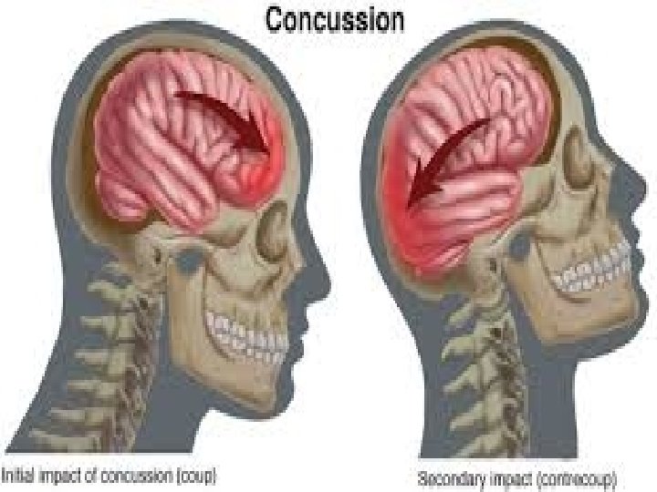
Brain Injury Closed (blunt) brain injury occurs when the head accelerates and then rapidly decelerates or collides with another object (e. g. , a wall, the dashboard of a car) and brain tissue is damaged but there is no opening through the skull and dura
Brain Injury Open brain injury occurs when an object penetrates the skull, enters the brain, and damages the soft brain tissue in its path (penetrating injury), or when blunt trauma to the head is so severe that it opens the scalp, skull, and dura to expose the brain. Injuries to the brain can be focal or diffuse. Focal injuries include contusions and hematomas. Concussions and diffuse axonal injuries are the major diffuse injuries
Types of Brain Injury: concussion A concussion after head injury is a temporary loss of neurologic function with no apparent structural damage to the brain. A concussion (also referred to as a mild TBI) may or may not produce a brief loss of consciousness. The mechanism of injury is usually blunt trauma from an acceleration-deceleration force, a direct blow, or a blast injury
Types of Brain Injury: concussion There are three grades of concussion grade 1 has symptoms of transient confusion, no loss of consciousness, and duration of mental status abnormalities on examination that resolve in less than 15 minutes. A grade 2 also has symptoms of transient confusion and no loss of consciousness, but the concussion symptoms or mental status abnormalities on examination last more than 15 minutes. In a grade 3, there is any loss of consciousness lasting from seconds to minutes
Management of concussion Monitoring includes observing the patient for a decrease in level of consciousness (LOC), worsening headache, dizziness, seizures, abnormal pupil response, vomiting, irritability, slurred speech, and numbness or weakness in the arms or legs
Types of Brain Injury: Contusion a moderate to severe head injury, the brain is bruised and damaged in a specific area because of severe acceleration-deceleration force or blunt trauma. The impact of the brain against the skull leads to a contusion. Contusions are characterized by loss of consciousness associated with stupor and confusion. Other characteristics can include tissue alteration and neurologic deficit without hematoma formation, alteration in consciousness, and hemorrhage into the tissue that varies in size and is surrounded by edema.
Types of Brain Injury : Diffuse Axonal Injury results from widespread shearing and rotational forces that produce damage throughout the brain-to axons in the cerebral hemispheres, corpus callosum, and brain stem. It is associated with prolonged traumatic coma; it is more serious and is associated with a poorer prognosis than a focal lesion or ischemia.
Types of Brain Injury : Diffuse Axonal Injury The patient with DAI in severe head trauma experiences, immediate coma, decorticate and decerebrate posturing and global cerebral edema. Diagnosis is made by clinical signs in conjunction with a CT or MRI scan. Recovery depends on the severity of the axonal injury.
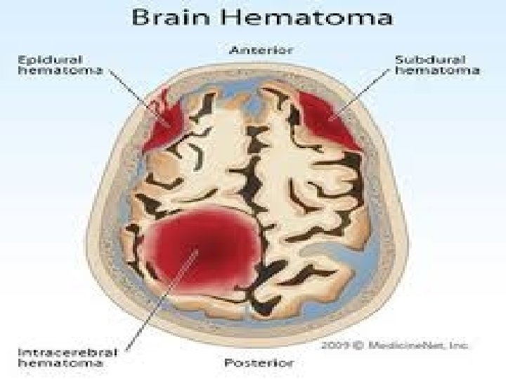
Types of Brain Injury : Intracranial Hemorrhage Hematomas are collections of blood in the brain that may be epidural (above the dura), subdural (below the dura), or intracerebral (within the brain). Major symptoms are frequently delayed until the hematoma is large enough to cause distortion of the brain and increased ICP.
Types of Brain Injury : Intracranial Hemorrhage The signs and symptoms of cerebral ischemia resulting from compression by a hematoma are variable and depend on the speed with which vital areas are affected and the area that is injured. In general, a rapidly developing hematoma, even if small, may be fatal, whereas a larger but slowly developing one may allow compensation for increases in ICP.
Assessment and diagnosis • Physical and neurologic examinations. • CT and MRI scans are the main neuroimaging diagnostic tools and are useful in evaluating the brain structure.
Management of Brain Injuries • Treatment of Increased Intracranial Pressure • Supportive Measures: that include ventilatory support, seizure prevention, fluid and electrolyte maintenance, nutritional support, and management of pain and anxiety. Patients who are comatose are intubated and mechanically ventilated to ensure adequate oxygenation and protect the airway.
Nursing Interventions • Maintaining The Airway: 1. Maintaining the unconscious patient in a position with the head of the bed elevated about 30 degrees to decrease intracranial venous pressure 2. Establishing effective suctioning procedures 3. Closely monitoring arterial blood gas values to assess the adequacy of ventilation. 4. Monitoring the patient who is receiving mechanical ventilation for pulmonary complications such as acute respiratory distress syndrome and pneumonia
Nursing Interventions • Monitoring Neurologic Function : 1. Level of Consciousness 2. Vital Signs 3. Motor Function. • Monitoring Fluid and Electrolyte Balance • Promoting Adequate Nutrition • Preventing Injury • Maintaining Body Temperature • Maintaining Skin Integrity