
ADRENAL GLAND PART 1 The adrenal glands are
- Slides: 16
ADRENAL GLAND PART 1
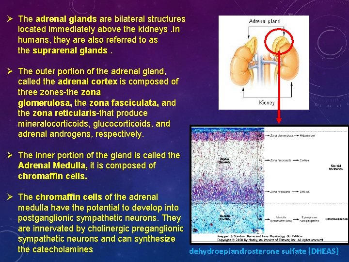
Ø The adrenal glands are bilateral structures located immediately above the kidneys. In humans, they are also referred to as the suprarenal glands. Ø The outer portion of the adrenal gland, called the adrenal cortex is composed of three zones-the zona glomerulosa, the zona fasciculata, and the zona reticularis-that produce mineralocorticoids, glucocorticoids, and adrenal androgens, respectively. Ø The inner portion of the gland is called the Adrenal Medulla, it is composed of chromaffin cells. Ø The chromaffin cells of the adrenal medulla have the potential to develop into postganglionic sympathetic neurons. They are innervated by cholinergic preganglionic sympathetic neurons and can synthesize the catecholamines dehydroepiandrosterone sulfate (DHEAS)
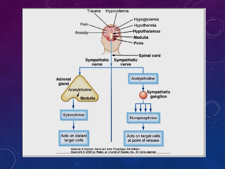
ADRENAL MEDULLA CATECHOLAMINES Instead of being secreted near a target organ and acting as neurotransmitters, adrenomedullary catecholamines are secreted into blood and act as hormones. About 80% of the cells of the adrenal medulla secrete epinephrine as a primary hormone, and the remaining 20% secrete norepinephrine. Although circulating epinephrine is derived entirely from the adrenal medulla, only about 30% of the circulating norepinephrine comes from the medulla. The remaining 70% is released from postganglionic sympathetic nerve terminals and diffuses into the vascular system. Because the adrenal medulla is not the sole source of catecholamine production, this tissue is not essential for lif e
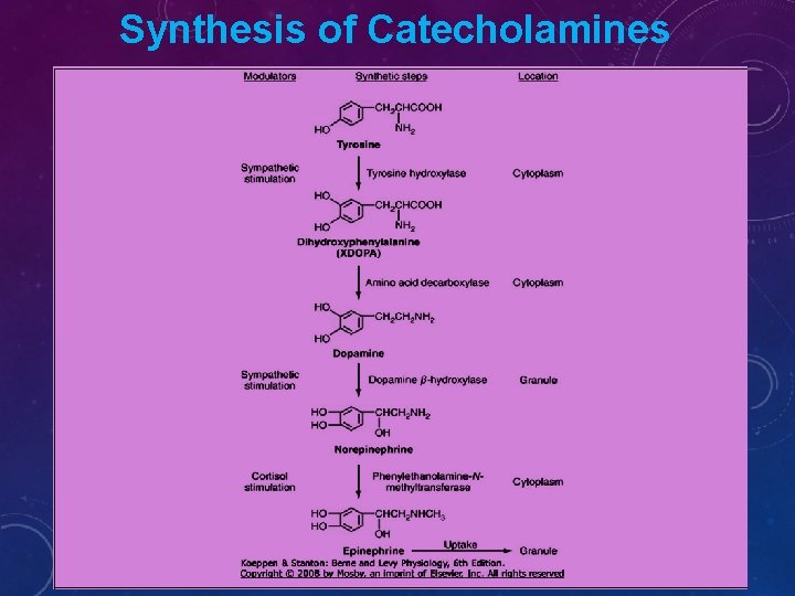
Synthesis of Catecholamines
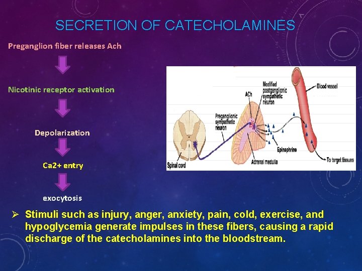
SECRETION OF CATECHOLAMINES Preganglion fiber releases Ach Nicotinic receptor activation Depolarization Ca 2+ entry exocytosis Ø Stimuli such as injury, anger, anxiety, pain, cold, exercise, and hypoglycemia generate impulses in these fibers, causing a rapid discharge of the catecholamines into the bloodstream.
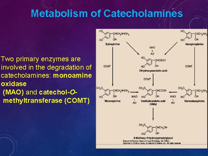
Metabolism of Catecholamines Two primary enzymes are involved in the degradation of catecholamines: monoamine oxidase (MAO) and catechol-Omethyltransferase (COMT)
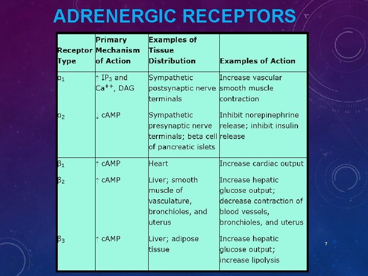
ADRENERGIC RECEPTORS 7
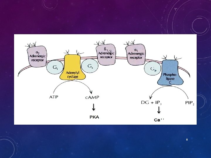
8
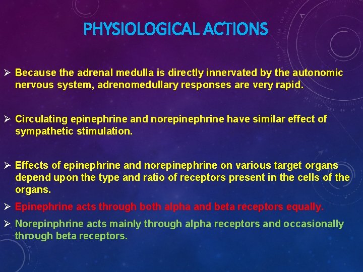
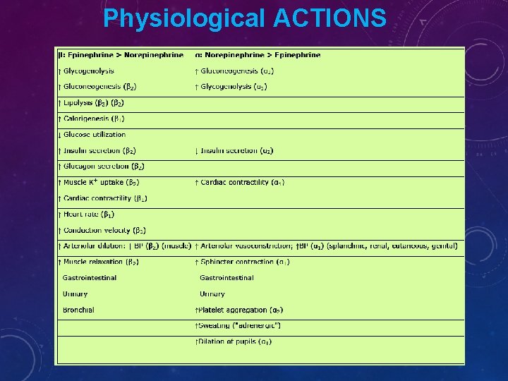
PHYSIOLOGICAL ACTIONS Ø Because the adrenal medulla is directly innervated by the autonomic nervous system, adrenomedullary responses are very rapid. Ø Circulating epinephrine and norepinephrine have similar effect of sympathetic stimulation. Ø Effects of epinephrine and norepinephrine on various target organs depend upon the type and ratio of receptors present in the cells of the organs. Ø Epinephrine acts through both alpha and beta receptors equally. Ø Norepinphrine acts mainly through alpha receptors and occasionally through beta receptors.
Physiological ACTIONS
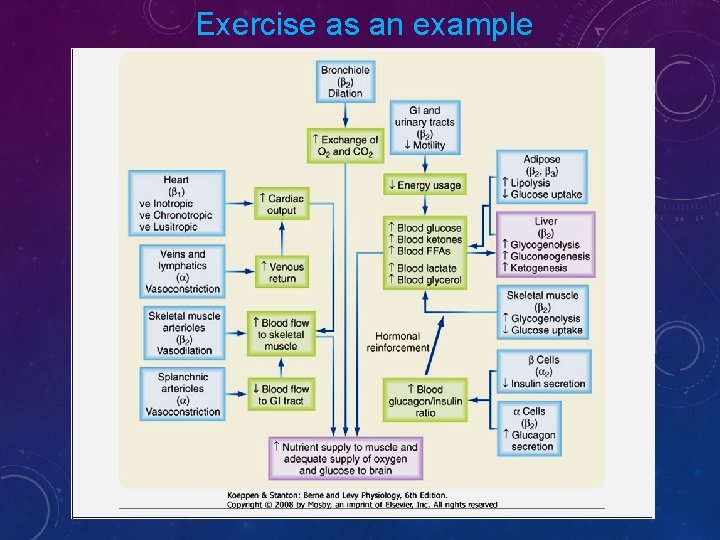
Exercise as an example
The overall goal of the sympathoadrenal system during exercise is to meet the increased energy demands of skeletal and cardiac muscle while maintaining sufficient oxygen and glucose supply to the brain. The response to exercise includes the following major physiological actions: 1. Increased blood flow to muscles is achieved by the integrated action of norepinephrine and epinephrine on the heart, veins and lymphatics, nonmuscular and muscular arteriolar beds.
Epinephrine promotes glycogenolysis in muscle. Exercising muscle can also utilize free fatty acids (FFAs), and epinephrine and norepinephrine promote lipolysis in adipose tissue. The actions just described increase circulating levels of lactate and glycerol, which can be used by the liver as gluconeogenic substrates to increase glucose. Epinephrine increases blood glucose by increasing hepatic glycogenolysis and gluconeogenesis. The promotion of lipolysis in adipose tissue is also coordinated with an epinephrineinduced increase in hepatic ketogenesis. The effects of catecholamines on metabolism are reinforced by the fact that they stimulate glucagon secretion (β 2 receptors) and inhibit insulin secretion (α 2 receptors). Efficient production of ATP during normal exercise (i. e. , a 1 -hour workout) also requires efficient exchange of gases with an adequate supply of oxygen to exercising muscle. Catecholamines promote this by relaxation of bronchiolar smooth muscle Catecholamines decrease energy demand by visceral smooth muscle. In general, a sympathoadrenal response decreases overall motility of the smooth muscle in the gastrointestinal (GI) and urinary tracts, thereby conserving energy where it is not needed
EPINEPHRINE VS. NOREPINEPHRINE Ø Epinephrine >> norepinephrine – in terms of cardiac stimulation leading to greater cardiac output ( stimulation). Ø Norepinephrine > epinephrine – in terms of constriction of blood vessels – leading to increased peripheral resistance – increased arterial pressure. Ø Epinephrine >> norepinephrine –in terms of increasing metabolism 15
CLINICAL INSIGHTS Pheochromocytoma is a tumor of chromaffin tissue that produces excessive quantities of catecholamines. These are commonly adrenal medullary tumors, but they can occur in other chromaffin cells of the autonomic nervous system. Although pheochromocytomas are not common tumors, they are the most common cause of hyperfunctioning of the adrenal medullary. The catecholamine most frequently elevated in pheochromocytoma is norepinephrine. For unknown reasons, the symptoms of excessive catecholamine secretion are often sporadic rather than continuous. Symptoms include hypertension, headaches (from hypertension), sweating, anxiety, palpitations, and chest pain. In addition, patients with this disorder may show orthostatic hypotension (despite the tendency for hypertension). This occurs because hypersecretion of catecholamines can decrease the postsynaptic response to norepinephrine as a result of down-regulation of the receptors. Consequently, the baroreceptor response to blood shifts that occurs on standing is blunted