Adaptations of lungs Starting activity Label as many

Adaptations of lungs Starting activity: Label as many of the different structures in the respiratory system as you can.

Match the definitions • Cell • Tissue • Organ • A group of similar cells that perform a particular function • Collection of tissues that work together to perform a specific overall function / set of functions within a multicellular organism • The smallest unit of an organism capable of surviving independently.

Lesson objectives By the end of this lesson pupils should be able to. . . . • Describe the features of the mammalian lung that adapt it to efficient gaseous exchange. • Describe, with the aid of diagrams and photographs, the distribution and functions of cartilage, ciliated epithelium, goblet cells, smooth muscle and elastic fibres in the trachea, bronchioles and alveoli of the mammalian gaseous exchange system.

Structure and function of the lungs The trachea, bronchi and bronchioles must be. . • large enough to allow sufficient air movement without obstruction. • Divide into smaller airways to deliver air to all the alveoli. • Strong enough to prevent collapse. • Flexible to allow movement. • Able to stretch and recoil.

The trachea and bronchi Have a similar structure. They differ only in size – the bronchi are narrower than the trachea. They have relatively thick walls that have several layers of tissue.
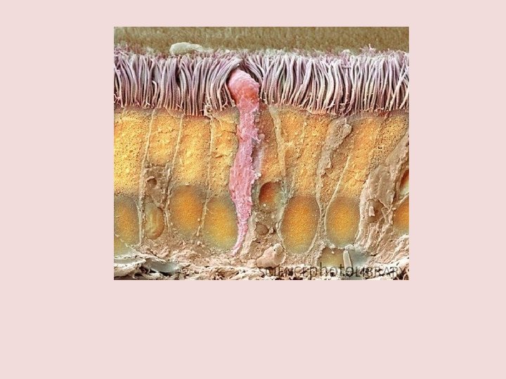

Transverse section through the human trachea Connective tissue Cartilage The gaps between the rings of cartilage are filled by the trachealis muscle Smooth muscle Sub mucosa Ciliated epithelium The sub-mucosa contains glands which are mixed sero-mucous glands. The watery secretions from the serous glands humidify the inspired air. The mucous, together with mucous from the goblet cells traps particles from the air which are transported upwards towards the pharynx by the cilia on the epithlium. This helps to keep the lungs free of particles and bacteria.

epithelium Goblet Ciliated cell Gland Blood vessel Smooth muscle Connective tissue Cartilage Connective tissue

Goblet cell

The bronchioles Label your diagrams on the sheet provided.

What is the role of each tissue? • Cartilage • Smooth muscle • Elastic fibres • Goblet cells and glandular tissue • Ciliated epithelium

Large bronchus Epithelium Ciliated Goblet cells Yes Cartilage Yes Glands Yes Smooth muscle Yes

Small bronchus Epithelium Ciliated Goblet cells Few Cartilage Little Glands Few Smooth muscle Yes

A bronchiole Epithelium Ciliated Goblet cells Very few Cartilage No Glands No Smooth muscle Yes

Tasks: • Colour code structure of the airways worksheet • Highlight lung structure and function worksheet

Smoker’s cough
 12 -15 mins Other exam")
Homework • Complete Q 5 Jan 05 (12 marks) 12 -15 mins Other exam questions include: June 07 Q 2, Jan 03 Q 2 a

Answers:
 • Colour in your mechanism of breathing worksheet • Answer the questions")
Ventilation (breathing) • Colour in your mechanism of breathing worksheet • Answer the questions about ventilation on the back of the sheet.

Inhalation

Exhalation
 Spend 30 minutes")
Homework: • Complete June 2004 question – all parts (11 marks) Spend 30 minutes on this to gain 100%!
- Slides: 23