ACUTE SINUSITIS Dr KCSUDEEP Sinusitis is an infection


 Exciting causes Nasal infection Swimming and diving Trauma Dental infection")


 Trephination of frontal sinus. 2)")


- Slides: 30
ACUTE SINUSITIS Dr. KCSUDEEP
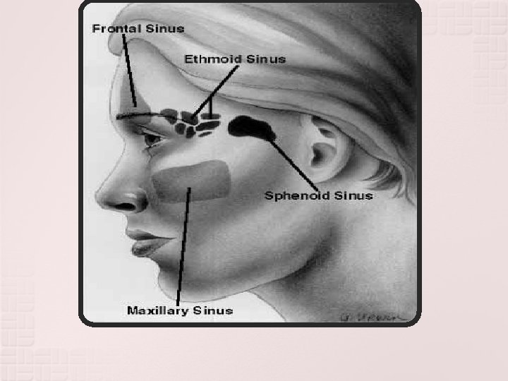
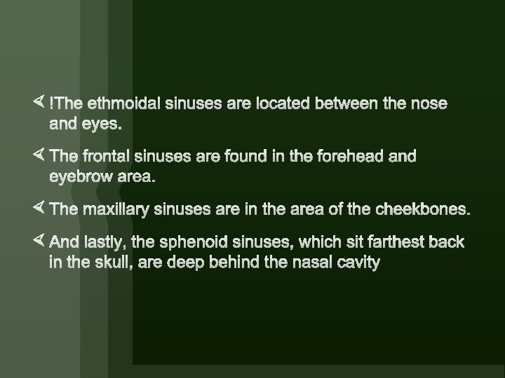
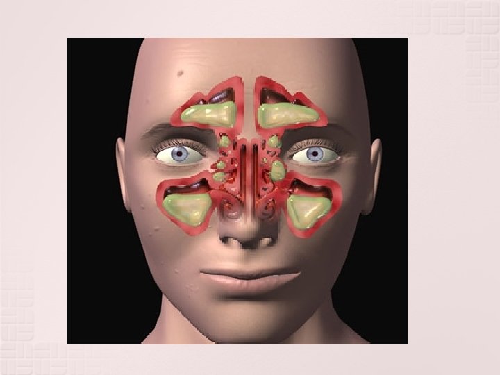
Sinusitis is an infection or inflammation that occurs in one or more of the four sets of paranasal sinuses. Sinuses are hollow, air filled cavities inside the head around the nasal cavity area. The sinuses all have openings into the nasal canal to allow for continuous air and mucous exchange and secretion. A common theory surrounding the need and purpose of sinuses is that they help to lighten the entire weight of the skull. Sinuses also function to help humidify air and capture inhaled or inspired dust particles. The four sets of paranasal sinuses include: Ethmoid Sinuses (green) Frontal Sinuses (checkered)
Most commonly involved is maxillary followed by ethmoidal , frontal and sphenoid. Multi sinusitis: more than one sinus is infected. Pansinusitis: all the sinus of one or both sides are involved simultaneously. Open type: inflammatory product of sinus cavity can drain freely into nasal cavity through natural ostia.
Acute sinusitis is the abnormal secretion and production of mucous. This is similar to cystic fibrosis. On x-rays, acute sinusitis is suggested by air fluid levels in the sinuses. Patients with immunodeficiency diseases, such as HIV or AIDS, are more likely to acquire acute sinusitis
AETIOLOGY OF SINUSITIS A) Exciting causes Nasal infection Swimming and diving Trauma Dental infection B)PREDISPOSING CAUSES: Obstruction to sinus ventilation and drainage. Stasis of secretions in nasal cavity. Previous attack of sinusitis.
GENERAL: Environment : Poor general health : BACTERIOLOGY: Most cases of acute sinusitis start as viral infections followed by bacterial invasion. Strepto. pneumoniae, H. influenza, Moraxella catarrhalis, strept. Pyogenes, Staph. aureus and kleb. Pneumoniae.
pathogenesis The paranasal sinuses all drain into the nasal cavity. All of the sinuses are lined with mucousal membranes. This is the area most commonly infected because of drainage problems. Sometimes a blockage can form from excessive build up of mucousal secretions. If a sudden blockage occurs, the sinuses can no longer drain in a normal fashion. This leads to an increase fluid in a sedentary space, which is a perfect breeding place for bacterial growth or viral infection. Once infection is set in place, the inflammation process of sinusitis begins. Air can also become trapped inside a sinus and cause increased pressure inside the paranasal sinuses leading to sinusitis
Failure of ostium to drain results in empyema of sinus and destruction of its bony walls leading to complications Drainage problems and air pressure are not the only causes of sinusitis. Any injury or infection that causes swelling of the face and nose can end up affecting the sinuses. Processes that are involved with changing anything in the sinuses can lead to serious problems and infections such as sinusitis.
ACUTE MAXILLARY SINUSITIS Aetiology Viral rhinitis is most common followed by bacterial invasion. Diving swimming in contaminated water. Dental infections. Trauma to sinus.
DIAGNOSIS The best way to diagnose sinusitis is through physical examination and good medical history including all symptoms. X-rays and CT (computed tomography) can also be performed. CT is the modality of choice because it shows the extent and severity of infection. If the maxillary sinuses are involved, a examination of the teeth may be performed to check for abscesses Radiographs of sinuses can show air fluid levels in the sinuses. They can also demonstrate nasal polyps, opacifications, cysts, and mucoceles. Sinusitis can also be seen on CT by demonstrating mucousal hyperplasia, bone lesions, fluid retention, and bullous concha. CT helps in imaging thickened mucousal membranes in the paranasal sinuses.
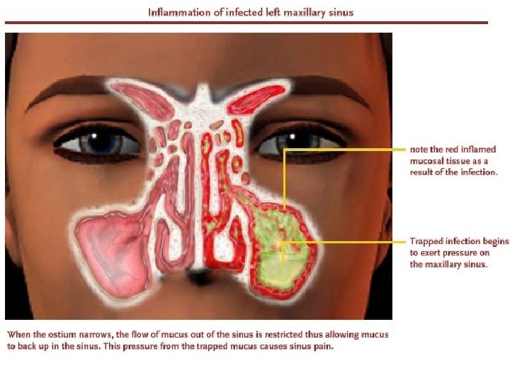
Fig-Right maxillary sinusitis
Diagnosis : x-ray will show either an opacity or a fluid level in the involved sinus. TREATMENT: Medical Antimicrobial drugs. Nasal decongestant drops. Steam inhalation after 15 to 20 minutes of decongestant drops. Analgesics. Hot fomentation.
Surgical Antral lavage COMPLICATIONS: Subacute or chronic sinusitis. Frontal sinusitis. Osteitis or osteomyelitis of maxilla. Orbital cellulitis or abscess.
ACUTE FRONTAL SINUSITIS Aetiology: Oedema of middle meatus, secondary to associated ipsilateral maxillary or ethmoid sinus infection. CLINICAL FEATURES: Frontal headache “office headache”. Tenderness. Oedema of upper eyelid with suffused conjunctiva and photophobia. Nasal discharge.
TREATMENT: MEDICAL: Same as acute maxillary sinusitis. SURGICAL: 1) Trephination of frontal sinus. 2) Antral lavage. COMPLICATIONS: Orbital cellulitis. Osteomyelitis of frontal bone and fistula formation. Meningitis, extradural or frontal lobe abscess. Ø Chronic frontal sinusitis.
ACUTE ETHMOID SINUSITIS AETIOLOGY: Acute ethmoiditis is often associated with infection of other sinuses. Ethmoid sinuses are more involved in infants and young children. CLINICAL FEATURES: Pain over bridge of nose. Oedema of lids with increased lacrimation. Nasal discharge. Swelling of middle turbinate.
TREATMENT: Visual deterioration and exopthalmos indicate abscess in posterior orbit, may require drainage of ethmoid sinuses into nose through external ethmoidectomy incision. COMPLICATIONS: Orbital cellulitis and abscess. Visual deterioration and blindness due to involvement of optic nerve. Cavernous sinus thrombosis. Extradural abscess, meningitis or brain abscess.
ACUTE SPHENOID SINUSITIS AETIOLOGY: n Isolated involvement of sphenoid sinus is rare. n It is often a part of pansinusitis or is associated with infection of posterior ethmoid sinuses.
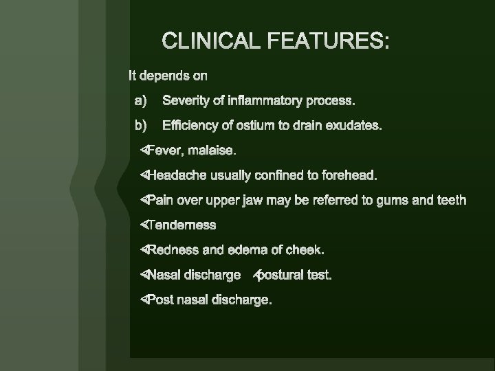
CLINICAL FEATURES: Headache usually localised to occiput or vertex may reffered to mastoid region. � Post nasal discharge on post. Rhinoscopy. � X-rays show opacity or fluid level in sphenoid sinus. Lateral view both in supine and prone position should be taken.
Prognosis Sinusitis is not a life threatening disease if properly treated under a physicians care. There is treatment available. If antibiotics and medications do not work. A specialists can be consulted on nasal and sinus surgery. These surgeries can improve drainage flow and remove infected mucousal materials. Removal of the adenoids in children can help clear up sinus problems. Opening of the nasal airway through removal of nasal polyps and correction of a deviated nasal septum can also be accomplished through surgery. This can also help eliminate or decrease sinus problems
§ Case 1: A 10 year old child was having a right mucopurulent otorhea for the last 4 years. A week ago he became dizzy with a whirling sensation, nausea, vomiting and nystagmus to the opposite side; his deafness became complete and his temperature was normal. Three days later he became feverish, irritable and continuously crying apparently from severe headache. Also he had some neck retraction. The child was not managed properly and died by the end of the week.
§ Case 2: A 50 year old male patient complained of right earache of 2 days duration. The pain was especially severe on chewing food and during speech. There was also marked edema of the right side of the face. On examination, pressure on the tragus was painful; and there was a small red swelling arising from the anterior external auditory meatal wall. Rinne test was positive in the right ear. The patient gave a history of 2 previous similar attacks in the same ear during the last six months but less severe.
§ Case 7: A 30 year old female complained of bilateral hearing loss more on the right side following the delivery of her first child; hearing loss was marked in quiet places but hearing improved in a noisy environment. Both tympanic membranes showed a normal appearance. Rinne tuning fork test was negative
§ Case 9: A 28 year old male has been complaining of hearing loss in the left ear for the last 6 years. The hearing loss was progressive in nature and accompanied by tinnitus. During the last 6 months there was swaying during walking to the left side, a change in his voice and an inability to close the left eye with deviation of the angle of the mouth to the right side. Otologic examination showed no abnormality. The corneal reflex was lost in the left eye.
Thank you have a nice day