
Acute Rhinosinusitis ARS Presented by Mohammad AlRamadneh Terminology
Acute Rhinosinusitis (ARS) Presented by: Mohammad Al-Ramadneh
Terminology and classification • Sinusitis or rhinosinusitis is defined as symptomatic inflammation of the paranasal sinuses and nasal cavity. • Nowadays the term rhinosinusitis has replaced the term sinusitis, as most patients often have symptoms of both rhinitis and sinusitis simultaneously. • Rhinosinusitis may be further classified according to the anatomic site (maxillary, ethmoidal, frontal, sphenoidal), pathogenic organism (viral, bacterial, fungal), presence of complication (orbital, intracranial), and associated factors (nasal polyposis, immunosuppression, anatomic variants)

Sinus Anatomy • Paranasal sinuses lined with pseudostratified ciliated columnar epithelium and goblet cells • Sinuses health depends on: 1. Mucous secretion of normal viscosity, volume, and composition. 2. Normal mucociliary flow to prevent mucous stasis and subsequent infection 3. Open sinus ostia to allow adequate drainage and aeration. 3
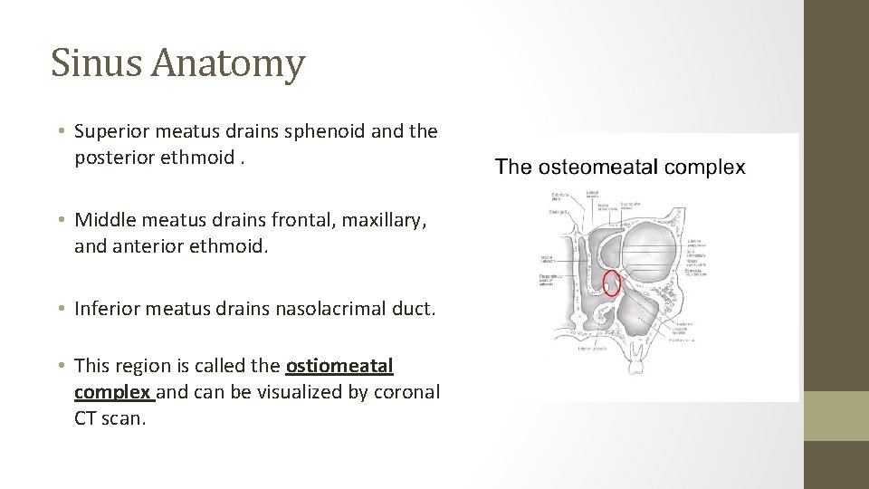
Sinus Anatomy • Superior meatus drains sphenoid and the posterior ethmoid. • Middle meatus drains frontal, maxillary, and anterior ethmoid. • Inferior meatus drains nasolacrimal duct. • This region is called the ostiomeatal complex and can be visualized by coronal CT scan.

Sinus normal physiology • The possible roles of the sinuses may include: 1. reducing the weight of the skull 2. dampening pressure; humidifying and warming inspired air 3. aiding in sound resonance • Obstruction of sinus drainage into nasal cavity → stagnation → inflammation (acute or chronic) of paranasal sinuses → pain over affected area
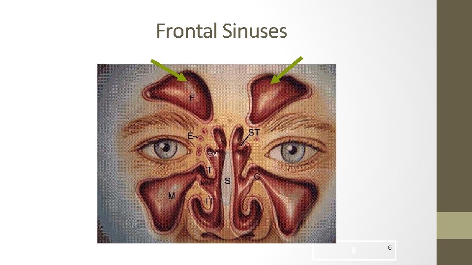
Frontal Sinuses 6 6
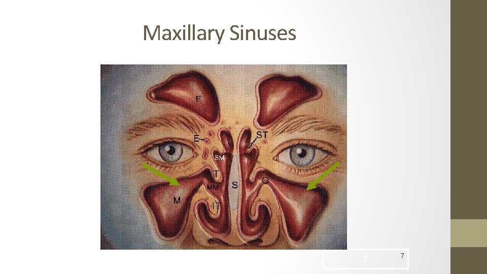
Maxillary Sinuses 7 7

Ethmoid Sinuses 8 8
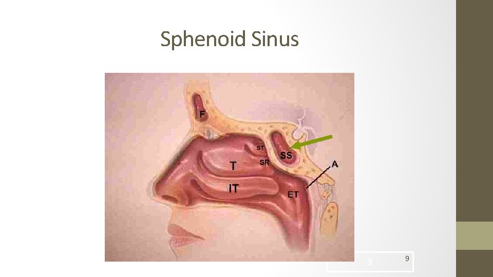
Sphenoid Sinus 9 9
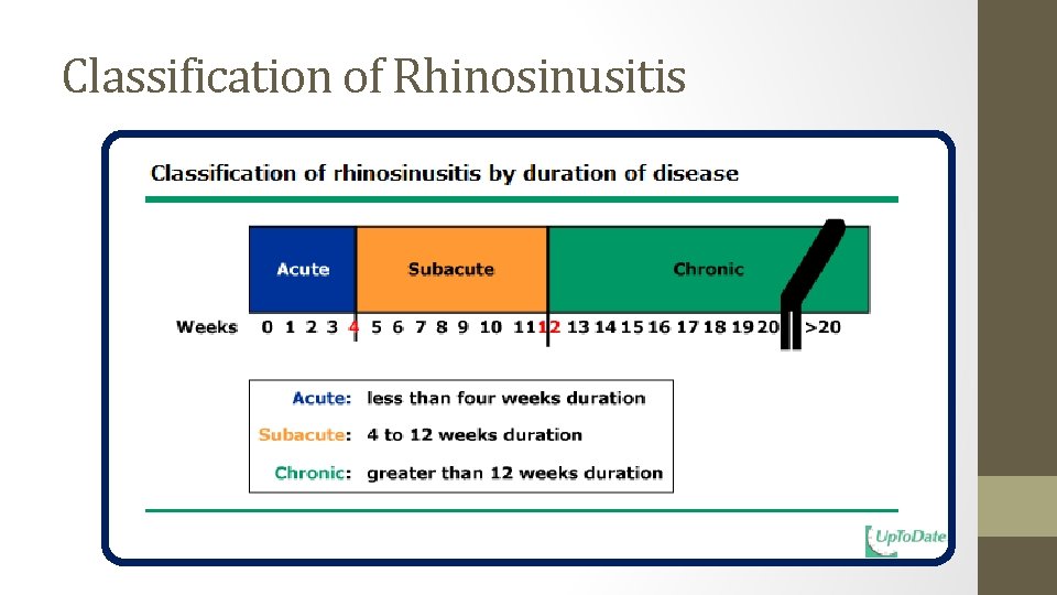
Classification of Rhinosinusitis
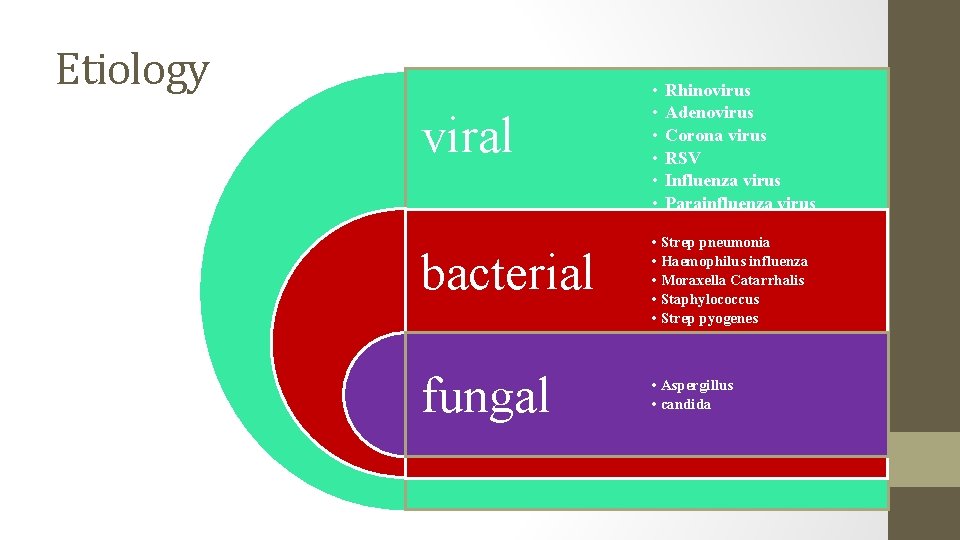
Etiology viral bacterial fungal • • • Rhinovirus Adenovirus Corona virus RSV Influenza virus Parainfluenza virus • Strep pneumonia • Haemophilus influenza • Moraxella Catarrhalis • Staphylococcus • Strep pyogenes • Aspergillus • candida
Acute viral rhinosinusitis ( common cold) • The vast majority ( up to 70%) of rhinosinusitis episodes are caused by viral infection. • Approximately 90% of patients who have viral upper respiratory tract infections have sinus involvement, but only 5 -10% of these patients have bacterial superinfection requiring antimicrobial treatment. • Most viral upper respiratory tract infections are caused by rhinovirus, coronavirus, influenza A and B, parainfluenza, respiratory syncytial virus, adenovirus, and enterovirus are also causative agents.
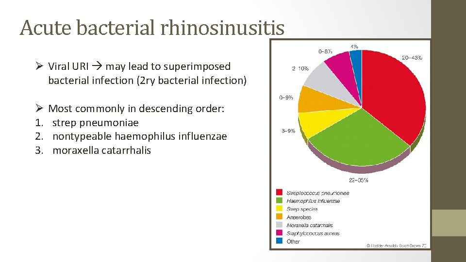
Acute bacterial rhinosinusitis Ø Viral URI may lead to superimposed bacterial infection (2 ry bacterial infection) Ø 1. 2. 3. Most commonly in descending order: strep pneumoniae nontypeable haemophilus influenzae moraxella catarrhalis
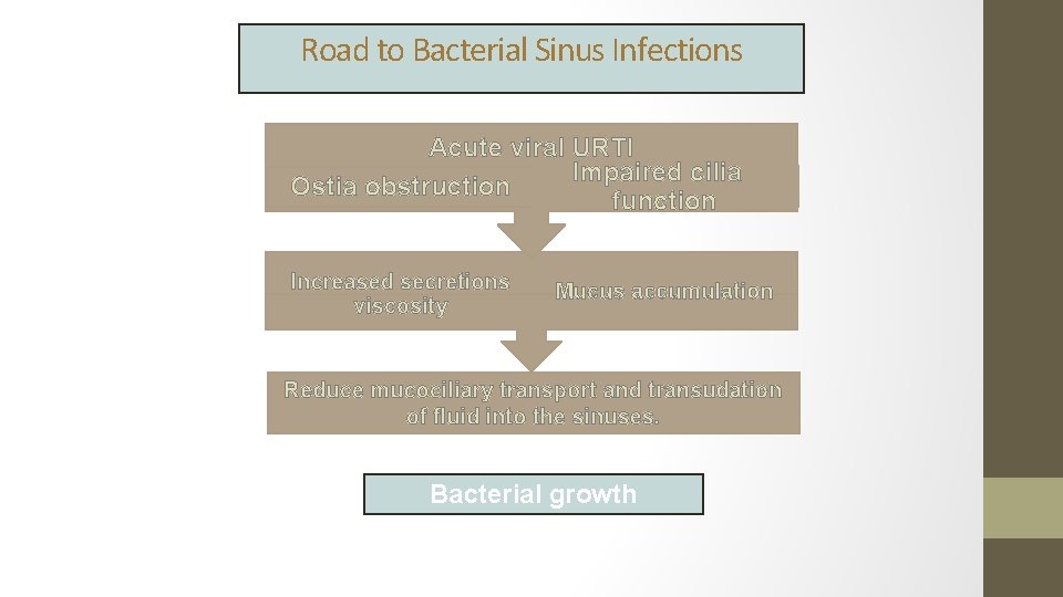
Road to Bacterial Sinus Infections Acute viral URTI Impaired cilia Ostia obstruction function Increased secretions viscosity Mucus accumulation Reduce mucociliary transport and transudation of fluid into the sinuses. Bacterial growth
Clinical course History and physical exam • Acute sinusitis is a clinical diagnosis; thus, an understanding of its presentation is of paramount importance in differentiating this entity from allergic or vasomotor rhinitis and common upper respiratory infections. • No specific clinical symptom or sign is sensitive or specific for acute sinusitis, so the overall clinical impression should be used to guide management. • A change in the color or characteristic of the nasal discharge is not a specific sign of bacterial rhinosinusitis.
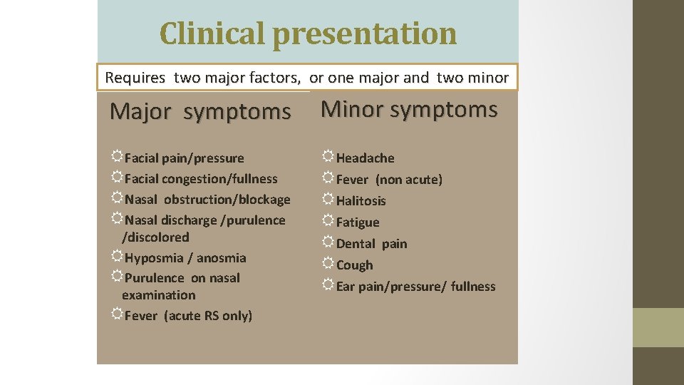
Clinical presentation Requires two major factors, or one major and two minor Major symptoms Minor symptoms RFacial pain/pressure RFacial congestion/fullness RNasal obstruction/blockage RNasal discharge /purulence RHeadache RFever (non acute) RHalitosis RFatigue RDental pain RCough REar pain/pressure/ fullness /discolored RHyposmia / anosmia RPurulence on nasal examination RFever (acute RS only)
Diagnosis ØClinically ØIf symptoms persisted • X-ray (sinus view) • CT scan (better than x-ray) • Direct visualization (nasal speculum) • Nasal endoscopy
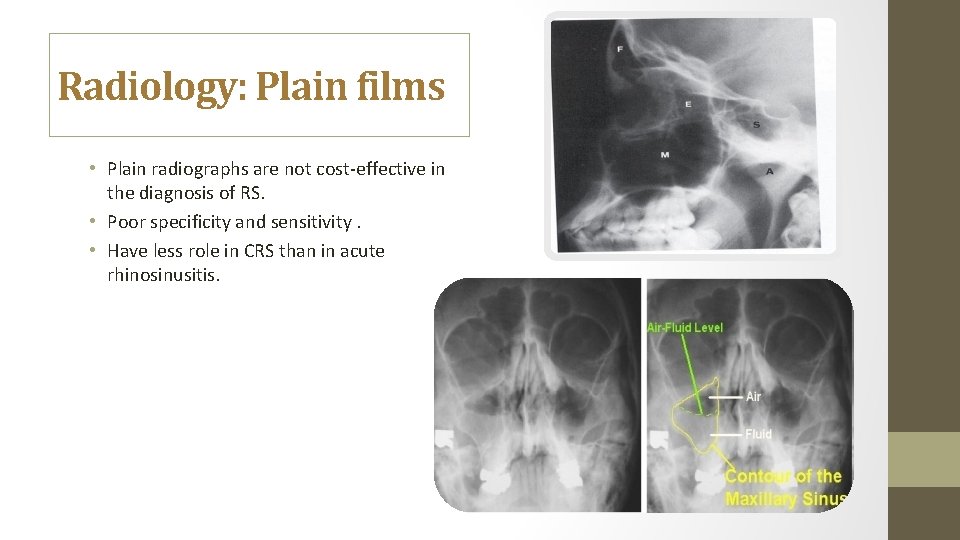
Radiology: Plain films • Plain radiographs are not cost-effective in the diagnosis of RS. • Poor specificity and sensitivity. • Have less role in CRS than in acute rhinosinusitis.
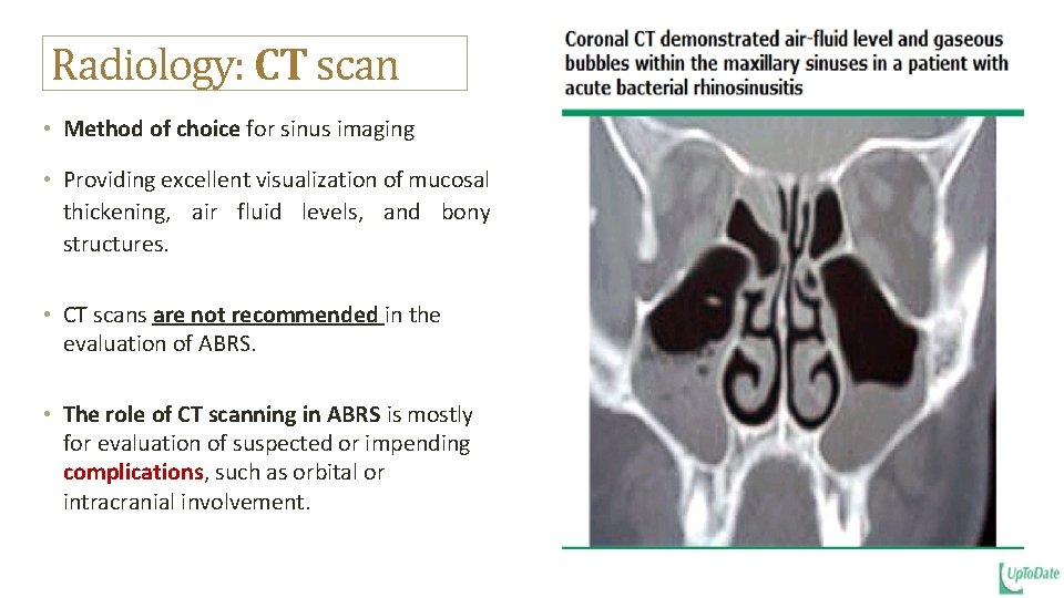
Radiology: CT scan • Method of choice for sinus imaging • Providing excellent visualization of mucosal thickening, air fluid levels, and bony structures. • CT scans are not recommended in the evaluation of ABRS. • The role of CT scanning in ABRS is mostly for evaluation of suspected or impending complications, such as orbital or intracranial involvement.
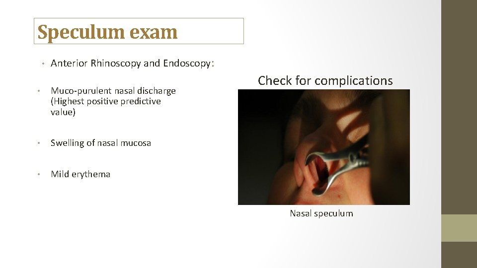
Speculum exam • Anterior Rhinoscopy and Endoscopy: • Muco-purulent nasal discharge (Highest positive predictive value) • Swelling of nasal mucosa • Mild erythema Check for complications Nasal speculum
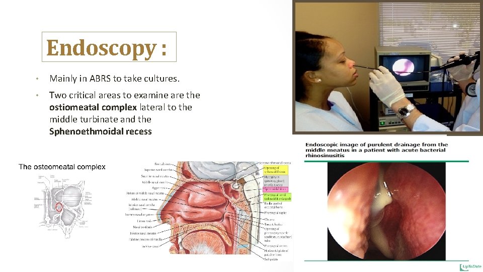
Endoscopy : • Mainly in ABRS to take cultures. • Two critical areas to examine are the ostiomeatal complex lateral to the middle turbinate and the Sphenoethmoidal recess
Management ØWatch and wait: most cases are caused by viruses (self-limiting, do not respont to antibiotics). ØIf persisted (probably bacterial or viral complicated by a secondary bacterial infection): • NSAIDs • Nasal steroids • Decongestant nasal sprays • Humidification/vaporizer, warm compresses, adequate hydration, smoking cessation, balanced nutrition.
Management ØIf symptoms persisted or worsens for more than 7 -10 days • Antibiotics: Amoxicillin alone, or with clavulanate. ØIf allergic to penicillin Macrolide (clindamycin). Fluoroquinolone. Tetracycline (doxycycline). ØIf child clindamycin plus a third-generation oral cephalosporin (cefixime or cefpodoxime)
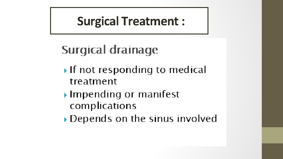
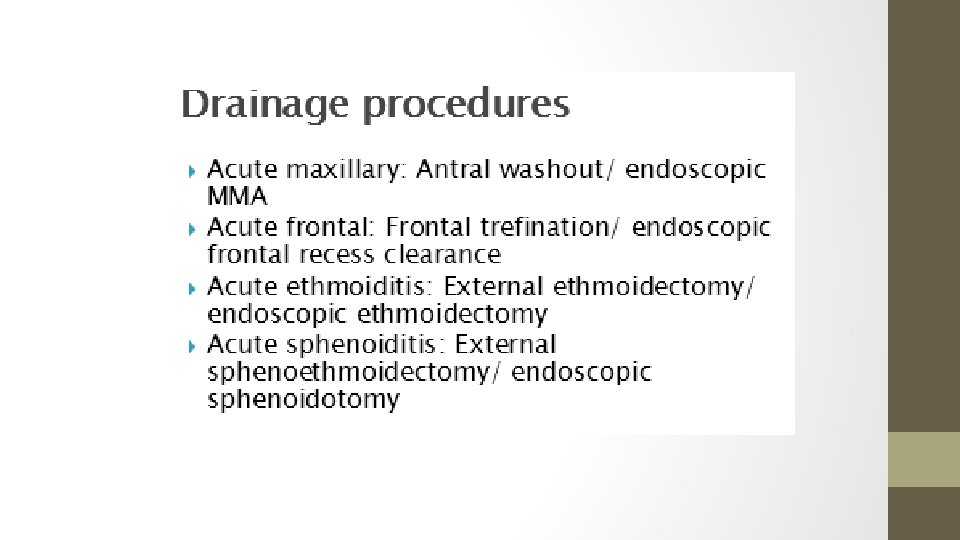
Surgical Treatment :
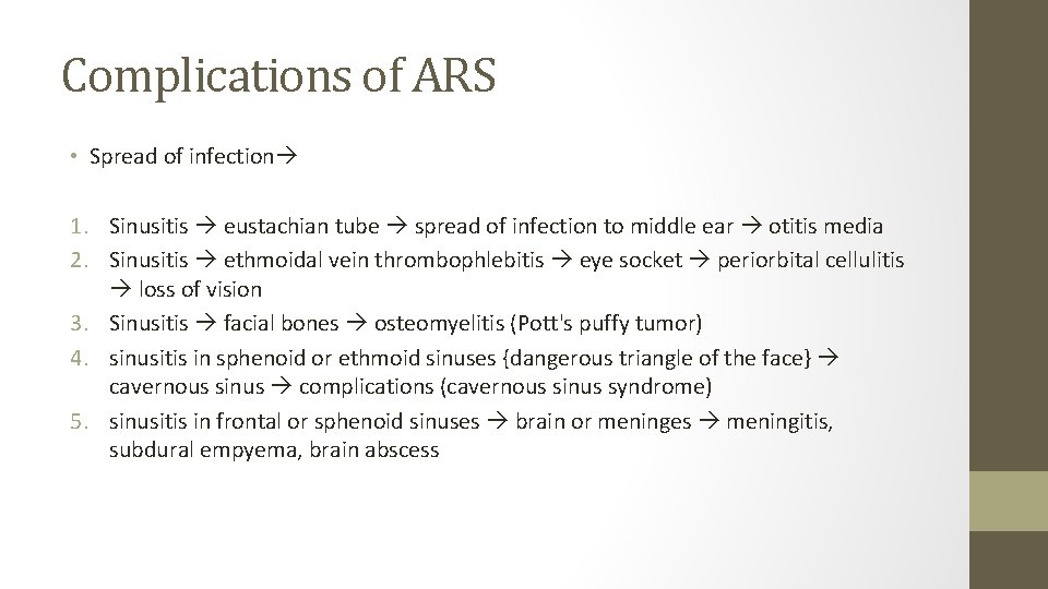
Complications of ARS • Spread of infection 1. Sinusitis eustachian tube spread of infection to middle ear otitis media 2. Sinusitis ethmoidal vein thrombophlebitis eye socket periorbital cellulitis loss of vision 3. Sinusitis facial bones osteomyelitis (Pott's puffy tumor) 4. sinusitis in sphenoid or ethmoid sinuses {dangerous triangle of the face} cavernous sinus complications (cavernous sinus syndrome) 5. sinusitis in frontal or sphenoid sinuses brain or meninges meningitis, subdural empyema, brain abscess
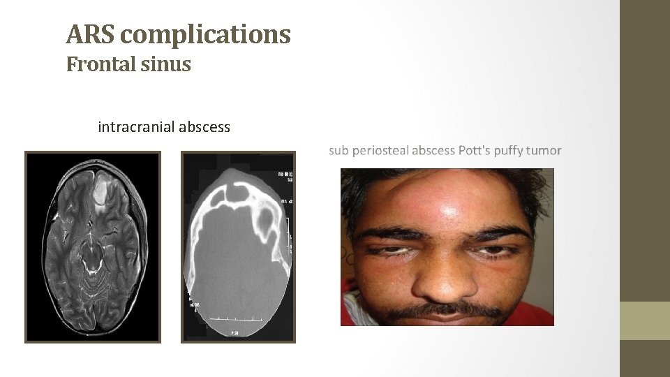
ARS complications Frontal sinus intracranial abscess
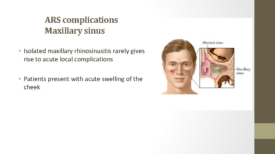
ARS complications Maxillary sinus • Isolated maxillary rhinosinusitis rarely gives rise to acute local complications • Patients present with acute swelling of the cheek
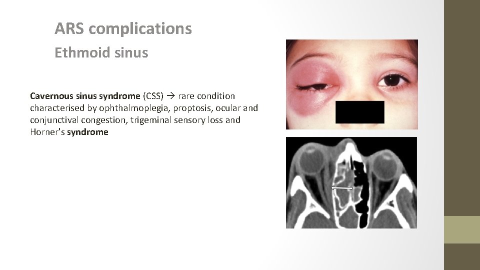
ARS complications Ethmoid sinus Cavernous sinus syndrome (CSS) rare condition characterised by ophthalmoplegia, proptosis, ocular and conjunctival congestion, trigeminal sensory loss and Horner's syndrome
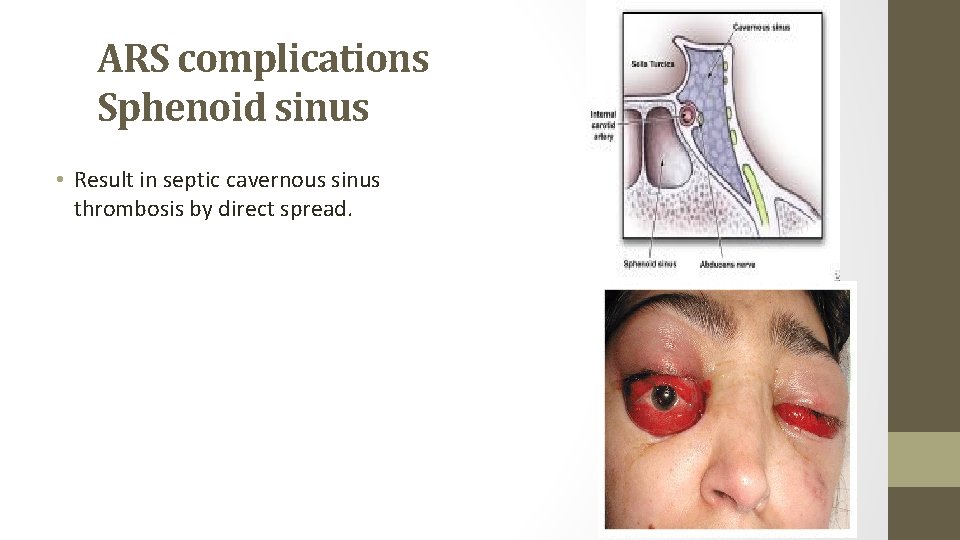
ARS complications Sphenoid sinus • Result in septic cavernous sinus thrombosis by direct spread.

- Slides: 31