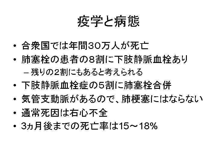
Acute Pulmonary Embolism NEJM 2008 358 1037 52

Acute Pulmonary Embolism NEJM 2008: 358: 1037 -52
で")
l Low: 1. 3% l Moderate: 16. 2% l High: 37. 5% l Low&D-dimer(-)で PEを完全否定 (99. 5%)
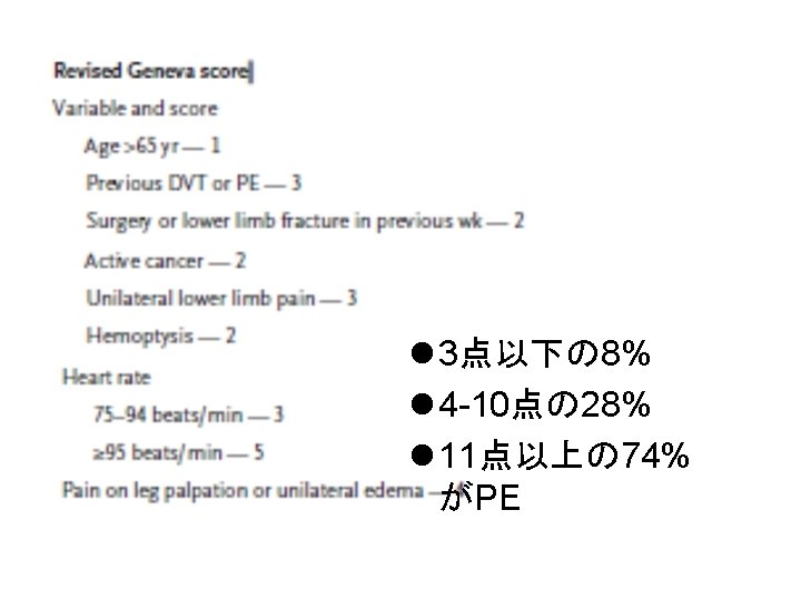
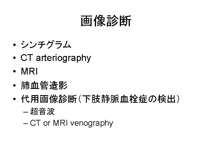

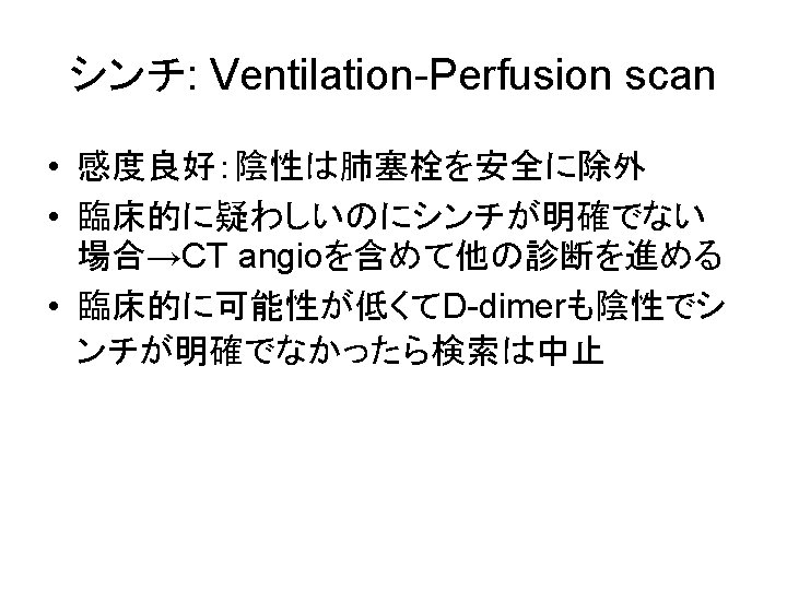
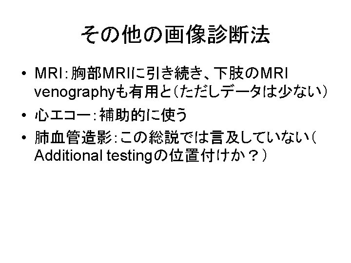

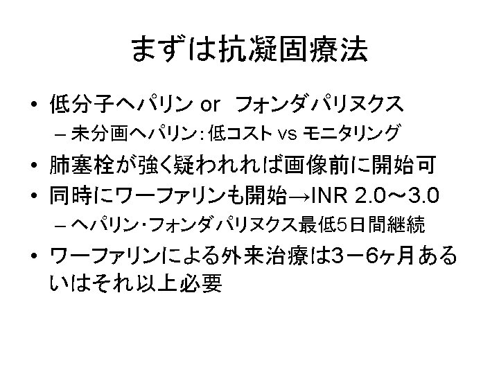
Figure 3. Diagnostic Approach to Suspected Acute Pulmonary Embolism. The use of prediction rules and d-dimer testing may reduce the need for imaging. If the risk of bleeding is deemed to be low, initiation of therapy before a proven diagnosis of pulmonary embolism should be considered. 40 At this juncture, the chest radiograph and other specific imaging may already be completed. A ventilation–perfusion (VQ) scan is more likely to yield a diagnosis when there is no associated cardiopulmonary disease. A scan indicating a high probability of pulmonary embolism is confirmatory except when there has been aprior pulmonary embolism, in which case a previous VQ scan may be useful in proving that defects are new. 34, 36 As with computed tomographic arteriography (CTA), the approach to a nondiagnostic scan includes evaluation of clinical probability as well as consideration of additional testing. Deep venous thrombosis discovered by leg ultrasonography, CT venography, or magnetic resonance venography suggests concomitant pulmonary embolism. 36, 37 Standard pulmonary arteriography or venography is rarely needed. Adding CT venography to CT arteriography enhances the overall sensitivity for detecting venous thromboembolism, 33 although an excellent outcome has beendemonstrated without additional testing when CTA is negative. 22 With the use of CTA or CT venography, caution is advised when the creatinine level rises above 1. 5 mg per deciliter; the patient’s age relative to the creatinine clearance should be considered. 36 ELISA denotes enzyme-linked immunosorbent assay.
- Slides: 17