Acute otitis Media And Otitis Media with Effusion


 Fullness in the ear")


")


- Slides: 23
Acute otitis Media And Otitis Media with Effusion By Prof. Ehab Taha Yaseen
Acute otitis media Definition: Collection of purulent fluid in the middle ear cleft for less than 3 weeks. Define the middle ear cleft ((Home work)) Sources of infection: 1. Upper airway Slide 11, Slide 12 2. Exanthemas 3. Perforation of the tympanic membrane. Slide 13
• Bacteriology Haemolytic streptococcus, strep pneumonia, Haemophilus influenzae and Branhamella cattaralis. • Pathology 1 - Tubal occlusion 2 - Engorgement and edema of the lining 3 - Exudation, serous then become purulent. This fluid results in bulging of the tympanic membrane which may cause rupture of the tympanic membrane by pressure necrosis usually posteroinferiorly. Slide 14
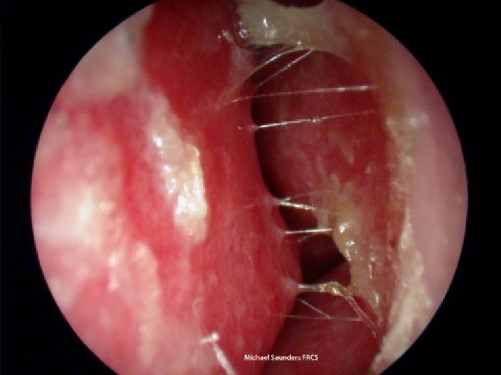
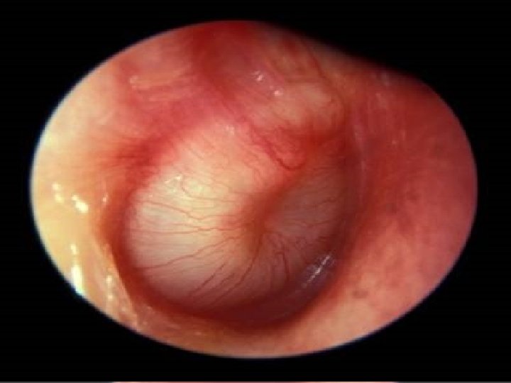
Clinical features: • Phase I: Acute tubal obstruction (acute salpingitis) Fullness in the ear with conductive deafness. • Phase II: Acute infection of the tympanic cavity (acute tympanitis) -- Before perforation: deafness increases, bubbling sounds are heared, otalgia and general symptoms especially seen in children and present as fever and malaise. On examination by otoscope: there are dilated vessels around the handle of the malleus and periphery of the tympanic membrane. This increases till the whole membrane is red, lusterless and bulging. -- After perforation: Otorrhea which may be serous, bloodstained, mucopurulent or purulent. The pain and tenderness are relieved immediately. • Phase III: Retention of pus in the mastoid (acute mastoiditis – rarely seen today) pain and tenderness over the mastoid region with edema of the posterosuperior wall of the deep meatus (a sign called sagging of meatal wall) with increase in the general symptoms. DD* ((Home work))
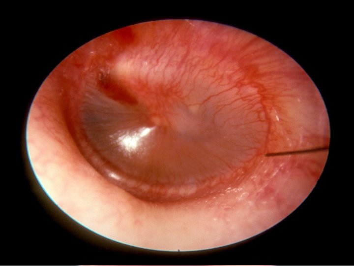
Treatment: • Symptomatic: rest, sedation, analgesia and local heat. • Medical: - Antibiotics amoxicillin, amoxicillin-calvulonic acid, cephalosporin (2 nd and 3 rd generation), macrolides as azithromycin, and clarithromycin - Nasal vasoconstrictor (xylometazolin drops, oral pesudoephedrin preparations) - Local treatment: - Before perforation of the TM myringotomy!! -When there is perforation • Surgical: - Myringotomy {site}
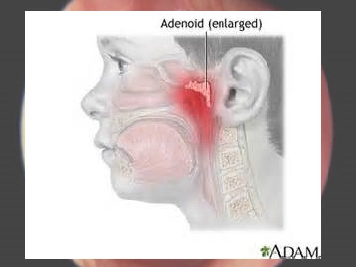
Recurrent Acute Otitis Media Definition: is recurrent acute otitis media (5 -6 times per year or 3 -4 times per 6 months) and in between there is complete resolution of symptoms and signs Predisposing factor: • Anatomical characteristics of the Eustachian tube • Bottle fed baby vs. breast fed • Parental smoking • Low socioeconomic • Crowded homes • Poor nutrition • Day care attendance • Low immunity • Nasal and postnasal problems (adenoid, allergy, polyps etc…) • Perforated tympanic membrane
Non-Suppurative Otitis Media (Otitis Media with Effusion)
Definition It is a clinical condition that is characterized by the presence of non- purulent fluid in the middle ear cleft. Acute vs. Chronic (duration, type of fluid) Aetiology 1. Obstruction of the Eustachian tube 2. Unresolved acute suppurative Otitis media. 3. Viral Otitis media 4. Allergy as hay fever 5. Cleft palate and its repair.
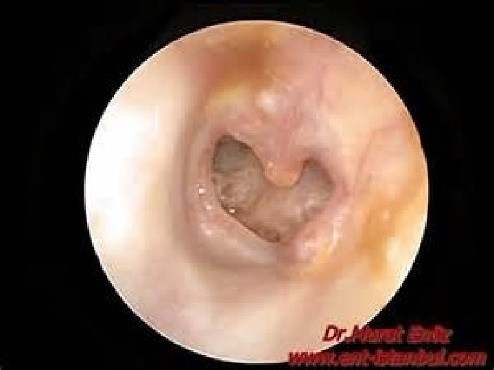
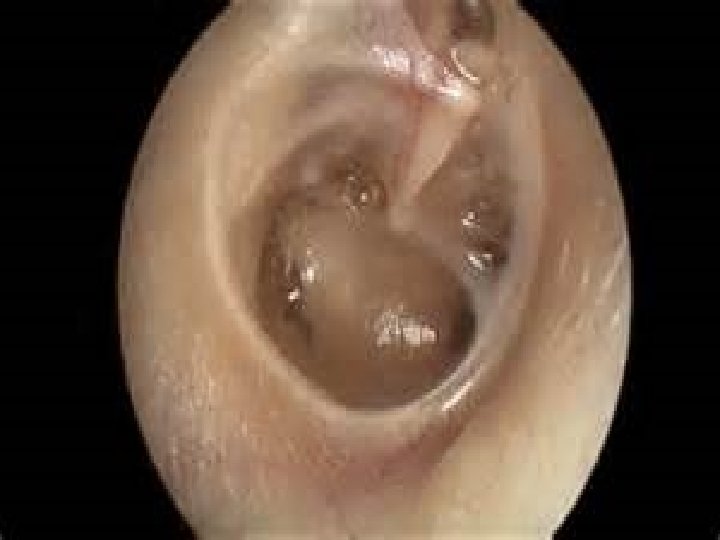
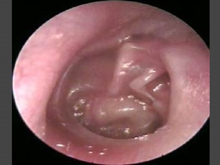
Clinical Features - Conductive deafness, - Otalgia - Otoscopic findings: Dull and retracted TM Handle of the malleus is prominent, foreshortened and more horizontal. Slide 16 The color of the TM is pale yellow, sometime grey or even blue. Sometime fluid level (hair line) or air bubbles are seen. The fluid is usually clear, yellow, serous and sterile (no bacteria or inactive bacteria).
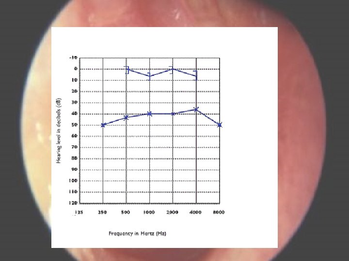
Diagnosis • History and examination • PTA shows conductive deafness. Slide 21 • Tympanometry (acoustic impedance measurements) show reduction in compliance and negative middle ear pressure). ((Home work))
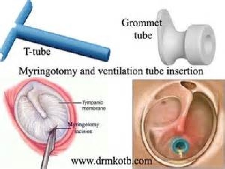
Treatment Medical treatment • Nasal decongestant • Antihistamines e. g. triprolidine and pseudoephedrine • Oral antibiotics: amoxicillin-calvulonic acid • Duration of treatment for up to one month. Surgical treatment: indicated when there is failure of medical treatment and may include • Myringotomy to restart ventilation of the middle ear. Slide 18 • Myringotomy and grommet • Surgery for the cause like adenoidectomy. END
Slide 19
THANK YOU DO NOT FORGET THE HOME WORK 1. Defination of middle ear cleft 2. DD. Of acute otitis media 3. Diagrams of tympanometry