Acute Leukemias Accumulation of blasts in the marrow


Acute Leukemias • Accumulation of blasts in the marrow

AML Delicate chromatin Much cytoplasm Fine granules/Auer rods ALL Clumped chromatin Scanty cytoplasm No granules
 Classification: (ALL-L 1; L 2; L 3) ALL-L 1 •")
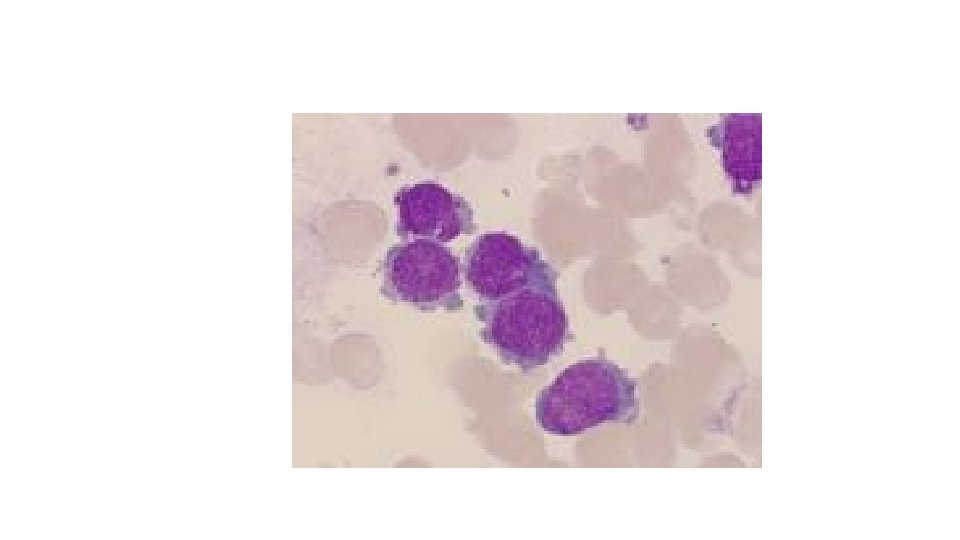
FAB (French American British) Classification: (ALL-L 1; L 2; L 3) ALL-L 1 • Commonest type • Better prognosis and response to therapy • Blast cells. Homogeneous population Small sized High N: C ratio Indistinct nucleoli • c. ALLa + 60% • PAS +

ALL- L 1 § § Homogenous Population Small sized blasts Scanty cytoplasm Round nuclei with dense chromatin.

ALL-L 2 • Comparatively poor prognosis and response to therapy • Blast cells Heterogeneous population Large Low N: C ratio Distinct Nucleoli

ALL-L 2 Heterogeneous Population Low N/C Ratio Irregular nuclear outlines and Prominent Nucleoli.
")
PAS Stain(ALL-L 2)

ALL-L 3 • Least common • Worst prognosis and response to therapy • B-cell in origin • Counterpart of Burkitt’s lymphoma • Blast cells • PAS Negative Large Abundant, bluish vacuolated cytoplasm Distinct nucleoli

ALL-L 3: Medium sized blasts of with High N/C ratio, Dark blue cytoplasm Cytoplasmic vacuolation

Prognosis in ALL Parameters Good Poor WBC Gender Age low Girls Child High (>50 x 10 9 /l) Boys Adult or infant. Time to clear < 1 week blast from blood Time to < 4 weeks remission > 1 week CNS disease at presentation Minimal residual disease. Absent Present Negative at 1 -3 months Still positive at 36 months. > 4 weeks

CD MARKERS & Stain for ALL CD 10, CD 19, CD 20 PAS, Sudan Black B+

Acute Myeloid Leukemia

AML • 80% of patients are adults • Amongst children > common < 2 years age • More aggressive manifestations than ALL

Clinical Features: Are Secondary to : Bone marrow Failure • WBC infection. (fever, malaise, mouth, throat, skin, respiratory , other infections) • Hb anemia (pallor, lethargy, dysnea) • platelets bleeding. (bruises, Purpura, bleeding gums, epistaxis, menorrhagia)
 ØHepatosplenomegaly ØLymphadenopathy less common ØBleeding is more sever and")
Manifestations ØConstitutional features (Fever; malaise) ØHepatosplenomegaly ØLymphadenopathy less common ØBleeding is more sever and more common Ø Tissue infiltrations are > common e. g. gum hyperplasia Skin infiltration

• • Diagnosis History /Examination Complete Blood Picture Peripheral Blood Examination Bone marrow aspiration - >30% blasts Subtyping Special Stains Immunophenotyping
 Platelets")
Complete Blood Picture Hemoglobin: Low TLC: Low/Normal/High(may be increased upto 200 x 109/l) Platelets : Decreased

Peripheral Film Examination Variable number of Blast cells Bone marrow Examination Hypercellular (in majority of cases , rarely can be hypocellular) with more than 30% Blast cells identified by morphology and Immunophenotyping

Myeloid maturation myeloblast promyelocyte metamyelocyte band neutrophil MATURATION Adapted and modified from U Va website

AML-M 1 • Minimal differentiation • Maturing cells <10% • Myeloblasts >90% • Auer rods are seen • Sudan Black positive (>03%) • Type I blasts (No granules)
")
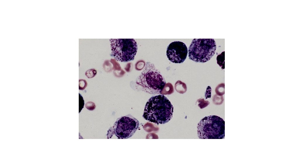
AML FAB Classification (AML-M 0 -M 7)

AML-M 1

AML-M 2 • With differentiation • Maturing cells >10% • Myeloblasts 30 -89% • Many Auer rods are seen • Sudan Black positive (>03%) • Type II blasts (with granules)

AML-M 2

Sudan Black B Stain
 • Hypergranular Promyelocytes (>30%) • Numerous Auer rods (Faggot")
AML-M 3 (Ac. Promyelocytic Leukemia) • Hypergranular Promyelocytes (>30%) • Numerous Auer rods (Faggot cells) • Sudan positive • Bleeding is sever and more common (DIC) • Infections are more common • T(15: 17) • Respond to ATRA (All-Trans-Retinoic Acid)

AML-M 3
 • Myeloblasts as well as monoblasts are seen •")
AML-M 4 (Acute Myelo-monocytic Leukemia) • Myeloblasts as well as monoblasts are seen • Maturing cells of both myeloid and monocytoid series are present • Auer rods are observed • Sudan Black positive • NAE Positive • Tissue infiltrations are more common • Gingival ‘hypertrophy’ is common

AML-M 4
 • Monoblasts >30% • M 5 a No maturation")
AML-M 5 (Acute Monoblastic leukemia) • Monoblasts >30% • M 5 a No maturation • M 5 b promonocytes and monocytes are seen • Tissue infiltrations are very common • Gingival “hypertrophy” is common • NAE positive • Poor prognosis

AML-M 5

AML-M 5 Esterase Stain
 • Normoblasts >50% • Myeloblasts >30% of non-erythroid cells •")
AML-M 6 (Acute Erythroleukemia) • Normoblasts >50% • Myeloblasts >30% of non-erythroid cells • Poor prognosis and response to therapy

AML-M 6
 • Megakaryoblasts >30% • Severe fibrosis (Acute Myelofibrosis) •")
AML-M 7 (Ac. Megakaryocytic Leukemia) • Megakaryoblasts >30% • Severe fibrosis (Acute Myelofibrosis) • Very aggressive leukemia • Blood tap is common • Trephine biopsy • Diagnosed by monoclonal markers (CD 41 and CD 61)

Bone Marrow Fibrosis in AML-M 7
 Sudan Black negative (<3%) Poor")
AML-M 0 Undifferentiated AML No maturation (100% blast cells) Sudan Black negative (<3%) Poor response to therapy

AML-M 0

- Slides: 42