Acute Inflammation Dr S Nishan Silva MBBS General
")
Acute Inflammation Dr. S. Nishan Silva (MBBS)

General Facture of Inflammation �In Cell Injury – various exogenous and endogenous stimuli can cause cell injury which involve the cells, nuclei and organelles of the cells. In Vascularized Tissue – same exogenous and endogenous stimuli produce inflammation. �

INFLAMMATION Inflammation: Inflammation is the reaction of blood vessels, leading to the accumulation of fluid (Serum) and leukocytes in extra vascular tissue.

Role Of Tissue And Cells In Inflammation � Many tissue and cells are involved in inflammation. The tissue & fluid are: � The fluid and proteins of plasma. � Blood vessels. � Cellular and extra cellular constituents of connective tissue (mast cells & fibroblast).

Role of tissue and cells in inflammation The circulating cells are: � Neutrophils. � Monocytes. � Eosinophils. � Lymphocytes. � Basophils. � Platelets.
. �")
Sign & Symptoms Of Inflammation These are: � Fever � Pain. (increase temperature). � Tissue damage. � Swelling of tissue. � Redness of tissue. � Loss of movements or restricted movement, if near joints.

Types Of Inflammation is divided into I - Acute inflammation, which occurs over seconds, minutes, hours, and days. II - Chronic inflammation, which occurs over longer times, days & months.

Acute Inflammation � Acute inflammation, begins within seconds to minutes following the injury of tissues. � The damage may be purely physical, or it may involve the activation of an immune response.

Chronic Inflammation Chronic inflammation is of longer duration and is associated histologically with the presence of: � Lymphocytes and macrophages. � The proliferation of blood vessels. � Fibrosis and tissue necrosis.

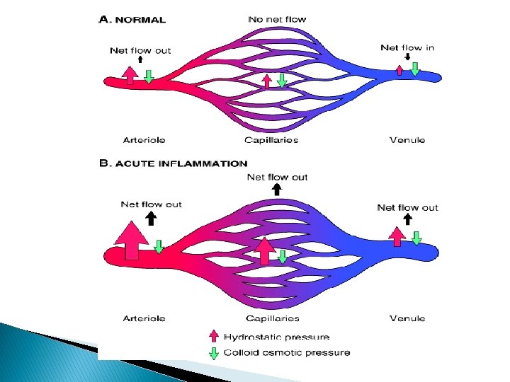
Response Of Inflammation The main processes are: I - Increased blood flow. II - Increased permeability. III - Migration of neutrophils. IV - Chemotaxis. V - Leucocytes recruitment & activation.

Response Of Inflammation The main processes are: I - Increased blood flow due to dilation of blood vessels (arterioles) supplying the region. II - Increased permeability of the capillaries, allowing fluid and blood proteins to move into the interstitial spaces
 out")
Response Of Inflammation III - Migration of neutrophils (and perhaps a few macrophages) out of the venules and into interstitial spaces.

Injury response

Response Of Inflammation IV - Chemotaxis Once outside the blood vessel, a neutrophil is guided towards an infection by various diffusing chemotactic factors. Examples include the chemokines and the complement peptide C 5 a, which is released when the complement system is activated either via specific immunity or innate immunity.

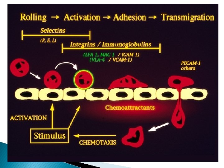
Response Of Inflammation V - Leucocytes recruitment & activation. � This is the first step is the binding of the neutrophils to the endothelium of the blood vessels. � The binding is due to molecules, called cell adhesion molecules (CAMs), found on the surfaces of neutrophils and on endothelial cells in injured tissue.
 The binding of")
Response of Inflammation V - Leucocytes recruitment & activation (contd. ) The binding of leukocytes occur in two steps: � In the first step, adhesion molecules called selectins tightly gather the neutrophil to the endothelium, so that it begins rolling along the surface.
. � In a second")
Response of Inflammation V - Leucocytes recruitment & activation (contd). � In a second step, a much tighter binding occurs through the interaction of ICAMs on the endothelial cells with integrins on the neutrophil.

Response of Inflammation Eosinophils. However, in some circumstances eosinophils rather than neutrophils predominate in acute inflammation. This tends to occur with parasites (worms), against which neutrophils have little success.

Response of Acute Inflammation � Increased Blood Flow, increased permeability and Edema in Inflammation: � The increased blood flow & increased permeability are readily visible within a few minutes following a scratch that does not break the skin.

Response of Acute Inflammation � At first, there is pale red line of scratch. � Later on there is accumulation of inflammatory cells lead swelling, (inflammation). � Finally, there is accumulation of interstitial fluid cause edema.
.")
Acute Inflammation (recruitment of neutrophils).

Acute Inflammation

Acute Inflammation

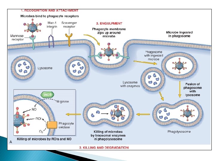
Acute Inflammation CELLULAR EVENTS: LEUKOCYTE EXTRAVASATION AND PHAGOCYTOSIS

Acute Inflammation

Acute Inflammation
")
Acute Inflammation (with pus)
")
Acute Inflammation (Acute Bronchitis)

Acute Inflammation

Acute Inflammation

Vasodilation Role of Mediators in Different Reactions of Inflammation Increased vascular permeability Chemotaxis, leukocyte recruitment and activation Fever Pain Tissue damage Prostaglandins Histamine Nitric oxide Vasoactive amines Bradykinin Leukotrienes C 4, D 4, E 4 PAF Substance P C 5 a Leukotriene B 4 Chemokines IL-1, TNF Bacterial products IL-1, TNF Prostaglandins Bradykinin Neutrophil and macrophage lysosomal enzymes Oxygen metabolites Nitric oxide

Objectives � Patterns � � � of Acute Inflammation Outcomes of Acute Inflammation Patterns of chronic Inflammation Defect in leukocyte function Complement deficiency Systemic manifestation

Morphologic Patterns of Acute Inflammation �Several types of inflammation vary in their morphology and clinical correlates. Why? ◦ ◦ The severity of the reaction specific cause the particular tissue site involved

Morphologic Patterns of Acute Inflammation �SEROUS INFLAMMATION �FIBRINOUS INFLAMMATION �SUPPURATIVE OR PURULENT INFLAMMATION �ULCERS

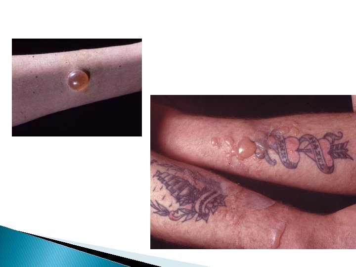
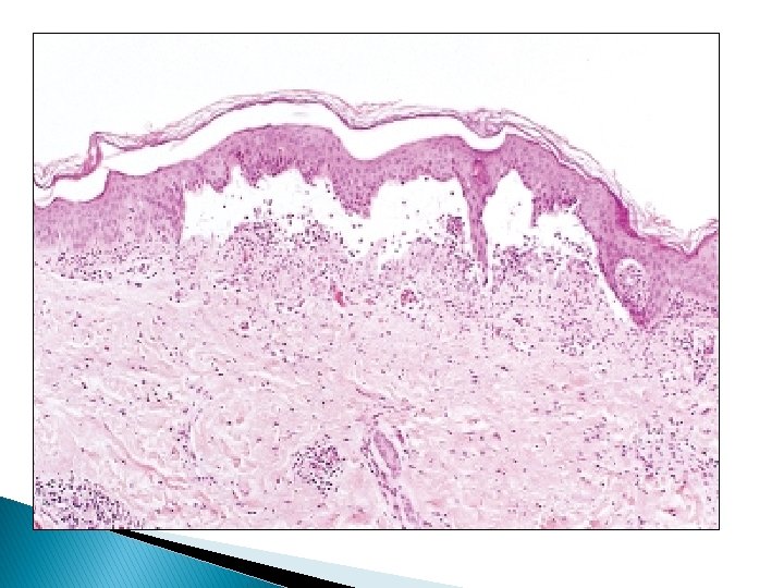
Patterns of Acute Inflammation �SEROUS INFLAMMATION: ◦ Serous inflammation is marked by the outpouring of a thin fluid ◦ e. g. the skin blister resulting from a burn or viral infection represents a large accumulation of serous fluid

Morphologic Patterns of Acute Inflammation � FIBRINOUS INFLAMMATION ◦ more severe injuries and more greater vascular permeability, larger molecules such as fibrinogen pass the vascular barrier, and fibrin is formed and deposited

Morphologic Patterns of Acute Inflammation FIBRINOUS INFLAMMATION ◦ A fibrinous exudate is characteristic of inflammation in the lining of body cavities, such as the meninges, pericardium and pleura

Morphologic Patterns of Acute Inflammation � FIBRINOUS INFLAMMATION ◦ Fibrinous exudates may be removed by fibrinolysis ◦ But when the fibrin is not removed, it may stimulate the ingrowth of fibroblasts and blood vessels and thus lead to scarring (organization)

Morphologic Patterns of Acute Inflammation SUPPURATIVE OR PURULENT INFLAMMATION ◦ characterized by the production of large amounts of pus or purulent exudate consisting of neutrophils, necrotic cells, and edema fluid ◦ Certain bacteria (e. g. , staphylococci) produce this localized suppuration and are therefore referred to as pyogenic (pusproducing) bacteria

Suppurative inflammation. A, A subcutaneous bacterial abscess with collections of pus. B, The abscess contains neutrophils, edema fluid, and cellular debris.

Purulent inflammation

Morphologic Patterns of Acute Inflammation SUPPURATIVE OR PURULENT INFLAMMATION ◦ Abscesses : localized collections of purulent inflammatory tissue caused by suppuration buried in a tissue, an organ, or a confined space

Brain Abscess

Liver abscess

Localized liquefactive necrosis liver abscess Removal of the dead tissue leaves behind a scar

Morphologic Patterns of Acute Inflammation ULCERS ◦ An ulcer is a local defect of the surface of an organ or tissue that is produced by the sloughing (shedding) of inflammatory necrotic tissue
 inflammatory necrosis of the mucosa")
Morphologic Patterns of Acute Inflammation ULCERS encountered in: 1) inflammatory necrosis of the mucosa of the mouth, stomach, intestines, or genitourinary tract 2) subcutaneous inflammation of the lower extremities in older persons who have circulatory disturbances

Ulceration can occur only when tissue necrosis and resultant inflammation exist on or near a surface Epithelial Defect Necrotic base Fibrinopurulent exudates Granulation tissue Fibrosis

Objectives � Patterns of Acute Inflammation �Outcomes � Patterns of Acute Inflammation of chronic Inflammation � Defect in leukocyte function � Complement deficiency � Systemic manifestation

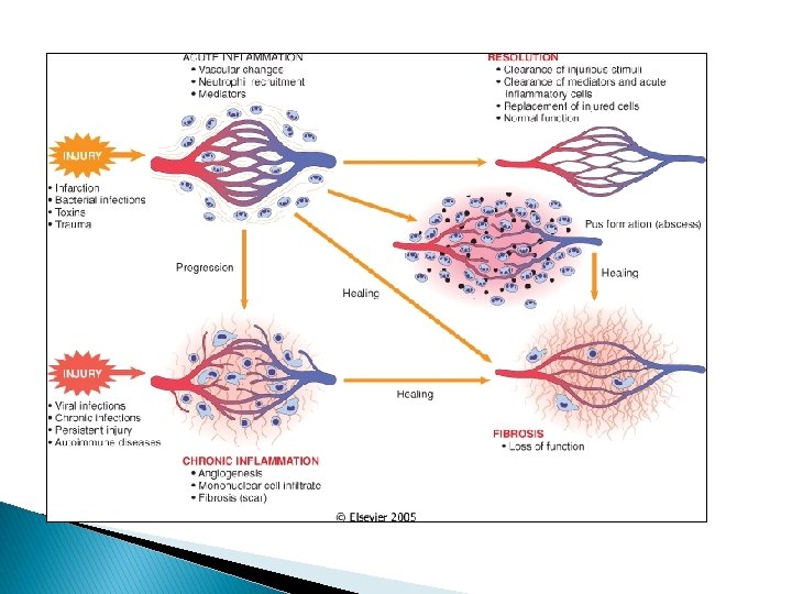
Outcomes of Acute Inflammation Acute inflammation may have one of the four outcomes: � ◦ ◦ Complete resolution Healing by connective tissue replacement (fibrosis) Progression of the tissue response to chronic inflammation Abcess formation
 the injury is limited or short-lived")
Outcomes of Acute Inflammation Complete resolution When? 1) the injury is limited or short-lived 2) there has been little tissue destruction 3) the damaged parenchymal cells can regenerate

Outcomes of Acute Inflammation Complete resolution Mechanism: �Neutralization and removal of chemical mediators �Normalization of vascular permeability �halting of leukocyte emigration �Clearance of edema (lymphatic drainage) , inflammatory cells and necrotic debris (macrophages).

Events in the resolution of inflammation
: � This occurs")
Outcomes of Acute Inflammation � Healing by connective tissue replacement (fibrosis): � This occurs after substantial tissue destruction ◦ the inflammatory injury involves tissues that are incapable of regeneration ◦ there is abundant fibrin exudation. v The destroyed tissue is resaorbed and eventually replaced by fibrosis.

Outcomes of Acute Inflammation � Progression of the tissue response to chronic inflammation: ◦ occurs when the acute inflammatory response cannot be resolved WHY? Due to: 1. the persistence of the injurious agent 2. some interference with the normal process of healing

A 36 -year-old man has had midepigastric abdominal pain for the past 3 months. An upper gastrointestinal endoscopy shows a 2 -cm, sharply demarcated, shallow ulceration of the gastric antrum. A biopsy specimen of the ulcer base shows angiogenesis, fibrosis, and mononuclear cell infiltrates with lymphocytes, macrophages, and plasma cells. Which of the following terms best describes this pathologic process? (A) Acute inflammation (B) Serous inflammation (C) Granulomatous inflammation (D) Fibrinous inflammation (E) Chronic inflammation

Systemic effects of Inflammation � Acute phase reaction/response - IL-1 and TNF - Fever - Malaise - Anorexia � Bone marrow - leukocytosis - IL-1 + TNF � Lymphoid organs � Liver -IL-6, IL-1, TNF -Acute phase proteins ◦ C-reactive protein ◦ Lipopolysaccharide binding protein ◦ Serum amyloid A ◦ a-2 macroglobulin ◦ Haptoglobin ◦ Ceruloplasmin ◦ fibrinogen

Systemic Effects of Inflammation Fever � Fever is produced in response to Pyrogens � What are pyrogens? ◦ act by stimulating prostaglandin synthesis in the vascular and perivascular cells of the hypothalamus. � Bacterial products (called exogenous pyrogens), stimulate leukocytes to release cytokines such as IL-1 and TNF (called endogenous pyrogens) that increase the enzymes (cyclooxygenases) that convert AA into prostaglandins.

Fever • In the hypothalamus, the prostaglandins, especially PGE 2, stimulate the production of neurotransmitters such as cyclic AMP, which function to reset the temperature set-point at a higher level. • NSAIDs, including aspirin , reduce fever by inhibiting cyclooxygenase and thus blocking prostaglandin synthesis. • fever may induce heat shock proteins that enhance lymphocyte responses to microbial antigens.

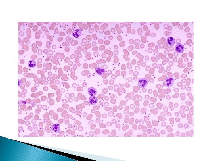
Inflammation Systemic Manifestations Leukocytosis: WBC count climbs to 15, 000 or 20, 000 cells/μl most bacterial infection Lymphocytosis: Infectious mononucleosis, mumps, German measles Eosinophilia: bronchial asthma, hay fever, parasitic infestations Leukopenia: typhoid fever, infection with rickettsiae/protozoa

Acute inflammation � Time 4 -6 hours to 3 -5 days � Vascular involvement ◦ Active hyperemia ◦ Edema, occ. fibrin thrombi � Neutrophils � Cardinal signs of inflammation � Lymphatics ◦ Role to remove exudate ◦ Can lead to inflammation. � Lymphangitis � Lymphadenitis


- Slides: 70