
ACUTE ABDOMEN DR D VINDHYA Dept of Emergency
ACUTE ABDOMEN DR. D. VINDHYA Dept of Emergency & Critical Care Medicine, Vinayaka Mission Medical College & Hospital, Salem
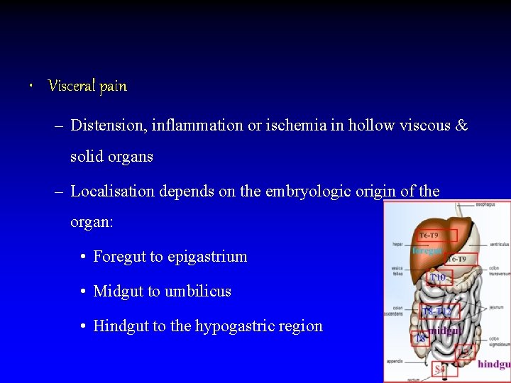
• Visceral pain – Distension, inflammation or ischemia in hollow viscous & solid organs – Localisation depends on the embryologic origin of the organ: • Foregut to epigastrium • Midgut to umbilicus • Hindgut to the hypogastric region
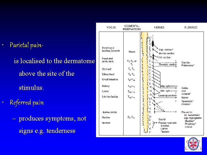
• Parietal painis localised to the dermatome above the site of the stimulus. • Referred pain – produces symptoms, not signs e. g. tenderness
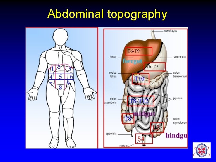
Abdominal topography
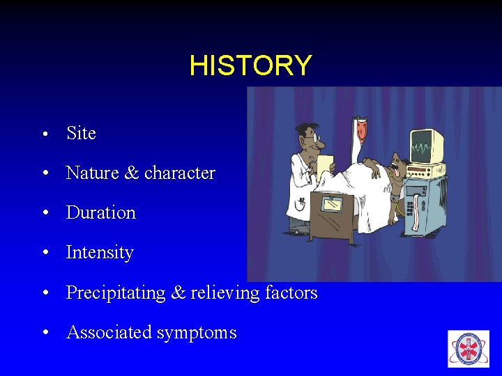
HISTORY • Site • Nature & character • Duration • Intensity • Precipitating & relieving factors • Associated symptoms
• Previous episodes of AP • Investigations • Chronic disease • Immunosuppression • Medications (NSAIDs) • surgeries
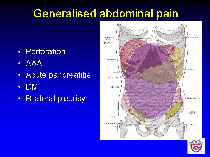
Generalised abdominal pain • • • Perforation AAA Acute pancreatitis DM Bilateral pleurisy
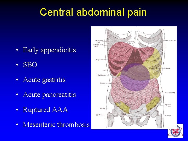
Central abdominal pain • Early appendicitis • SBO • Acute gastritis • Acute pancreatitis • Ruptured AAA • Mesenteric thrombosis
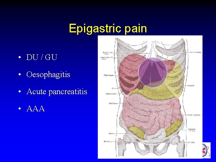
Epigastric pain • DU / GU • Oesophagitis • Acute pancreatitis • AAA
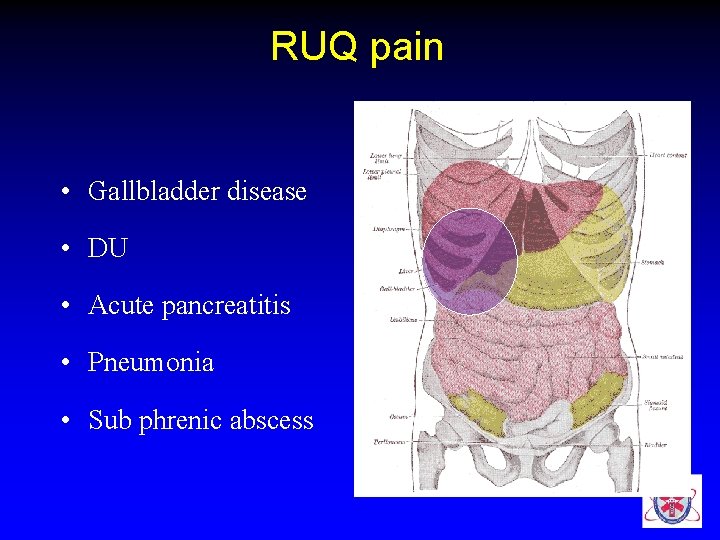
RUQ pain • Gallbladder disease • DU • Acute pancreatitis • Pneumonia • Sub phrenic abscess

Differential diagnosis of RUQ pain CONDITION CLUES Biliary colic, acute cholecystitis Recurrent attacks, tender over gall bladder area Acute hepatitis Alcohol h/o, medications, icterus Acute pyelonephritis Dysuria, fever, costovertebral angle tenderness CCF Edema, dyspnoea, elevated JVP Retrocaecal appendicitis Shift of pain, tenderness Right lower lobe pneumonia Fever, tachypnoea, bronchial
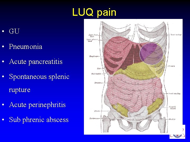
LUQ pain • GU • Pneumonia • Acute pancreatitis • Spontaneous splenic rupture • Acute perinephritis • Sub phrenic abscess
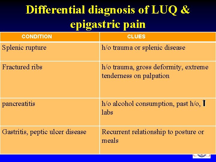
Differential diagnosis of LUQ & epigastric pain CONDITION CLUES Splenic rupture h/o trauma or splenic disease Fractured ribs h/o trauma, gross deformity, extreme tenderness on palpation pancreatitis h/o alcohol consumption, past h/o, labs Gastritis, peptic ulcer disease Recurrent relationship to posture or meals
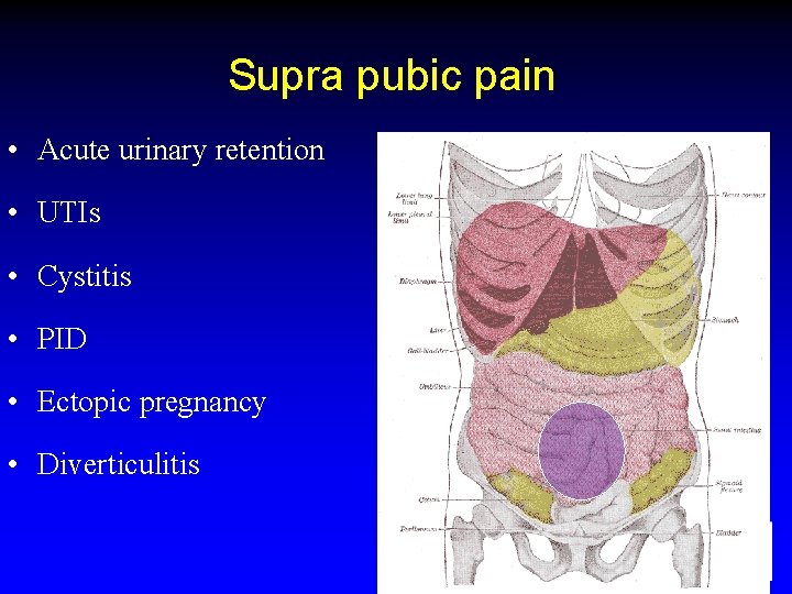
Supra pubic pain • Acute urinary retention • UTIs • Cystitis • PID • Ectopic pregnancy • Diverticulitis
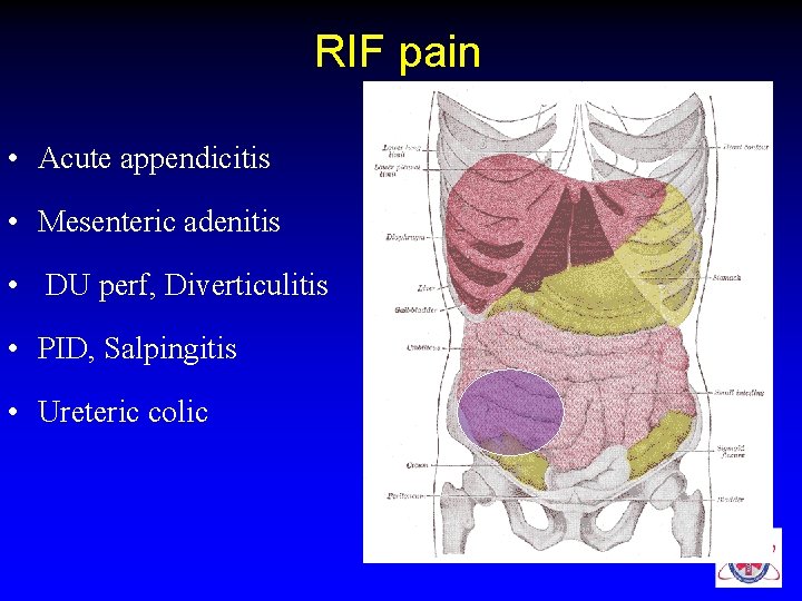
RIF pain • Acute appendicitis • Mesenteric adenitis • DU perf, Diverticulitis • PID, Salpingitis • Ureteric colic
• Meckel’s diverticulum • Ectopic pregnancy • Crohn’s disease • Biliary colic (low-lying gall bladder)

Differential diagnosis of RLQ pain CONDITIONS CLUES Mesentric adenitis Fever, inconstant signs Rt renal colic Colic pain , haematuria Rt. testis torsion Tender swollen testis Crohns disease Recurrent h/o diarrhoea, colicky pain, wt loss
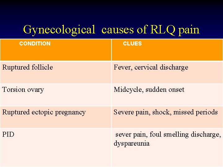
Gynecological causes of RLQ pain CONDITION CLUES Ruptured follicle Fever, cervical discharge Torsion ovary Midcycle, sudden onset Ruptured ectopic pregnancy Severe pain, shock, missed periods PID sever pain, foul smelling discharge, dyspareunia
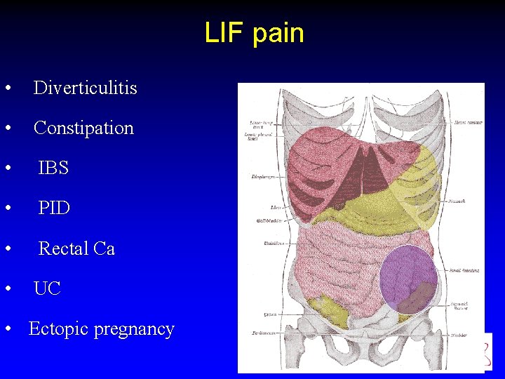
LIF pain • Diverticulitis • Constipation • IBS • PID • Rectal Ca • UC • Ectopic pregnancy
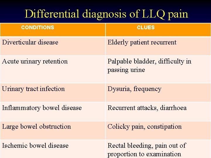
Differential diagnosis of LLQ pain CONDITIONS CLUES Diverticular disease Elderly patient recurrent Acute urinary retention Palpable bladder, difficulty in passing urine Urinary tract infection Dysuria, frequency Inflammatory bowel disease Recurrent attacks, diarrhoea Large bowel obstruction Colicky pain, constipation Ischemic bowel disease Rectal bleeding, pain out of proportion to examination
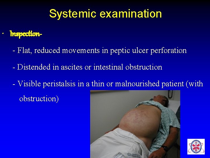
Systemic examination • Inspection- Flat, reduced movements in peptic ulcer perforation - Distended in ascites or intestinal obstruction - Visible peristalsis in a thin or malnourished patient (with obstruction)
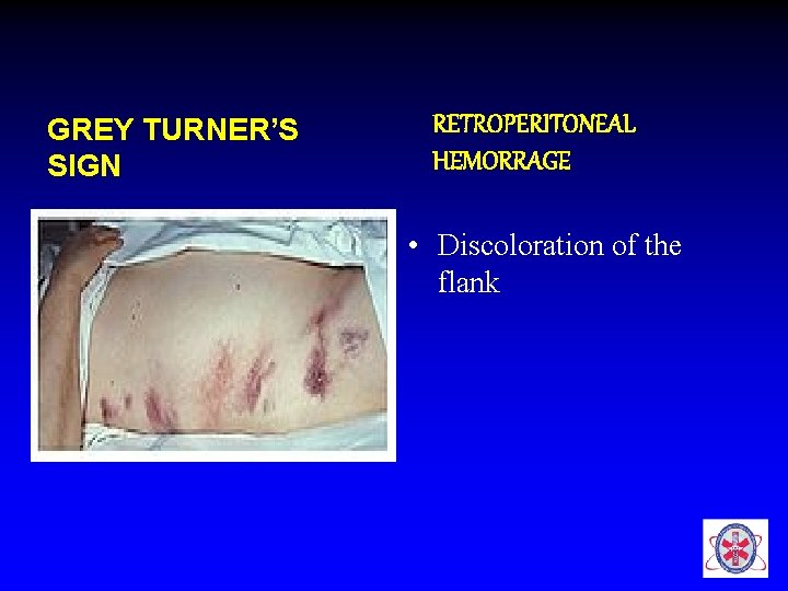
GREY TURNER’S SIGN RETROPERITONEAL HEMORRAGE • Discoloration of the flank
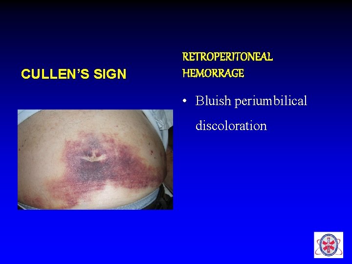
CULLEN’S SIGN RETROPERITONEAL HEMORRAGE • Bluish periumbilical discoloration
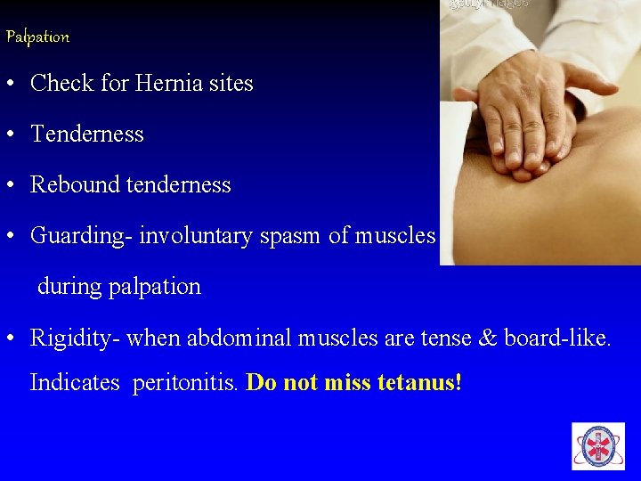
Palpation • Check for Hernia sites • Tenderness • Rebound tenderness • Guarding- involuntary spasm of muscles during palpation • Rigidity- when abdominal muscles are tense & board-like. Indicates peritonitis. Do not miss tetanus!
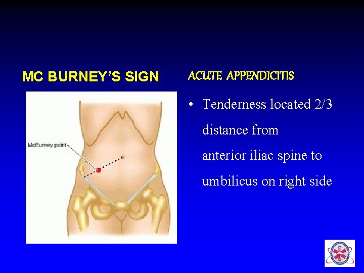
MC BURNEY’S SIGN ACUTE APPENDICITIS • Tenderness located 2/3 distance from anterior iliac spine to umbilicus on right side
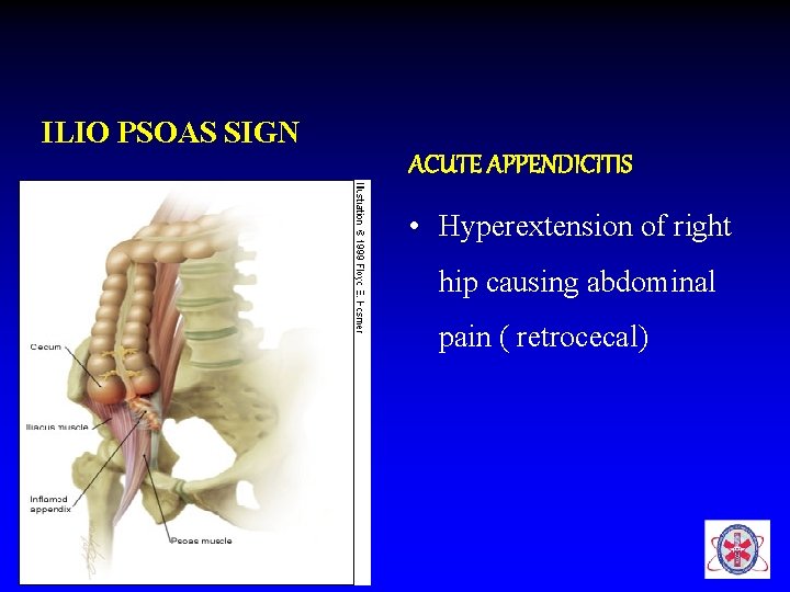
ILIO PSOAS SIGN ACUTE APPENDICITIS • Hyperextension of right hip causing abdominal pain ( retrocecal)
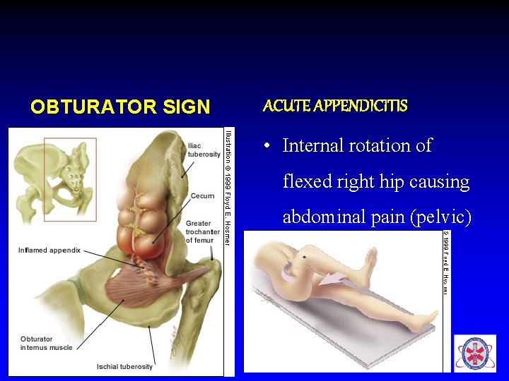
OBTURATOR SIGN ACUTE APPENDICITIS • Internal rotation of flexed right hip causing abdominal pain (pelvic)
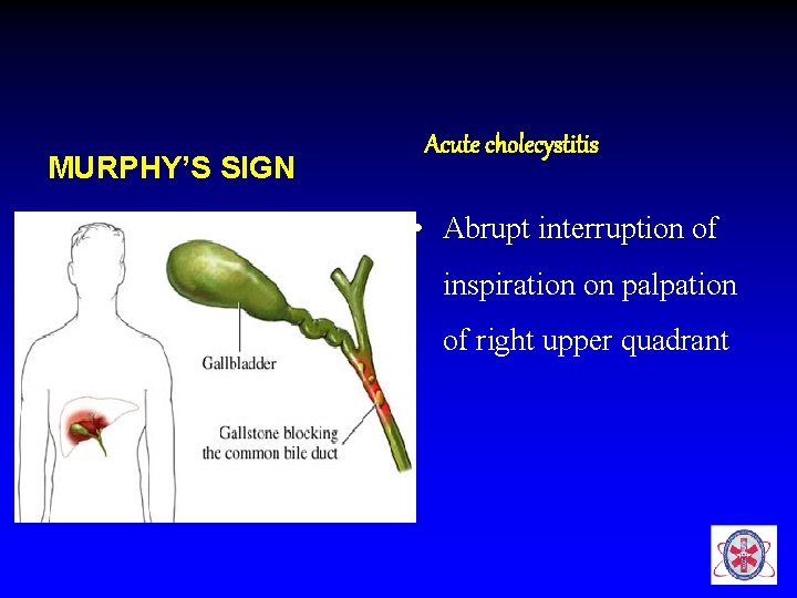
MURPHY’S SIGN Acute cholecystitis • Abrupt interruption of inspiration on palpation of right upper quadrant
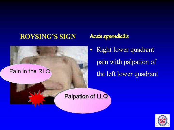
ROVSING’S SIGN Acute appendicitis • Right lower quadrant pain with palpation of Pain in the RLQ the left lower quadrant Palpation of LLQ
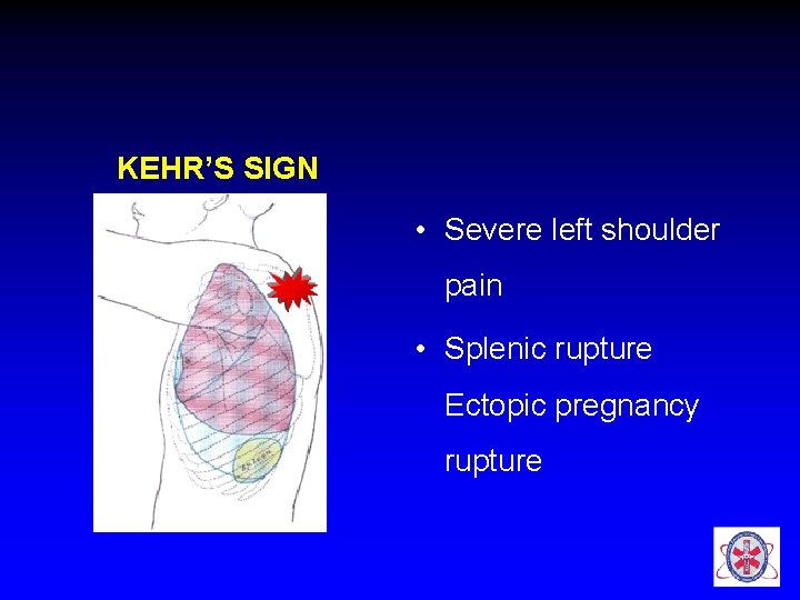
KEHR’S SIGN • Severe left shoulder pain • Splenic rupture Ectopic pregnancy rupture
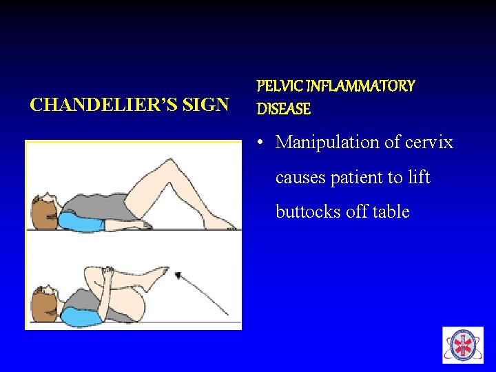
CHANDELIER’S SIGN PELVIC INFLAMMATORY DISEASE • Manipulation of cervix causes patient to lift buttocks off table
• Auscultation • BS – > 2 min to confirm absent – High pitched, hyperactive or tinkling – Bruit in epigastrium
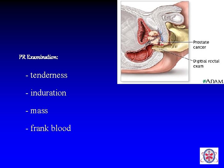
PR Examination: - tenderness - induration - mass - frank blood
Investigations • CBC • Amylase & lipase • Erect & supine abdominal XRay • stool & Urine analysis, • pregnancy test, USG, CT scan • If severe, unrelenting pain urgent surgical referral
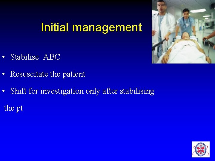
Initial management • Stabilise ABC • Resuscitate the patient • Shift for investigation only after stabilising the pt
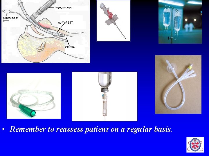
• Remember to reassess patient on a regular basis.
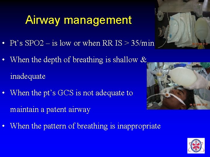
Airway management • Pt’s SPO 2 – is low or when RR IS > 35/min • When the depth of breathing is shallow & inadequate • When the pt’s GCS is not adequate to maintain a patent airway • When the pattern of breathing is inappropriate
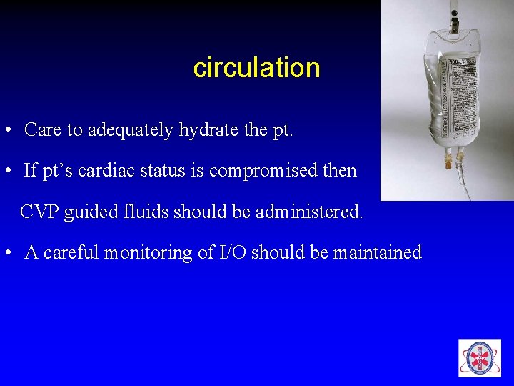
circulation • Care to adequately hydrate the pt. • If pt’s cardiac status is compromised then CVP guided fluids should be administered. • A careful monitoring of I/O should be maintained
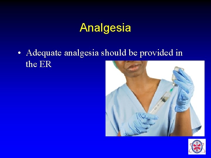
Analgesia • Adequate analgesia should be provided in the ER
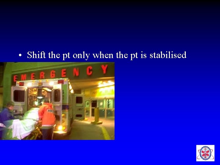
• Shift the pt only when the pt is stabilised
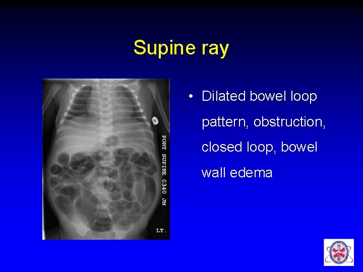
Supine ray • Dilated bowel loop pattern, obstruction, closed loop, bowel wall edema
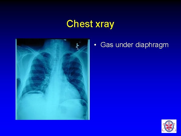
Chest xray • Gas under diaphragm
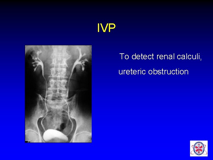
IVP To detect renal calculi, ureteric obstruction
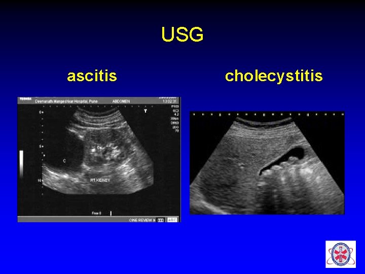
USG ascitis cholecystitis
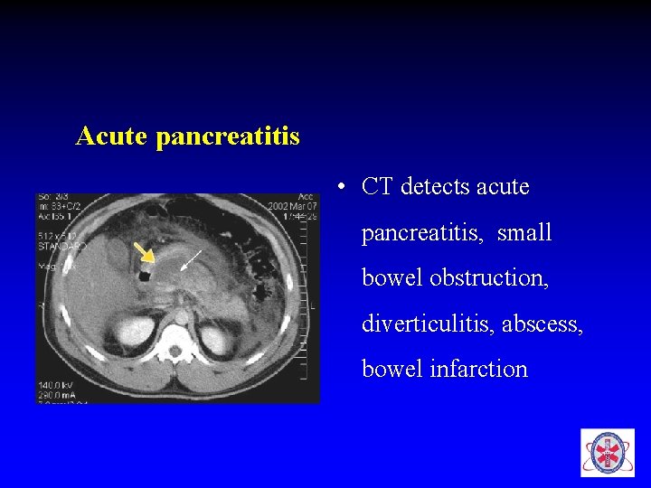
Acute pancreatitis • CT detects acute pancreatitis, small bowel obstruction, diverticulitis, abscess, bowel infarction
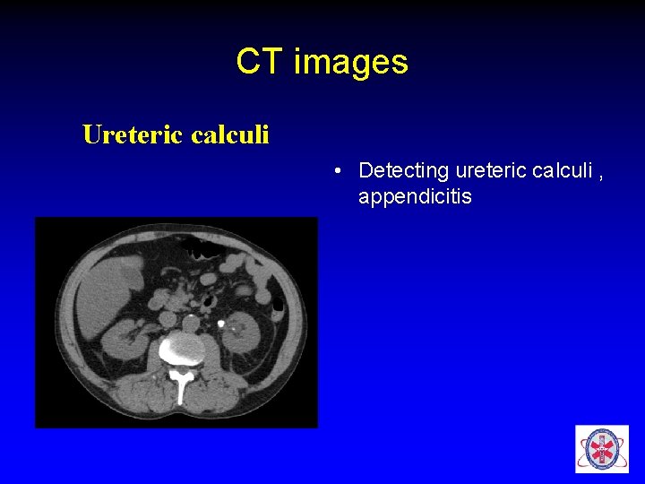
CT images Ureteric calculi • Detecting ureteric calculi , appendicitis
CASE DISCUSSIONS
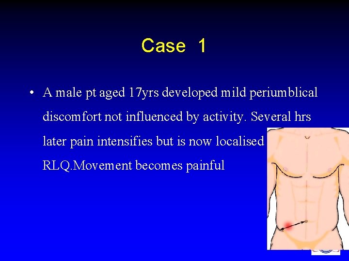
Case 1 • A male pt aged 17 yrs developed mild periumblical discomfort not influenced by activity. Several hrs later pain intensifies but is now localised to RLQ. Movement becomes painful
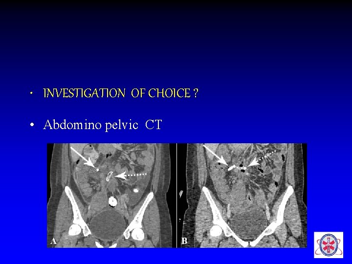
• INVESTIGATION OF CHOICE ? • Abdomino pelvic CT
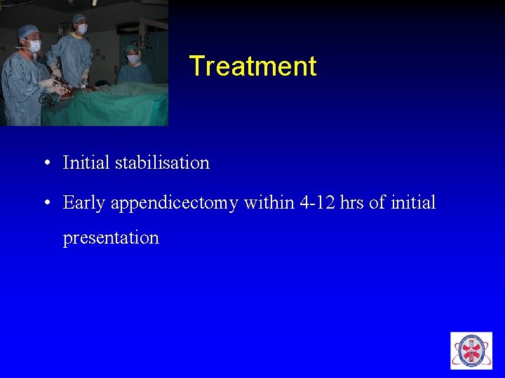
Treatment • Initial stabilisation • Early appendicectomy within 4 -12 hrs of initial presentation
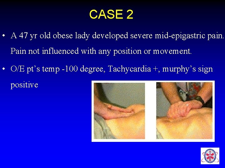
CASE 2 • A 47 yr old obese lady developed severe mid-epigastric pain. Pain not influenced with any position or movement. • O/E pt’s temp -100 degree, Tachycardia +, murphy’s sign positive
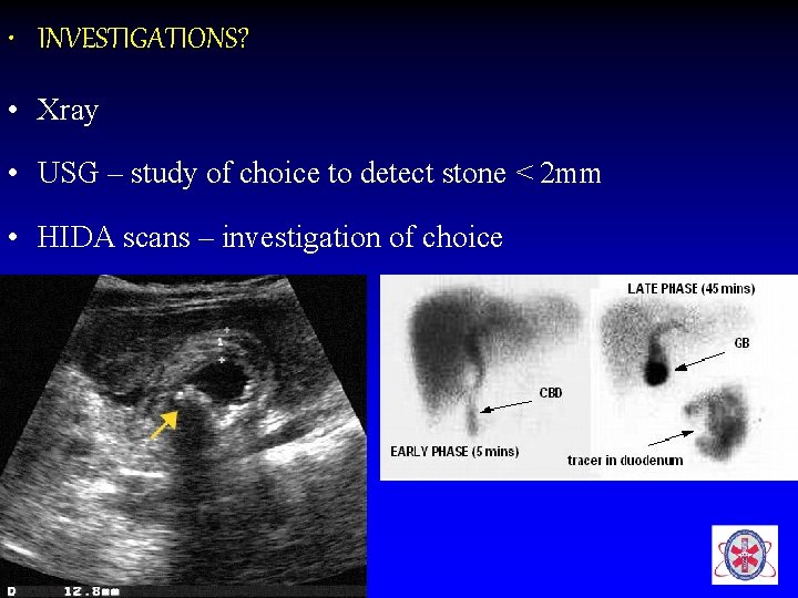
• INVESTIGATIONS? • Xray • USG – study of choice to detect stone < 2 mm • HIDA scans – investigation of choice
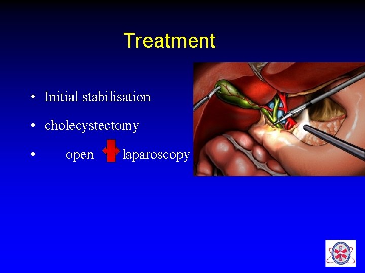
Treatment • Initial stabilisation • cholecystectomy • open laparoscopy
CASE 3 • A 62 yr old man C/O severe abdominal pain – generalised in nature. H/O consumption of NSAIDS. He also c/o lt shoulder pain. He feels more comfortable sitting than lying. • O/E pt conscious , afebrile, sweating profusely • HR-120/min, BP-120/90 mm hg
• Abd- rigid, tenderness , rebound tenderness & guarding present in all quadrants. percussion –absence of liver dullness • What is the likely diagnosis? • Investigations ?
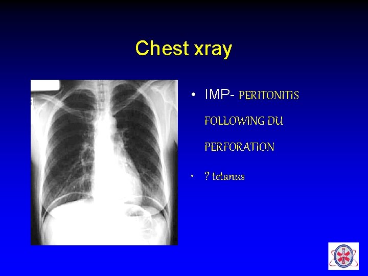
Chest xray • IMP- PERITONITIS FOLLOWING DU PERFORATION • ? tetanus
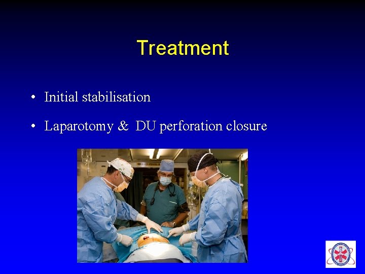
Treatment • Initial stabilisation • Laparotomy & DU perforation closure
Case 4 • A 34 y old female pt rushed to ER in shock. O/E • HR-120/min, BP- 90/60 mmhg, • RR-26/min, SPO 2 -94% on RA • CVS, RS – NAD • ABD – LLQ tenderness + • Next ?
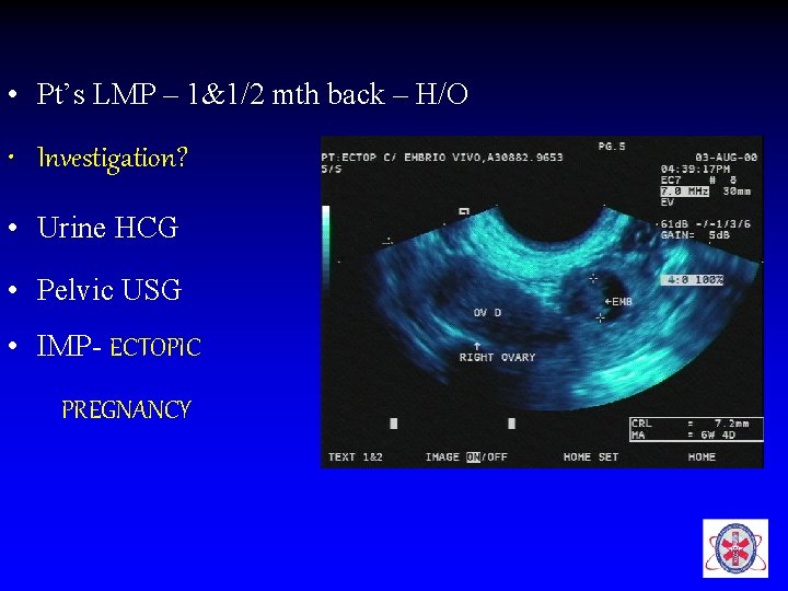
• Pt’s LMP – 1&1/2 mth back – H/O • Investigation? • Urine HCG • Pelvic USG • IMP- ECTOPIC PREGNANCY
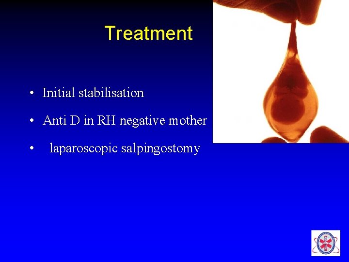
Treatment • Initial stabilisation • Anti D in RH negative mother • laparoscopic salpingostomy
Case 5 • A 23 yr old student brought to ER writhing with pain radiating from lt lumbar to groin associated with vomiting • Next ?
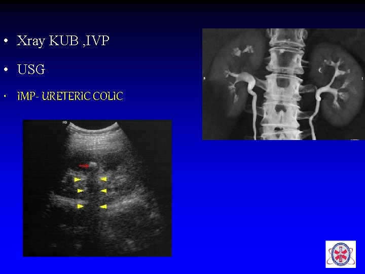
• Xray KUB , IVP • USG • IMP- URETERIC COLIC
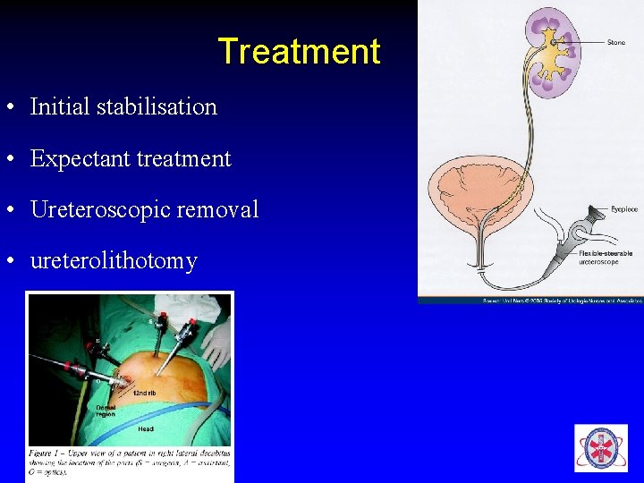
Treatment • Initial stabilisation • Expectant treatment • Ureteroscopic removal • ureterolithotomy
Case 6 • A 65 yr old male, known diabetic admitted at 9 pm with h/o abdominal pain associated with profuse sweating & vomiting since evening 7 pm O/E HR- 68/min, BP – 90/70 mmhg. What next?
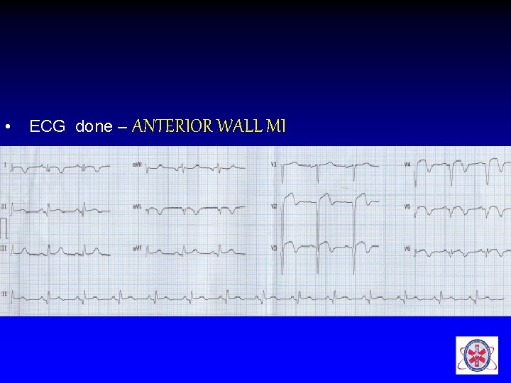
• ECG done – ANTERIOR WALL MI
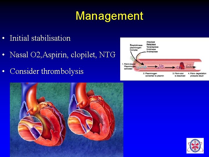
Management • Initial stabilisation • Nasal O 2, Aspirin, clopilet, NTG • Consider thrombolysis
Case 7 • A 42 yr old male pt, known alcoholic presented to our ER with H/O persistent epigastric pain improving on bending forward & worsens with lying down. • O/E vitals are stable except for tachycardia • Systemic examination – NAD except for tenderness in the epigastric region
• What is the likely diagnosis? • What are the investigations to be done?
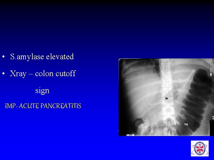
• S. amylase elevated • Xray – colon cutoff sign IMP- ACUTE PANCREATITIS
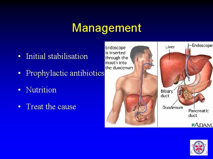
Management • Initial stabilisation • Prophylactic antibiotics • Nutrition • Treat the cause
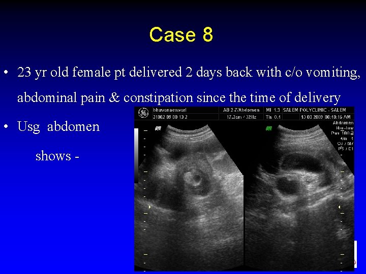
Case 8 • 23 yr old female pt delivered 2 days back with c/o vomiting, abdominal pain & constipation since the time of delivery • Usg abdomen shows -
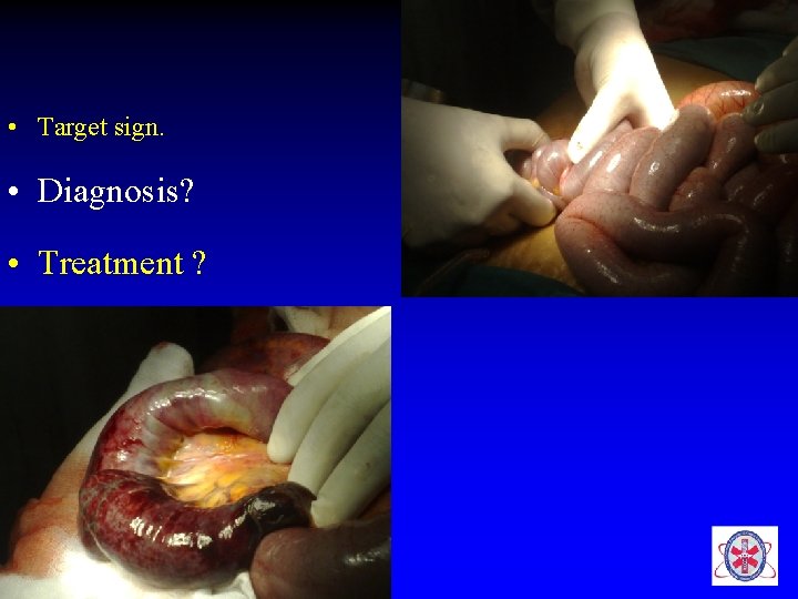
• Target sign. • Diagnosis? • Treatment ?
Carry home message • Our priority- ABC ECG CBG ABG • All abdominal aches need not arise from the abdomen • Adequate hydration, adequate analgesia, appropriate antibiotic coverage at ER

THANQ
- Slides: 74