Acupuncture for Neurological Disorders It matters not whether



- Slides: 154

Acupuncture for Neurological Disorders It matters not whether medicine is new or old, it only matters that it is applied for the benefit of the patient.

Neurologic Assessment • Is it a neurologic disease? – Seizures – Intention tremor – CN deficits • Head tilt • Nystagmus – CP deficits – Dysmetria – Paralysis

Minimum Database • CBC • Chemistry – Bile acids – Cholinesterase • Urinalysis • Chest and abdominal radiographs • Abdominal ultrasound • Heartworm test • Fecal

Ancillary Neurologic Tests • Electrodiagnostics • Radiographs • CSF tap & analysis • Muscle Analysis – – – – EEG EMG BAER Cells & protein Pressure Cholinesterase Titers – – – – Skull & spinal films Myelography CT scan MRI Enzymes 2 M antibody Anti-ACH receptor antibody – Biopsy

When all else fails… Look at the patient!!!

Seizures in Small Animals • It is estimated that the overall incidence of seizure disorders in dogs and cats is around 1% • In pure breed dogs, this incidence may increase to 1520%, due to the presence of inherited, primary epilepsy in those breeds

Lesion Localization in Seizures • Cerebral Cortex • Diencephalon – Thalamus – Hypothalamus • Mesencephalon

Seizure Diagnoses SEIZURES Primary Epilepsy Secondary Epilepsy Inactive Reactive Epilepsy Active

Seizure Diagnosis • Minimum Database • CT or MRI Scan • CSF tap & analysis • EEG – Abnormal in Reactive Epilepsy – Abnormal in Active Secondary Epilepsy – Abnormal in Secondary Epilepsy • All test are normal in Primary Epilepsy

Asymmetrical Seizures

Licking Seizure

Fly-Biting Seizure

Seizures and Signalment • Primary Epilepsypurebred dogs 1 -3 years of age • Secondary Epilepsyany age but especially under 6 months and over 3 years

Seizure Treatment • Acupuncture alone • Acupuncture in conjunction with drugs • Traditional Chinese herbs • Western herbal medicine – – – Valerian Root Kava Oats Hops Passionflower • Western drugs

Phenobarbital • Cheap and effective • Dose 2 -4 mg/kg for a serum level 20 -40 mcg/ml • Takes 3 -5 days to reach steady state • Safe but can effect the liver in a few cases • Controls 80% of seizures

KBr (Bromide) • Compounded by pharmacist dissolved in H 2 O at 250 mg/ml • Dose at 22 mg/kg q 12 h • Blood level in 2 months between 1500 -4000 µg/ml • Use alone or in conjunction with Phenobarbital • Bypass liver • Good for cluster seizures

Other Anticonvulsants With Efficacy • Diazepam – 1 mg/kg q 12 h • Primidone – 15 -22 mg/kg q 12 h • Felbamate – 15 -45 mg/kg q 8 h No Efficacy • • Valproic acid Nimodapine Chorazepate Lamotrigine • Phenytoin – 3 -5 mg/kg q 8 h • Gabapentin – 30 -60 mg/kg q 8 -12 h • Topiramate – 15 -25 mg/kg q 8 -12 h Note: TOXIC to Dogs

Seizures- -TCM • Represent various aspects of the Liver (Wood) system • Excess (3 types) • Deficiency (3 types)

Seizures- -TCM • Excess – Wind-Phlegm • Tongue pale & greasy • Pulse wiry & slippery – Phlegm-Fire • Tongue red & greasy • Pulse rapid, wiry & slippery – Blood Stagnation • Tongue & Pulse like Wind-Phlegm • History of head trauma • Deficiency – Liver Blood Def. • Tongue pale & dry • Pulse weak & thready – Liver & Kidney Yin Def. • Tongue red & dry • Pulse weak & thready – Kidney Jing Def. • Tongue pale or red & dry • Pulse weak & thready • < 1 year of age

Seizures- -TCM • Excess – Wind-Phlegm • expel phlegm, extinguish the wind, open the orifice and stabilize the seizures • Ding Xian Wan – Phlegm-Fire • clear the liver, drain the heat, transform phlegm and open the orifices • Di Tang and Long Dan Xie Gan Tang – Blood Stagnation • expel phlegm, extinguish the wind, open the orifice, stabilize the seizures and invigorate blood • Ding Xian Wan and Tao Hong Si Wu San • Deficiency – Liver Blood Def. • tonify Qi and Blood and quiet the wind • Bu Xue Xi Feng San or Di Tang plus Rehmannia 8 – Liver & Kidney Yin Def. • nourish Yin and extinguish wind • Yang Yin Xi Feng San or Di Tang and Left Side Replenished (Zuo Gui Wan) or Tian Ma Gou Teng plus – Kidney Jing Def. • extinguish the wind astringe or nourish the kidney jing • Di Tang and Epimedium Powder

Epilepsy -- TCM • Internal heat leading to generation of wind • Clear wind & heat and calm the Shen • Points – Constitutional points – Clear wind & heat • GB 20, LI 4, LI 11, GV 14, LIV 3 – Calm the shen • PC 6, HT 7 – Local points • GV 17, GV 20, GV 21, Long hui, GB 9, GB 13, BL 5, GV 1, ST 40 • TCM Herbals – Di Tang (TCM phenobarbital) – Specific herbs for excesses or deficiencies present

Basic Antioxidants Dogs • Vitamin E 10 IU/lb daily • Vitamin C 5 -10 mg/lb twice a day • Selenium 2 µg/lb daily • Beta carotene 250 IU/lb daily • B Complex 2 mg/kg twice a day Cats • Vitamin E 100 -400 IU daily • Vitamin C 100 -250 mg twice a day • Selenium 50 µg daily • Vitamin A 1000 -5000 IU daily • B Complex 10 mg twice a day

Additional Considerations • Probably safe parasite control – – – Interceptor Frontline Top Spot Revolution • Should avoid – – – – Heartgard Proheart 6 Program Sentinel Frontline Spray Advantage Advantix

Additional Considerations • Diet – Low-carbohydrate food • Supplements – Ginkgo biloba • 2 -4 mg/kg q 8 -12 h • Ginkoba or Publix brand – Tofu or Lecithin • 20 mg/kg daily – Acetylcysteine • 25 mg/kg q 8 h qod

Meningoencephalomyelitis • Infectious Diseases – Species Specific • Steroid Response ME (SRME) • Necrotizing Vasculitis (SRMA) • Necrotizing ME (NME) • Granulomatous ME (GME)

Meningoencephalomyelitis • Pain to paresis to plegia • Dx with CSF tap • Spinal radiographs normal • Myelography normal (might be contraindicated)

Meningoencephalomyelitis

CSF Tap • Collection site for seizures is at the cisterna magnum. • Allows analysis for cells, protein and pressure. • Cytology and titers for infectious organisms can be obtained.

Meningoencephalomyelitis • CSF Analysis – may be normal or show increased pressure, protein and/or cells. • CSF Titers – species specific tests – many must be paired with serum titers. CSF cytology form a dog exhibiting a mixed reaction with neutrophils, lymphocytes and macrophages.

Meningoencephalomyelitis • Infection – – – virus rickettsia protozoa fungus bacteria – – GME NME SRMA • Inflammation

GME • Can be: – – – peracute & progressive chronic • In brainstem, tends to be a multifocal inflammatory disorder • Responds temporarily to steroids. Patient with GME presenting with vertical nystagmus, long tract signs, and circling with incoordination.

GME histologically causes multifocal meningoencephalitis due to proliferation of reticulohistiocytic cells. Lesions also show multinucleated giant cells.

Treatment of ME • Depends upon whether infectious or inflammatory • Prednisolone – Find minimum daily dose and then used 2 times MDD QOD • Primor (activated sulfadimethoxine) – 15 mg/kg BID • Doxycycline – 5 -10 mg/kg QD • Herbal Support – Bromelain/Curcumin • 2. 5/5 mg/kg TID

Menigoencephalomyelitis – Wind-Phlegm • expel phlegm, extinguish the wind, open the orifice and stabilize the seizures • Ding Xian Wan – Phlegm-Fire • clear the liver, drain the heat, transform phlegm and open the orifices • Di Tang and Long Dan Xie Gan Tan

Brain Abscess in a Foal

Brain Abscess in a Foal

Vestibular Disease • Cardinal Signs – Head Tilt – Nystagmus • • Horizontal Rotatory Vertical Positional – Circling (tight) – Imbalance & Incoordination

Vestibular Disease 8 th Nerve only Idiopathic V. D. 8 th Nerve, 7 th Nerve & Horner’s Syndrome Inner Ear Disease Anything Else Central V. D.

Idiopathic Vestibular Disease • Acute Onset of Vestibular Signs – Head tilt – Horizontal or Rotatory nystagmus with fastphase away from head tilt – Nothing else • Can Be Very Severe • Acute, regressive disease

Idiopathic Vestibular Disease -- TCM • Wind (heat) invasion • Clear wind & heat and calm the Shen • Points – Constitutional points – Clear wind & heat • GB 20, LI 4, LI 11, GV 14 – Calm Shen • PC 6, HT 7, GV 17, GV 20, GV 21 – Local points • TH 17, TH 18, TH 21, SI 19, GB 2, Er jian, An shen

Inner Ear Disease • 8 th Nerve Signs • 7 th Nerve Signs – ear & lip droop – lack of palpebral reflex – nose turn – nostril flaring • Horner’s Syndrome

Horner’s Syndrome • Small Animals – – – Ptosis Myosis Enophthalmos • Large Animals – Facial sweating (horse) – Lack of muzzle sweating (cow)

Inner Ear Disease • Most cases are secondary to bacterial infection (otitis media & interna) – extension from otitis externa – pharyngitis with extension up the Eustachian tube – hematogenous spread

Ear Polyps in Cats • Benign growth in the external ear canal which causes signs by extension. • Can also be pharyngeal mass which grows into middle ear via the Eustachian tube.

Ear Polyps in Cats • Treatment is surgical removal. • Damage can be permanent, if pressure necrosis has destroyed the inner ear structure.

Inner Ear Disease -- TCM • Invasion of external pathogen leading to wind, heat, damp. • Heat boils the fluids leading to the accumulation of phlegm. • Quiet the wind, reduce heat, disperse damp and activate the blood to dissolve stagnation.

Inner Ear Disease -- TCM • Points – Constitutional points – Clear wind & heat • GB 20, LI 4, LI 11, GV 14 – Calm the shen • PC 6, HT 7, GV 17, GV 20, GV 21 – Eliminate damp • SP 9 – Activate Qi & blood • ST 36, ST 40, Xin shu – Local points • TH 17, TH 18, TH 21, SI 19, GB 2, Er jian, An shen

Central Vestibular Disease • Postural Changes – CP Deficit – Dysmetria • Reflex Changes – hyperactive reflexes – crossed-extensor reflexes – Babinski’s sign Conscious proprioceptive deficit may be on the same or opposite side of the lesion.

Central Vestibular Disease • CSF Analysis – may be normal or show increased pressure, protein and/or cells. • CSF Titers – species specific tests – many must be paired with serum titers. CSF cytology form a dog exhibiting a mixed reaction with neutrophils, lymphocytes and macrophages

Central Vestibular Disease • Inflammatory or Infectious Diseases – canine distemper – toxoplasmosis and neosporiosis – fungal – rickettsial – GME – SRME

Central Vestibular Disease • Trauma or Vascular – remember dogs don’t get atherosclerosis ! • Neoplasia – meningiomas – choroid plexus papillomas – oligodendrogliomas – astrocytomas – metastatic neoplasia

Central Vestibular Disease MRI of Cerebellar Meningioma

Central Vestibular Disease • Infectious Diseases – – FIP Fe. LV toxoplasmosis cryptococcosis • Trauma • Metabolic – thiamine deficiency • Toxicity – organophosphates • Neoplasia – meningiomas

Central Vestibular Disease -- TCM • Can be wind, heatdamp or wind cold based upon the causative factor involved. • Points – – – Constitutional 8 Principle Zang-Fu

IVD- -TCM Diagnosis • Represents a “bi” syndrome often accompanied by “wei” syndrome • Under domain of KID (bones) & LIV (joints & free flow of qi & blood)

IVD- - TCM Patterns • Excess types • Deficient types – Wind-Cold-Damp – Yang deficiency – Blood stagnation – Yin deficiency – Yin & Yyang deficiency

Fibrocartilagenous Emboli • Vascular occlusion from IVD material – IVD herniates into the venous sinus or the vertebral body – the venous sinuses have no valves – increased pressure forces material into spinal cord

FCE • Generally affects a radicular penetrating branch which leads to a quadrant (wedge) of infarction • Many will improve with time

Schatzie

IVD- -Wind-Cold-Damp • Acute invasion of external pathogen leading to stagnation (cold slows blood flow which is worsened by accumulation of damp) • Tongue – Greasy • Pulse – Slow & soft • Rx principle – Dispel W-C-D, activate blood & relieve stagnation • TCM herbal – Xiao Huo Luo Dan • Acupuncture – Hua-tuo-jia-ji, BL 23, BL 67, GB 39, GV 1, & GV 14

Acute Spinal Cord Injury • Damage affects the vascular supply leading to ischemia • The ischemia leads to lactic acidosis and lipid peroxidation which furthers the injury

Pathology of Spinal Injury • Within 5 minutes there are petechiations in the grey matter • Progresses to complete hemorrhagic necrosis of the grey matter by 4 hours

Pathology of Spinal Injury • From 4 -24 hours there is progressive local extension to involve the white matter. • If force is great enough, then progresses up & down spinal cord

Treatment of Acute SPI • Antioxidant steroids (Solu Medral or Solu Delta Cortef) – 30 mg/kg – 15 mg/kg every 8 hours for 24 -48 hours • Surgical correction Acupuncture needle in wei jian (tip of tail)

Intervertebral Disc Disease: chondrodystrophic dogs • Collagen fibers of the nucleus pulposus metamorphs into hyalin cartilage • IVD looses elasticity and leads to damage of annulus fibrosus

IVD- -chondrodystrophy • Annulus ruptures extruding degenerate nuclear material into the neural canal • This leads to pain, paresis or paralysis

IVD- -Pain Only • Cage Rest for 30 days or 3 weeks after patient becomes clinically normal. • Acupuncture • Oral steroids and diazepam only under supervision

IVD- -Paresis • Hospitalize – Prednisolone (2 mg/kg divided 2 -3 times a day) – Misoprostol 3 -4 µg/kg twice a day – Diazepam 0. 25 -0. 5 mg/kg TID • Should improve in first 5 -7 days

IVD- -Paralysis with Deep Pain • Emergency – Give Solu Medral or Solu Delta Cortef 30 mg/kg – Refer • May observe for 24 hours to see if dramatic improvement – If none, Emergency

IVD- -Paralysis No Deep Pain • Emergency – Give 30 mg/kg Solu Medral or Solu Delta Cortef – Refer • 75% respond in first 24 hours • 50% in first 72 hours • 25% after that

Integrative Therapy of IVD Disease • Acute IVD Disease is a surgical emergency – After 72 hours with no deep pain, the chances are no different – Even with no deep pain there is a 75% • Chronic IVD chance of success Disease may within the 1 st 24 respond poorly to hours & 50% chance in the 1 st surgery 72 hours

Hemilaminectomy • The thinned lamina is further removed and the laminectomy expanded with rongeurs exposing the spinal cord • The area is probed for the problem IVD material

IVD • After surgery, healing is needed • Physical therapy – – – Passive movements Massage Standing exercises Hydrotherapy Walking • Acupuncture – Control pain – Stimulate nerves • Magnet therapy – North pole magnet stimulates nerve regeneration • Healing touch

IVD- -Diet • Basic antioxidants – Vitamin E, vitamin C, vitamin B complex, selenium, beta carotene • Anti-inflammatory membrane stabilizers – Omega-3 -fatty acids, gamma linolenic acid, coenzyme Q-10 • Lecithin to help support myelination • Herbal medications to help immune system – Astragalus, cordyceps mushroom, garlic • Dietary cartilage

IVD- -Prevention • Diet & weight control – Low carbohydrate diet – Basic antioxidants • Chiropractic care • Massage • Exercise

Hans

Hans • Routine radiographs showed a narrowed IVD space at T 11 -12 with a cloudy IV foramen • Incidentally there was calcification of T 13 -L 1

Hans

IVD- -Blood Stagnation • Most common type in chondrodystrophic dogs – KID Jing deficiency leads to failure to nourish LIV leads to joint problems & stagnation • Tongue – Purple • Pulse – Wiry or Fast • Rx Principle – Activate blood, dissipate stagnation and resolve stasis • TCM herbal – Da huo luo dan (Double P formula #2) • Acupuncture – Hua-tuo-jia-ji, BL 23, BL 11, GB 39, GV 14, Wei jian, GV 6, GV 1, & LIV 3

Cervical Spondylomyelopathy • Young Great Danes and older Doberman Pinchers – Young dogs is due to misarticulation and spondylolithesis – Older dogs is due to IVD disease and ligamentous hypertrophy

Diagnosis of CSM • Plain Radiographs – mild changes which suggest problem • CSF tap – normal • EMG • Myelography – dynamic views

Myelograpgy- -dynamic views Ventral Flexion Dorsal Flexion

CSM- -Treatment • Surgery is designed to remove IVD protrusion and to lessen ligamentous compression • Best accomplished with distraction techniques – “Screw and Washer” – methylmethacrylate

LS Stenosis A Pain in the Butt! DVM

LS Stenosis- -Cardinal Signs • LS Back Pain – pain on palpation at LS junction – pain on raising the tail head • Diminished tail movement • Urinary and Fecal continency problems

LS Stenosis

LS Stenosis- -Diagnosis • EMG – fibrillation potentials and positive sharp waves caudal to LS junction, distal limb and tail • Imaging techniques – CT Scan – MRI Scan

LS Stenosis- -Treatment • Medical management – corticosteroids & diazepam – carprofen & diazepam – acupuncture & TCM herbs • Surgery – dorsal laminectomy ± stabilization

IVD- -Yang Deficient • Old age leads to KID deficiency – General weakness & cold back • Tongue – Pale & wet with teeth • Pulse (swollen marks) – Deep & weak • Rx principle – Nourish Yang & warm KID • TCM herbal – Sang ji sheng san (lorathus powder) • Chronic IVD

IVD- -Yin Deficiency • Chronic illness or old age consumes KID Yin • Rx principle – Nourish Yin & tonify KID – Weakness in back worse at night • TCM herbal – Red & dry • Chronic IVD • Tongue • Pulse – Deep, thready & weak – Di gu pi san

Discospondylitis • Infection of the intervertebral space • Common causes – – – Staph. aureus Strep. sp. Corynebactrium • Signs – Pain (can be extreme) – Ataxia to plegia

Discospondylitis • Diagnosis can be made on plain radiographs – May initially be normal, until 2 -3 weeks of incubation • Find organism via – Blood culture – Urine culture

Discospondylitis • Also consider – Nocardia or other fungal cause (aspergillosis) – Brucella canis – Spirocerca lupi • Treatment (6 -8 wk) – Cephalosporins – Sulfa drugs

Moose • 9 year old M/C Labrador • HBC 4 months ago – Recovered • Chronic, progressive paresis over 2 weeks

Moose- -Myelogram

Moose- -Surgical Observation Abnormal articular process at T 12 Epidural mass

Moose- -Cytology • Impression smears from both the articular process and the epidural mass revealed PMN with intracellular bacteria

Moose- -CT scan

Moose- -Post OP • Antibiotics – Sulfadimethoxine (Primor) 15 mg/kg q 12 h – Cephalexin 22 mg/kg every 8 hours – Use for 6 -8 weeks

IVD- -Yin & Yang Def. • Aging leads to KID • Rx Principle Yang & Yin – Nourish Yin & tonify KID deficiency – decreases • TCM herbal resistance & allows low grade infection to start • Tongue – Pink or pale • Pulse – Deep & weak – Double P #1 (hindquarter formula) • Very chronic

CNS Neoplasia Today’s Frontier

CNS Neoplasia- -Dogs • Common Types – – Meningioma Astocytoma Oligodendroglioma Choroid plexus papilloma – Lymphoma – Neuroectodermal tumors – Metastatic tumor

CNS Neoplasia- -Dogs • Dog has 1 -18 months average survival time – – – 1 -3 with nothing 6 -9 with surgery 12 -18 with radiation treatment • All tumors are invasive (malignant)

CNS Neoplasia- -Cats • Most common is meningioma • Usually extra-axial and benign • Seen in aged cats • Present with depression and dementia

Elvis

Elvis • MRI revealed mass in the cerebral cortex

Elvis

Treatment • Prednisolone (0. 25 -0. 5 mg/kg q 12 h) • • – Treats secondary effects – Cover with gastroprotectants Chemotherapy Radiation therapy

Chemotherapy • Lomustine – – – 60 mg/M 2 given Monitor CBC weekly If WBC and platelets okay in 5 weeks, then give 80 mg/M 2 – Repeat every 5 -8 weeks • Procarbazine – 2 -4 mg/kg/day for 1 week, then 4 -6 mg/kg/day – Monitor CBC – Continue as long as WBC levels are > 4000/µl & platelet count is > 100, 000/µl

Chemotherapy • 5 -hydroxyurea – – Meningiomas 50 mg/M 2 given Monitor CBC weekly Repeat every 3 -4 weeks • Melatonin – Gliomas – 0. 1 -0. 2 mg/kg once a day in the evening – Reduces growth of many tumors by 50%

Treatment • Diet – Low carbohydrate • Supplements – Antioxidants & membrane stabilizers • Herbal medications – Western- -Canine Cancer formula – Traditional Chinese medicine- -Stasis in the Mansion of the Mind

Radiosurgery (3 D radiation therapy) • Mc. Knight Brain Institute at UF • High, single dose radiotherapy • 5 arcs of radiation provide sphere of death – Based upon the focal size and tissue treated

Radiosurgery- -MRI • Tumors are identified with MRI • Fusion studies are performed

Radiosurgery- -Bite plate • A molded “bite plate” is made for the patient and secured in position • Can be taken off and re-applied for later treatment

Radiosurgery- -3 D alignment • Special orientation system is applied to bite plate before CT scan • Infrared cameras are used to align device for radiosurgery

Radiosurgery- -CT scan • Fusion CT scan is obtained is biteplate and alignment guide in place • CT guided biopsy is obtained for tissue type

Image fusion snapshots

Radiosurgery- -Treatment plan • Fused MRI and CT images provide target • Provides anatomic detail of MRI with precision of CT • Tumor margins outlined with combined spheres of radiation

Treatment plan • Treatment plan generated & evaluated in transverse, dorsal and sagittal planes

Radiosurgery- -Treatment • Treatment is applied with LINAC unit at UFBI • Cost of radiosurgery – procedure is around $3000. 00 – plus workup & conventional surgery

Radiosurgery Success • Overall ED 50 is around 13 months • Some patients survive much longer • Prognosis for meningiomas is better than others

Cervical Neoplasia • Acute or chronic progressive quadriparesis • Usually has neck pain • Often in older animals

Cervical Neoplasia

Cervical Neoplasia

Spinal Deformities • Hemivertebra – Typical butterfly vertebra – May be incidental or cause progressive neurologic disease

Hemivertebra • CT reconstruction can help determine nature of defects

Louis • 9 month old GSD • Progressive posterior paresis • Also, poor foreleg conformation

Hemivertebra • May correct by altering the vertebral body (floor of the vertebral canal) using CT-guided surgery

Hemivertebra • Surgery is performed to stabilize condition and prevent progression • Best by dropping the floor of the spinal canal • Limitation is angle of deformity

Louis

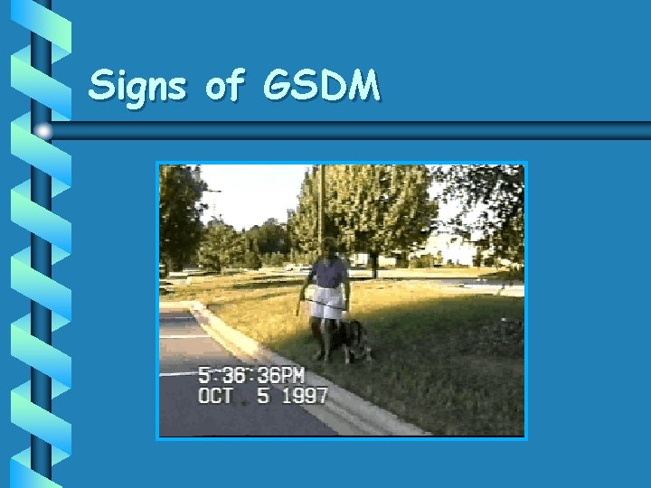
German Shepherd Degenerative Myelopathy (GSDM) • A chronic, progressive neurodegenerative disease • Initial signs are due to TL spinal cord disease • Represents an autoimmune disorder

Signalment • Breeds – – – • Sex German Shepherd dogs Belgium Shepherds • Old English Sheepdogs Rhodesian Ridgebacks • Weimaraner Probably Great Pyrenes • Age – > 5 years old (usually 8 -9) – Equal Onset – 1 month to 1 year Clinical Course – Paralysis within 3 to 6 month without treatment

Early Clinical Signs • Mild Spinal Ataxia – – Diminished Proprioception Slight Hyper-reflexia in Rear Legs • Rear Leg Weakness – Slight Muscle Atrophy • Occasionally, Atypical LMN Dysfunction

Late Clinical Signs • Severe Spinal Ataxia – Conscious Proprioceptive Deficits – Unconscious Proprioceptive Deficits – Crossed-extensor Reflex – Babinski’s Sign

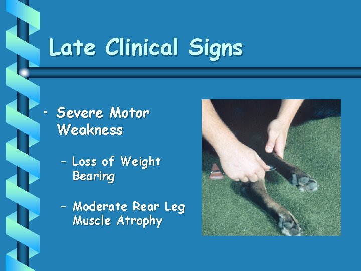
Late Clinical Signs • Severe Motor Weakness – Loss of Weight Bearing – Moderate Rear Leg Muscle Atrophy

Signs of GSDM

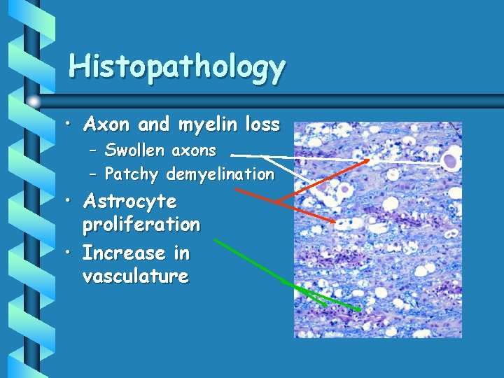
Histopathology • Axon and myelin loss – Swollen axons – Patchy demyelination • Astrocyte proliferation • Increase in vasculature

Diagnosis • Physical and Neurologic Examination – History of chronic progressive posterior paresis in susceptible breed – TL (non-localized) dysfunction

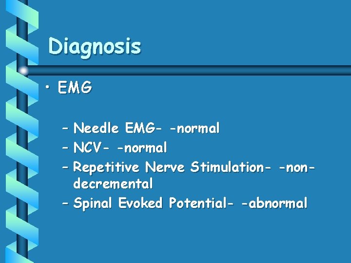
Diagnosis • EMG – Needle EMG- -normal – NCV- -normal – Repetitive Nerve Stimulation- -nondecremental – Spinal Evoked Potential- -abnormal

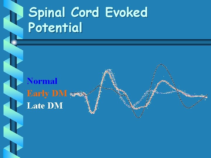
Spinal Cord Evoked Potential Normal Early DM Late DM

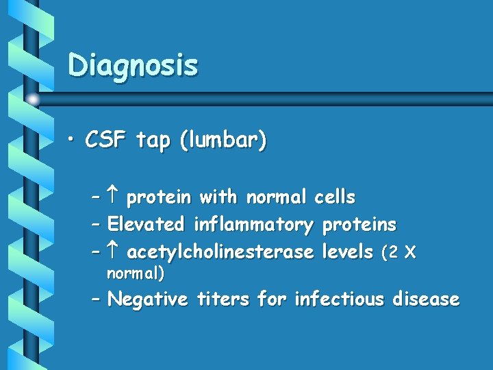
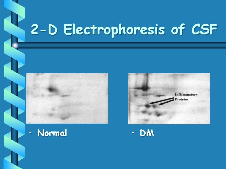
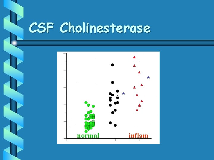
Diagnosis • CSF tap (lumbar) – protein with normal cells – Elevated inflammatory proteins – acetylcholinesterase levels (2 X normal) – Negative titers for infectious disease

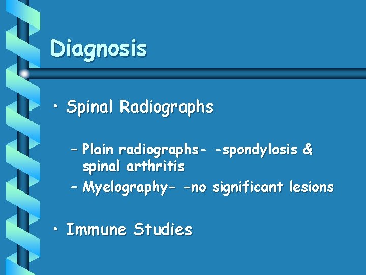
Diagnosis • Spinal Radiographs – Plain radiographs- -spondylosis & spinal arthritis – Myelography- -no significant lesions • Immune Studies

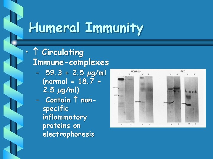
Humeral Immunity • Circulating Immune-complexes – 59. 3 + 2. 5 µg/ml (normal = 18. 7 + 2. 5 µg/ml) – Contain nonspecific inflammatory proteins on electrophoresis

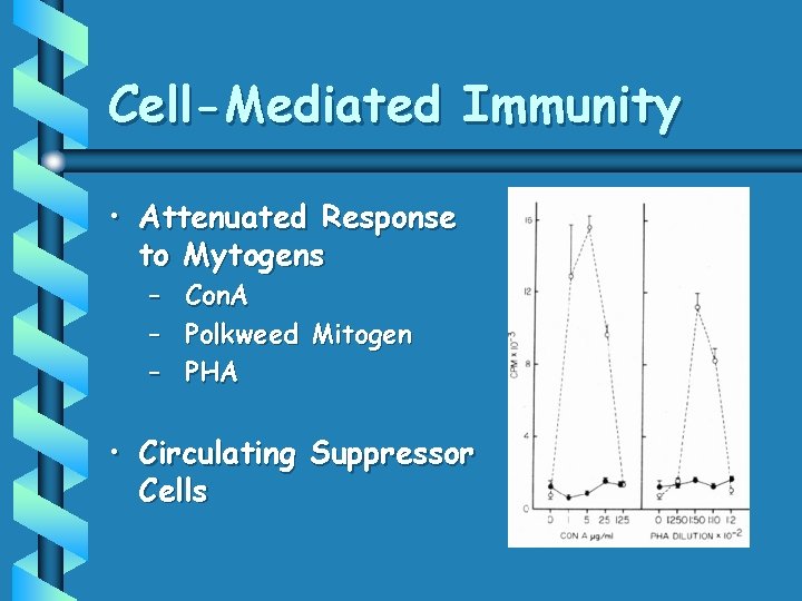
Cell-Mediated Immunity • Attenuated Response to Mytogens – – – Con. A Polkweed Mitogen PHA • Circulating Suppressor Cells

2 -D Electrophoresis of CSF • Normal • DM

CSF Cholinesterase * * normal DM inflam

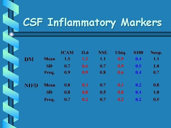
CSF Inflammatory Markers

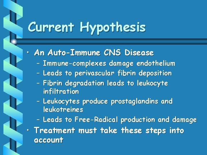
Current Hypothesis • An Auto-Immune CNS Disease – – – Immune-complexes damage endothelium Leads to perivascular fibrin deposition Fibrin degradation leads to leukocyte infiltration – Leukocytes produce prostaglandins and leukotreines – Leads to Free-Radical production and damage • Treatment must take these steps into account

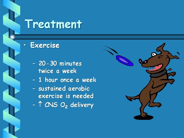
Treatment • Exercise – 20 -30 minutes twice a week – 1 hour once a week – sustained aerobic exercise is needed – CNS O 2 delivery

Treatment • Dietary Considerations – Tofu – Fresh vegetables • • carrots greens peppers broccoli – Ginger, garlic & mustard

Treatment • Supplements – – Antioxidants Membrane stabilizers Tonics Anti-inflammatory

Treatment • Medication – Aminocaproic acid (500 mg TID) – n-Acetylcysteine (25 mg/kg TID QD for 2 weeks, then TID QOD)

Conclusion • Degenerative Myelopathy appears to be an Autoimmune Disease and Treatment must be directed at this Process. – – Exercise Diet Supplements Medication Things that CNS O 2 Availability

Conclusions • Acupuncture can help treat or control a number of neurologic diseases • Point selection depends upon the constitution of the animal and the nature and location of the disease • Patience is still a virtue with neurologic conditions