ACTINOMYCOSIS Actinomyces Are gram positive anaerobic nonacidfast rods

ACTINOMYCOSIS

Actinomyces � Are gram positive, anaerobic, non-acid-fast rods. � Many of which are filamentous or branching. (Branches are <1 μm in diameter, as opposed to fungal filaments, which are >1 μm in diameter. ) � Although they are normal flora of the oral and nasopharyngeal membranes, several associated with diseases in animals. species are


Important species � A. actinoides is occasionally found as a secondary invader in enzootic pneumonia of calves and seminal vasculitis in bulls. � A. israelii is primarily associated with chronic granulomatous infections in people but has also been isolated rarely from pyogranulomatous lesions in pigs and cattle. � A. naeslundii has been isolated from suppurative infections in several animal species, the most common being aborted porcine fetuses.

� A. suis causes pyogranulomatous porcine mastitis, characterized by small abscesses containing thick, yellow pus. Yellow “sulfur granules” may be scattered throughout the pus. � A. hordeovulneris is a rare cause of canine actinomycosis, which can present with either localized abscesses or systemic infections. � A. viscosus causes cutaneous actinomycosis in dogs, which appears as localized subcutaneous abscesses. � A. bovis is the etiologic agent of lumpy jaw in cattle. Infrequently from infections in sheep, pigs, dogs, and other mammals, including chronic fistulous withers and chronic poll evil in horses.

LUMPY JAW � A localized, chronic, progressive, granulomatous abscess that most frequently involves the mandible, the maxillae, or other bony tissues in the head caused by Actinomyces bovis and characterized by rarefying osteomyelitis and chronic granulomatous inflammation in cattles. � Association of bacteria like Corynebacterium pyogenes and Staphylococcus are also seen.

Etiology � Actinomyces � Gram bovis +ve rod shaped anaorobes pleomorphic, non sporulating, non motile � Branching � Present � It forms (Ray fungus) in the mucosa of the mouth and pharynx is reported from most countries of the world and prevalent in India

Transmission � Actinomycosis generally affects cattle between 2 to 5 years and it is a sporadic disease and animal to animal transmission occurs rarely. � Wounds in oral cavity caused by awns, coarse hay hulls cutting teeth or lesion caused by FMD predisposes for actinomycosis, where it is introduced to underlying soft tissue and may set up infection. � Transmission of infection through dental alveoli at the time of eruption is noted. � Necrosis produced by penetrating objects, set up necessary anaerobic conditions for the organism to grow and thrive.

Pathogenesis � Organisms are inflammation. lodged, it incites a granulomatous � Around the colony of organisms, Indian club like structure is thought to be a product of immune reaction of host to invading organisms probably from macrophages. � Peripheral to these neutrophils surrounds and beyond these histiocytes and epithelioid cells found � Around this layer, lymphocytes, plasma cells and eosinophils accumulates and vascular fibrous tissue forms � Purulent centres surrounded by granulation tissue displaces normal tissue � The yellowish pus from abscess are referred to as 'sulphur granules' ( Bigger in size) � Central part containing the Gram positive organisms

SYMPTOMS � Swelling � There of lower jaw or around the mandibular region. is foul breath from the mouth known as halitosis. � Loose teeth induce hypersalivation and dysphagia (difficulty in feeding). � Involvement of adjacent bone results in facial distortion, loose teeth (making chewing difficult), and dyspnea from swelling into the nasal cavity. � Abscess may extend and may produce sinus to the skin surface where from, the purulent discharges are drained. � There health. is impairment of digestion resulting to loss of general
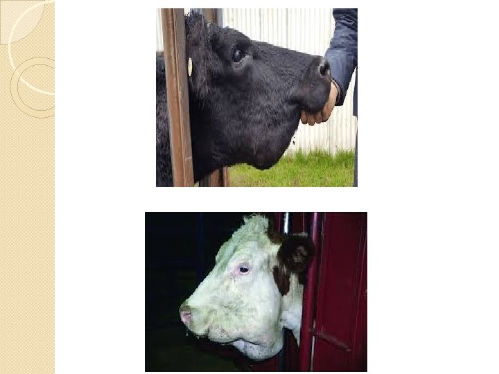

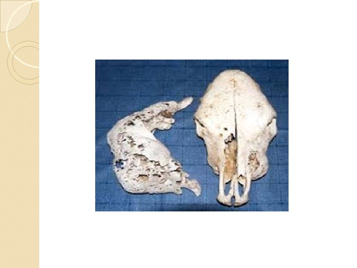
Gross lesions � The lesions appear initially as a slow-growing hard, painless, circumscribed protuberance usually at the level of central molar teeth of the mandible or maxilla. � Any part of the head can be affected; however, the alveoli around the roots of the cheek teeth are frequently involved. � The invasion damages the bony tissues and in some cattle, large granulomatous mass appear on the surface of the jaw followed by development of sinus tracts. � Ulceration forms in some cases, with or without fistulous tracts, and drainage of purulent exudate may occur. � Periosteum is irritated to form new subperiosteal bone (Involucrum) and leaving cavities and Fistulae. � The adjacent lymph nodes are not affected and the disease does not spread through lymphatic channel.

Microscopic lesions � Around the colony of organisms, Indian club like structure is seen � Accumulation of neutrophils, histiocytes and epithelioid cells are noticed. � Sometimes, lymphocytes, plasma cells and eosinophils and vascular fibrous tissue are seen
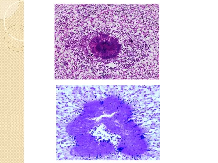

Diagnosis § Clinical signs. § Culture of the organism from the lesion. § A Gram stain of purulent material will reveal grampositive, club-shaped rods and filaments (sulfur granules). § Radiology of the head is also useful § Biopsy sample can be taken with a trephine and submitted for histopathology.

Control measures � There is no vaccine against this disease. � Isolation of infected animals and their treatment are to be rendered. � Removal of contaminated materials and disposal of animals with discharging foci may be made. � This condition should be consulted with qualified veterinarian for antibiotic treatment.
- Slides: 17