Acromioclavicular joint Coracoacromial arch coracoacromial ligament coranoid process


: • 骨盆下口Pelvic outlet : 尾骨尖、骶结节韧带、 坐骨结节、坐骨支、 耻骨下支和耻骨联合")


 外侧半月板lateral meniscus (O-shaped) • Movements: flexion and extension; flexed")

 • 关节面: lower ends of tibia and")
 – Medial lig. 外侧韧带Lateral lig. • • • 距腓前韧带Anterior")


- Slides: 40
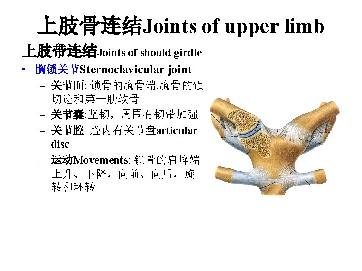
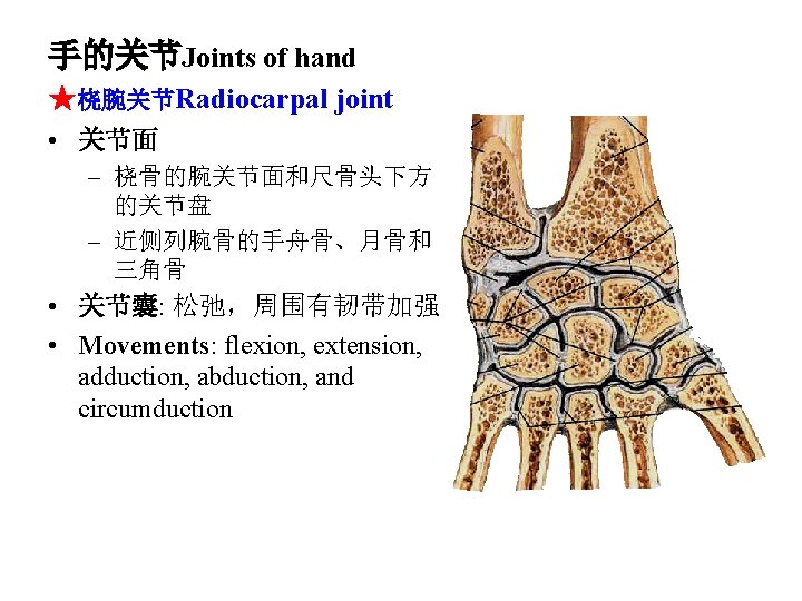
• 肩锁关节 Acromioclavicular joint – 关节面: 肩峰和锁骨 的肩峰端 – 运动: 肩胛骨以锁骨 为支点旋转 • 喙肩弓Coracoacromial arch 由喙肩韧带coracoacromial ligament, 喙突coranoid process, 和肩峰 acromion 构成 acromion coracoacromial ligament coranoid process
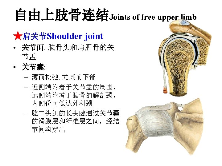
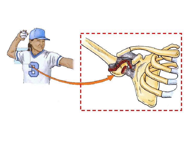
• 辅助结构Accessory structures – 盂唇Glenoid labrum: – 喙肱韧带Coracohumeral ligament :起于喙突 coracoid process 止于肱 骨的大结节 greater tubercle • 运动Movements: flexion, extension, adduction, abduction, medial and lateral rotation, circumduction
★肘关节Elbow joint • 关节面: 肱骨下端和尺、桡骨上端 – 肱尺关节Humeroulnar joint : 肱骨滑车 trochlear of humerus 和滑车 切迹 troclear noch – 肱桡关节Humeroradial joint: 肱骨小头 capitulum of humerus 和桡骨 头上方的关节凹 – 桡尺近侧关节Proximal radioulnar joint: 桡 骨头的环状关节面 articular circumference of radius 和尺骨的桡切 迹 radial notch of ulna • 关节囊: 前后壁薄而松弛,内、外侧 有侧副韧带加强
• 肘关节的韧带Ligaments: – 桡侧副韧带Radial collateral ligament: – 尺侧副韧带Ulnar collateral ligament: – 桡骨环状韧带Annular ligament of radius: 附着于尺骨桡切迹的前、后缘, 位于桡骨头的周围 • 运动Movements: flexion and extension, pronation and supination
Dislocation
尺、桡骨之间的连结Joints between radius and ulna • 桡尺近侧关节Proximal radioulnar joint • 桡尺远侧关节Distal radioulnar joint: 尺 骨头、桡骨的尺切迹和尺骨头下方的 关节盘 • 前臂骨间膜Interosseous membrane of forearm
• 腕骨间关节Intercarpal joints • 腕掌关节Carpometacarpal joints ★拇指腕掌关节 Carpometacarpal joint of thumb – 关节面: 大多角骨的远 侧面和第一掌骨底 – Movement: flexion, extension, adduction, abduction, and 对掌运 动opposition • 掌骨间关节Intermetacarpal joints • 掌指关节 Metacarpophalangeal joints • 指间关节Interphalangeal joints
髋骨和脊柱间的韧带连结 Vertebropelvic ligaments • 髂腰韧带Iliolumbal ligament: 第五腰椎的横突至髂嵴的后 上 ★骶结节韧带Sacrotuberous ligament ★骶棘韧带Sacrospinous ligament • These two ligaments convert the sciatic notches the坐骨大、 小孔greater and lesser sciatic foramina
• 耻骨联合Pubic symphysis – 构成Articulation: symphysial surface and interpubic disc – 韧带Ligaments: 耻骨上 韧带superior pubic ligament 和耻骨弓状韧 带 arcuate pubic ligament • 闭孔膜Obturator membrane 闭膜管obturator canal
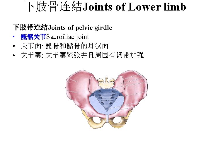
骨盆Bony pelvis 构成Composition: 双侧的髋骨、骶尾 骨和它们之间的连结 • 骨盆的正常解剖学方位the normal anatomical position • 界线Terminal line: 骶岬promontory of sacrum, 弓状线arcuate line, 耻骨 梳pectin of pubis, 耻骨结节 pubic tubercle, 耻骨联合上缘upper border of pubic symphysis • Two portions: a greater pelvis and a lesser pelvis
小骨盆Lesser pelvis • 骨盆上口pelvic inlet (terminal line): • 骨盆下口Pelvic outlet : 尾骨尖、骶结节韧带、 坐骨结节、坐骨支、 耻骨下支和耻骨联合 下缘 • Pelvic cavity • Pubic arch, 耻骨下角 subpubic angle
Main difference between male and femal pelvis
Main difference between male and femal pelvis Male Female Pelvic inlet Pelvic outet Pelvic cavity Pubic arch 90~1000 70~750
Main difference between male and femal pelvis Male Female Overall Narrow and long Wide and short Iliac ala More vertical More horizontal Inlet Oval or heart shaped Round Subpubic angle Acute angle (about 70~750) Right angle (about 90~1000) Pelvic cavity Deep narrow Shallower, wide Outlet Larger Small
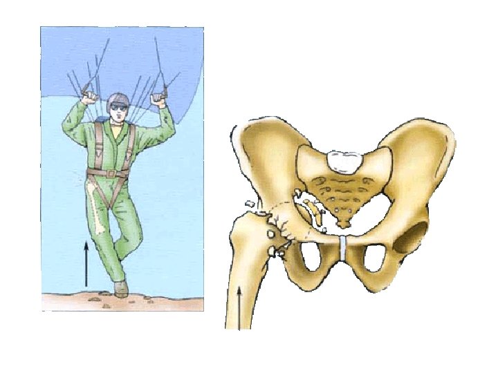
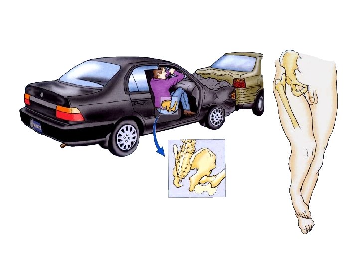
自由下肢的连结Joints of free lower limb ★髋关节Hip joint • 关节面: 髋臼acetabulum and 股骨头femoral head • 关节囊Articular capsule – Above: margins of acetabulum and transverse acetabular ligament – Below: in front to intertrochanteric line; behind, to the neck of femur above 1 cm above the intertrochanteric crest
• 辅助结构Accessory structures Acetabulum labrum; 髋臼横韧带 – 髋臼唇 transverse acetebular ligament – Ligaments • • • Acetabulum labrum Ligament of head of femur 髂股韧带Iliofemoral lig. 股骨头韧带Ligament of head of femur 耻股韧带Pubofemoral lig. 坐股韧带Ischiofemoral ligament 轮匝带Zona orbicularis • Movement: flexion, extention, adduction, abduction, medial and lateral rotation, circumduction Transverse acetebular lig.
Pubofemoral lig. Iliofemoral lig. Ischiofemoral lig. Zona orbicularis
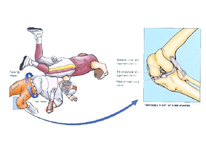
★膝关节Knee joint • 关节面: lower end of femur, upper end of tibia and patella • 关节囊: 髌上囊suprapatellar bursa, 髌下深囊deep infrapatellar bursa, 翼状襞alar folds
• 膝关节的辅助结构Accessory structures – ligaments • 髌韧带Patellar lig. • 腓侧副韧带Fibular collateral lig. • 胫侧副韧带Tibial collateral lig. Patellar lig. Fibular collateral lig. Tibial collateral lig.
popliteal ligament • 前交叉韧带Anterior cruciate ligment • 后交叉韧带Posterior cruciate ligament • 腘斜韧带Oblique
– 内侧半月板Medial – meniscus (C-shaped) 外侧半月板lateral meniscus (O-shaped) • Movements: flexion and extension; flexed knee joint may be passively rotated through 700 lateral Medial
胫腓连结Tibiofibular syndesmosis • 胫腓关节Tibiofibular joint • 小腿骨间膜Crural interosseous membrane • 胫腓前、后韧带Anterior and posterior tibiofibular ligaments
足的连结Joint of foot 距小腿关节Talocrural joint 踝关节(ankle joint) • 关节面: lower ends of tibia and fibula, trochlea of talus • 关节囊: 前后薄而松弛, 两侧有侧副韧带加强
• Ligments – 内侧韧带(三角韧带) – Medial lig. 外侧韧带Lateral lig. • • • 距腓前韧带Anterior talofibular lig. 跟腓韧带Calcaneofibular lig. 距腓后韧带Posterior talofibular lig. • Movements: 背屈(伸) dosiflexion (extension) and 趾屈(屈)plantar flexion (flexion); when the ankle joint is fully plantar flexed, small amounts of abduction, and adduction are possible
• 跗骨间关节Intertarsal joints – 距跟关节Talocalcaneal – 距跟舟关节 – joint Talocalcaneonavicular joint 跟骰关节Calcaneocuboid joint • 跗跖关节Tarsometatarsal joints • 跖骨间关节Intermetatarsal joints • 跖趾关节Metatarsophalangeal joints • 趾间关节Interphalangeal joints 跗横关节transverse tarsal joint
足弓Arches of foot • 内侧纵弓Medial longitudinal arch: 跟骨 calcaneus, 距骨talus, 足舟骨navicular, 3块楔骨和 1 -3跖骨, 距骨头是内侧纵弓的最高点
• 外侧纵弓Lateral longitudinal arch: 跟骨calcaneus, 骰骨cuboid, 4 -5跖骨metatarsals; 骰骨是 外侧纵弓的最高点
• 横弓Tranverse arch: 骰骨cuboid, 3块楔骨three cuniforms and all 跖骨 metatarsals; the intermediate cuneiform is the keystone of this arch • Function: give to foot strength stability and resilience; protect plantar vessels and nerves
Normal arch Flatfoot