
Acidbase status assessment Stanislav Matouek Ji Kofrnek Elementary
Acid-base status assessment Stanislav Matoušek, Jiří Kofránek

Elementary terms
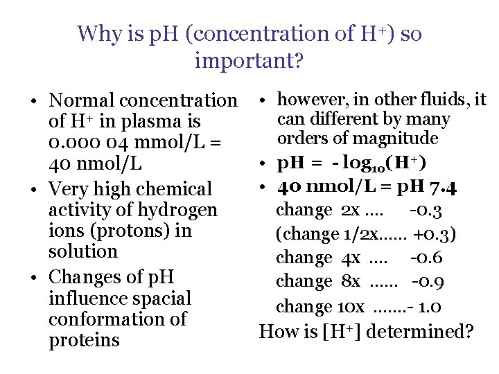
Why is p. H (concentration of H+) so important? • Normal concentration of H+ in plasma is 0. 000 04 mmol/L = 40 nmol/L • Very high chemical activity of hydrogen ions (protons) in solution • Changes of p. H influence spacial conformation of proteins • however, in other fluids, it can different by many orders of magnitude • p. H = - log 10(H+) • 40 nmol/L = p. H 7. 4 change 2 x …. -0. 3 (change 1/2 x…… +0. 3) change 4 x …. -0. 6 change 8 x …… -0. 9 change 10 x ……. - 1. 0 How is [H+] determined?
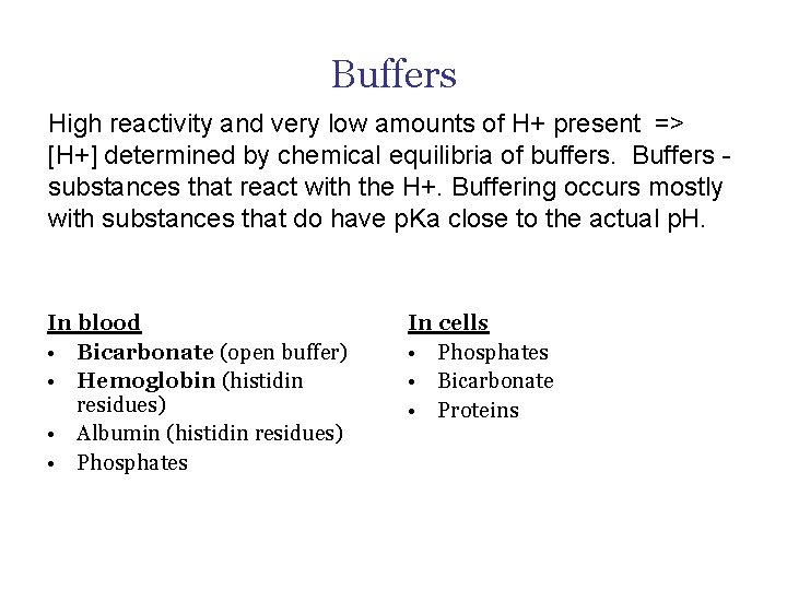
Buffers High reactivity and very low amounts of H+ present => [H+] determined by chemical equilibria of buffers. Buffers substances that react with the H+. Buffering occurs mostly with substances that do have p. Ka close to the actual p. H. In blood • Bicarbonate (open buffer) • Hemoglobin (histidin residues) • Albumin (histidin residues) • Phosphates In cells • Phosphates • Bicarbonate • Proteins
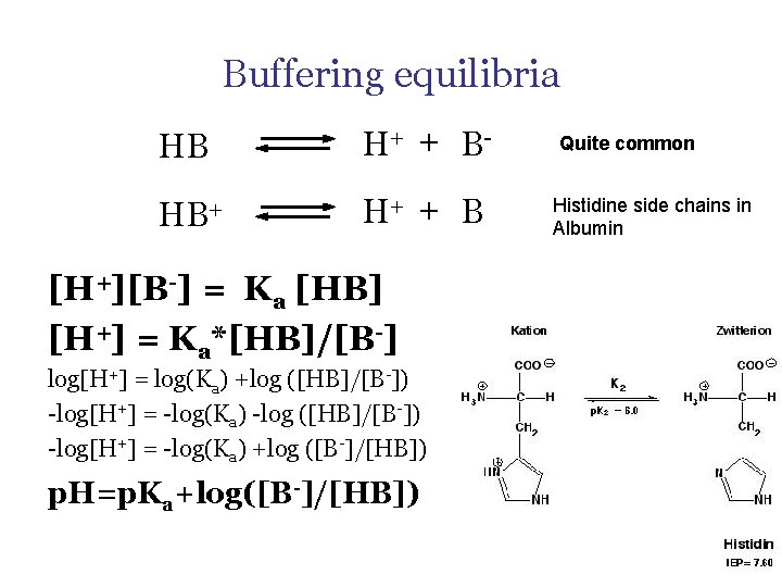
Buffering equilibria HB H+ + B- HB+ H+ + B [H+][B-] = Ka [HB] [H+] = Ka*[HB]/[B-] log[H+] = log(Ka) +log ([HB]/[B-]) -log[H+] = -log(Ka) -log ([HB]/[B-]) -log[H+] = -log(Ka) +log ([B-]/[HB]) p. H=p. Ka+log([B-]/[HB]) Quite common Histidine side chains in Albumin

Buffering HB and B-
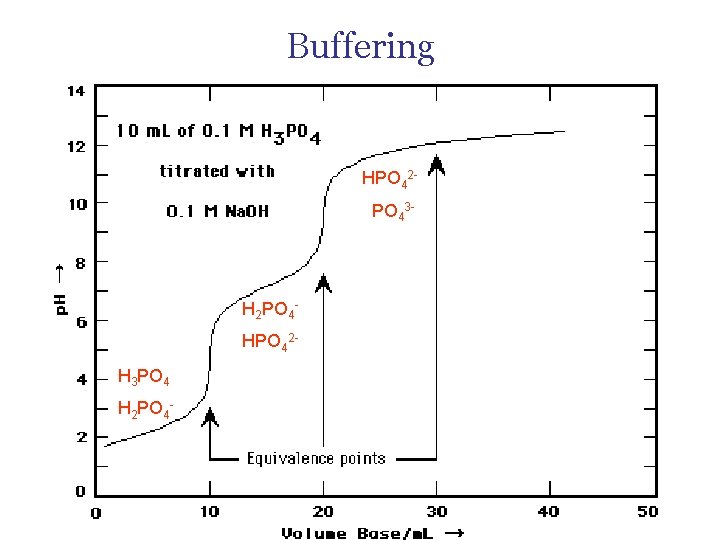
Buffering HPO 42 PO 43 - H 2 PO 4 HPO 42 H 3 PO 4 H 2 PO 4 -
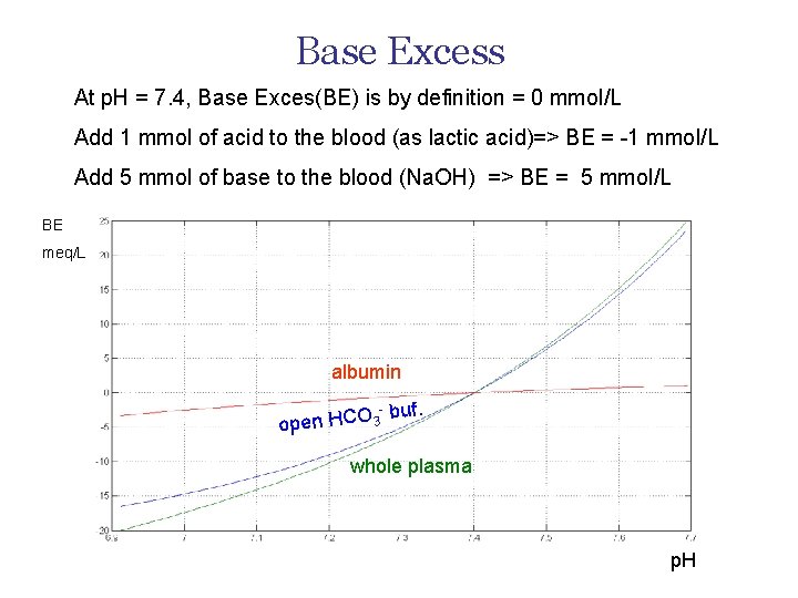
Base Excess At p. H = 7. 4, Base Exces(BE) is by definition = 0 mmol/L Add 1 mmol of acid to the blood (as lactic acid)=> BE = -1 mmol/L Add 5 mmol of base to the blood (Na. OH) => BE = 5 mmol/L BE meq/L albumin - open HCO 3 buf. whole plasma p. H
How to assess acid-base
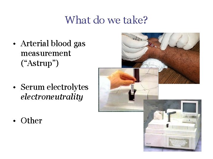
What do we take? • Arterial blood gas measurement (“Astrup”) • Serum electrolytes electroneutrality • Other
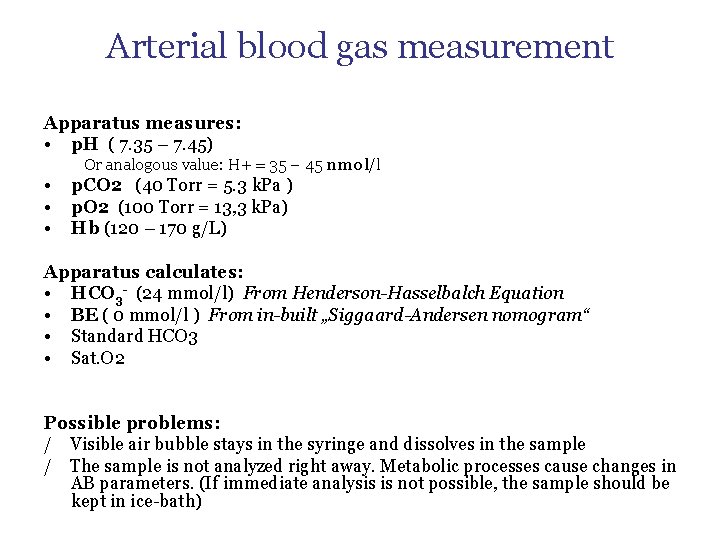
Arterial blood gas measurement Apparatus measures: • p. H ( 7. 35 – 7. 45) Or analogous value: H+ = 35 – 45 nmol/l • • • p. CO 2 (40 Torr = 5. 3 k. Pa ) p. O 2 (100 Torr = 13, 3 k. Pa) Hb (120 – 170 g/L) Apparatus calculates: • HCO 3 - (24 mmol/l) From Henderson-Hasselbalch Equation • BE ( 0 mmol/l ) From in-built „Siggaard-Andersen nomogram“ • Standard HCO 3 • Sat. O 2 Possible problems: / Visible air bubble stays in the syringe and dissolves in the sample / The sample is not analyzed right away. Metabolic processes cause changes in AB parameters. (If immediate analysis is not possible, the sample should be kept in ice-bath)
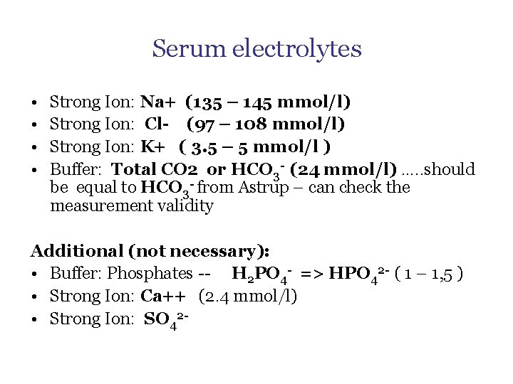
Serum electrolytes • • Strong Ion: Na+ (135 – 145 mmol/l) Strong Ion: Cl- (97 – 108 mmol/l) Strong Ion: K+ ( 3. 5 – 5 mmol/l ) Buffer: Total CO 2 or HCO 3 - (24 mmol/l) …. . should be equal to HCO 3 - from Astrup – can check the measurement validity Additional (not necessary): • Buffer: Phosphates -- H 2 PO 4 - => HPO 42 - ( 1 – 1, 5 ) • Strong Ion: Ca++ (2. 4 mmol/l) • Strong Ion: SO 42 -
Other Buffers: Albumin (35 – 50 g/l) “Strong Ion”, Acid: Lactate (0. 5 – 2. 5) “Strong Ions”, Acids: Ketoacids (0) Toxic substances, acids: Salycilates, methanol etc.

Change in buffer equilibria = change in p. H
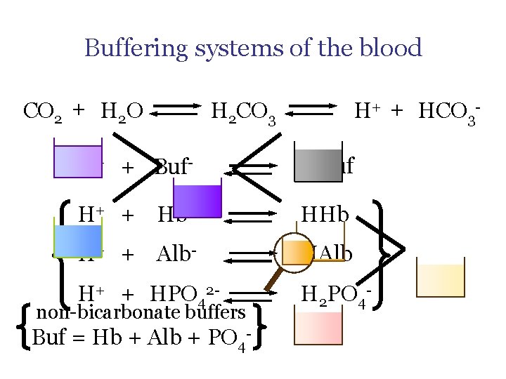
Buffering systems of the blood CO 2 + H 2 O H+ + HCO 3 - H 2 CO 3 H+ + Buf- HBuf H+ + Hb- HHb H+ + Alb- HAlb H+ + HPO 42 - H 2 PO 4 - non-bicarbonate buffers Buf = Hb + Alb + PO 4 -
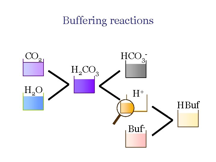
Buffering reactions HCO 3 - CO 2 H 2 CO 3 H 2 O H+ HBuf Buf-
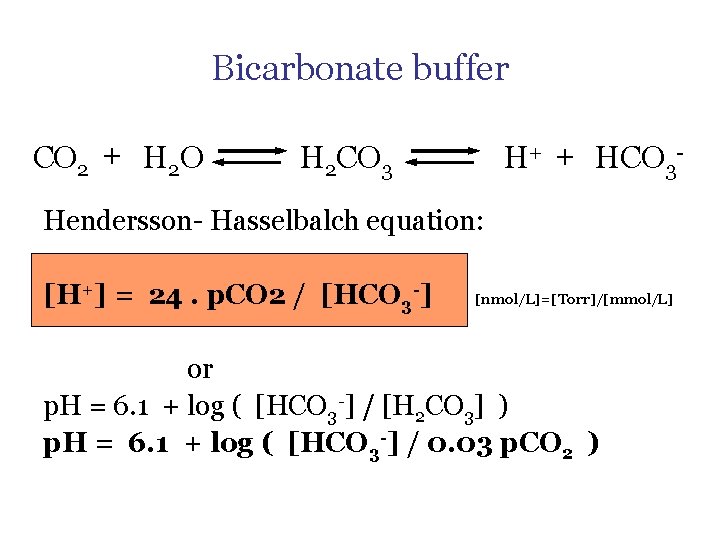
Bicarbonate buffer CO 2 + H 2 O H+ + HCO 3 - H 2 CO 3 Hendersson- Hasselbalch equation: [H+] = 24. p. CO 2 / [HCO 3 -] [nmol/L]=[Torr]/[mmol/L] or p. H = 6. 1 + log ( [HCO 3 -] / [H 2 CO 3] ) p. H = 6. 1 + log ( [HCO 3 -] / 0. 03 p. CO 2 )
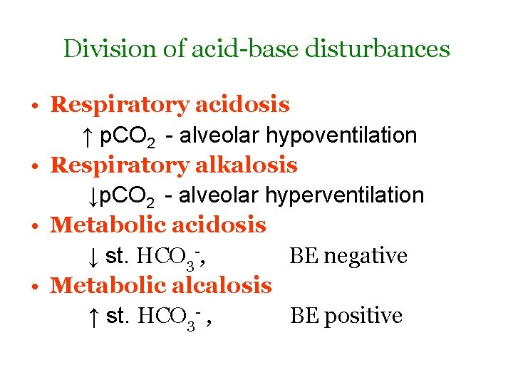
Division of acid-base disturbances • Respiratory acidosis ↑ p. CO 2 - alveolar hypoventilation • Respiratory alkalosis ↓p. CO 2 - alveolar hyperventilation • Metabolic acidosis ↓ st. HCO 3 -, BE negative • Metabolic alcalosis ↑ st. HCO 3 - , BE positive
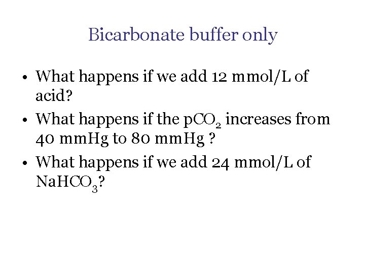
Bicarbonate buffer only • What happens if we add 12 mmol/L of acid? • What happens if the p. CO 2 increases from 40 mm. Hg to 80 mm. Hg ? • What happens if we add 24 mmol/L of Na. HCO 3?
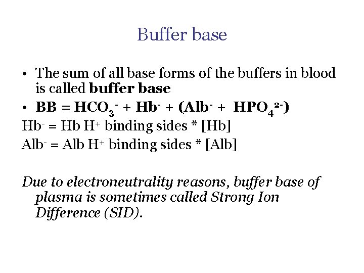
Buffer base • The sum of all base forms of the buffers in blood is called buffer base • BB = HCO 3 - + Hb- + (Alb- + HPO 42 -) Hb- = Hb H+ binding sides * [Hb] Alb- = Alb H+ binding sides * [Alb] Due to electroneutrality reasons, buffer base of plasma is sometimes called Strong Ion Difference (SID).
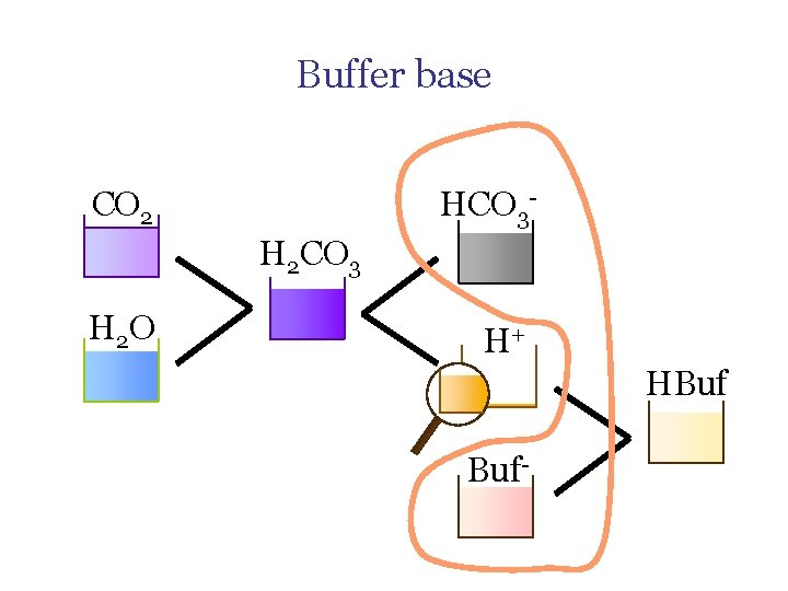
Buffer base HCO 3 - CO 2 H 2 CO 3 H 2 O H+ HBuf Buf-
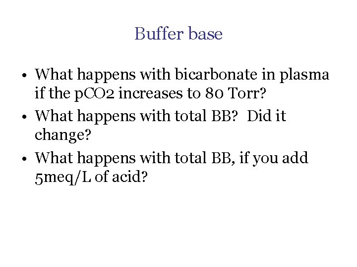
Buffer base • What happens with bicarbonate in plasma if the p. CO 2 increases to 80 Torr? • What happens with total BB? Did it change? • What happens with total BB, if you add 5 meq/L of acid?
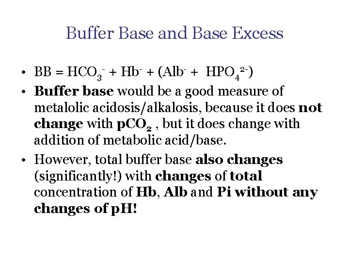
Buffer Base and Base Excess • BB = HCO 3 - + Hb- + (Alb- + HPO 42 -) • Buffer base would be a good measure of metalolic acidosis/alkalosis, because it does not change with p. CO 2 , but it does change with addition of metabolic acid/base. • However, total buffer base also changes (significantly!) with changes of total concentration of Hb, Alb and Pi without any changes of p. H!
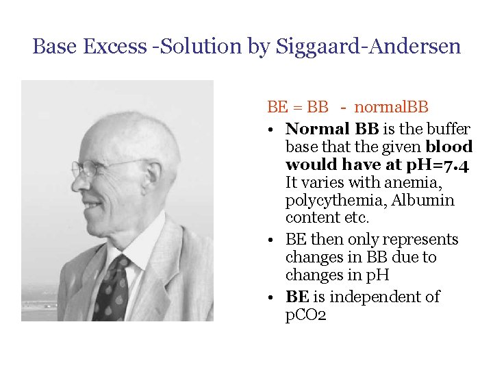
Base Excess -Solution by Siggaard-Andersen BE = BB - normal. BB • Normal BB is the buffer base that the given blood would have at p. H=7. 4 It varies with anemia, polycythemia, Albumin content etc. • BE then only represents changes in BB due to changes in p. H • BE is independent of p. CO 2
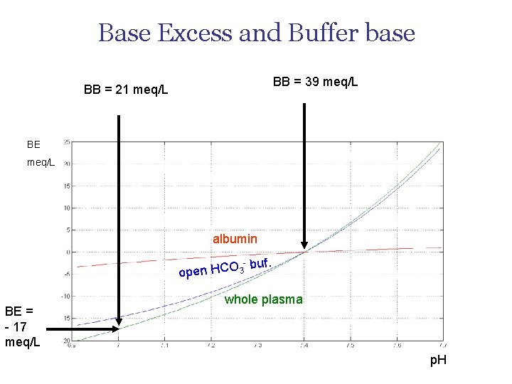
Base Excess and Buffer base BB = 39 meq/L BB = 21 meq/L BE meq/L albumin - open HCO 3 BE = - 17 meq/L buf. whole plasma p. H
Measure of metabolic disturbances – Americans: Standard Bicarbonates – the value of bicarbonates, when p. CO 2= 40 mm. Hg – Europeans: Base Excess – measure derived from BB, thus independent of p. CO 2
Compensation of Acid Base Disorders
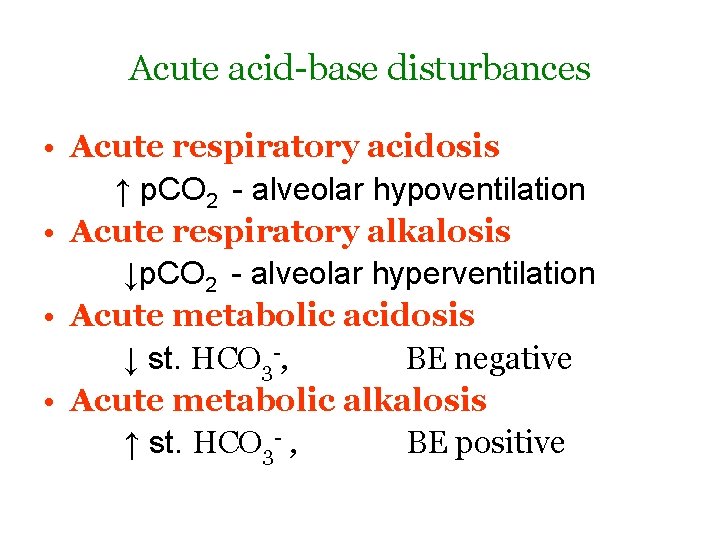
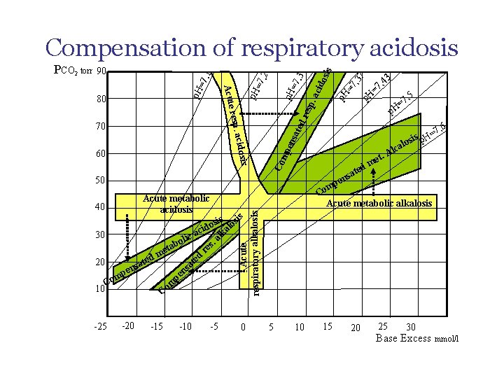
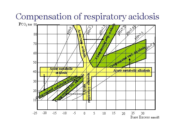
Acute acid-base disturbances • Acute respiratory acidosis ↑ p. CO 2 - alveolar hypoventilation • Acute respiratory alkalosis ↓p. CO 2 - alveolar hyperventilation • Acute metabolic acidosis ↓ st. HCO 3 -, BE negative • Acute metabolic alkalosis ↑ st. HCO 3 - , BE positive
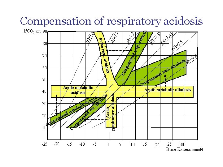
s 40 7 p. H esp = p. H , 5 7 H= dr ate 7, 6 = sis p. H mp ens alo sa pen 50 Acute metabolic acidosis 3 4 7, p Co cidosi 60 . ac =7 p. H resp. a 70 =7 , 3 , 2 =7 p. H 80 Acute =7, 1 90 p. H ted Alc. t me m Co Acute metabolic alkalosis s sis losi o cid lka a 30 c a li bo s. a e t r me d d e e t 20 sat sa n e n mp pe o C m o 10 C -25 -20 -15 -10 -5 Acute respiratory alkalosis PCO 2 torr ido sis Compensation of respiratory acidosis 0 5 10 15 20 25 30 Base Excess mmol/l
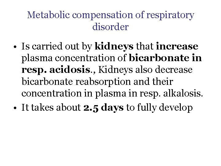
Metabolic compensation of respiratory disorder • Is carried out by kidneys that increase plasma concentration of bicarbonate in resp. acidosis. , Kidneys also decrease bicarbonate reabsorption and their concentration in plasma in resp. alkalosis. • It takes about 2. 5 days to fully develop
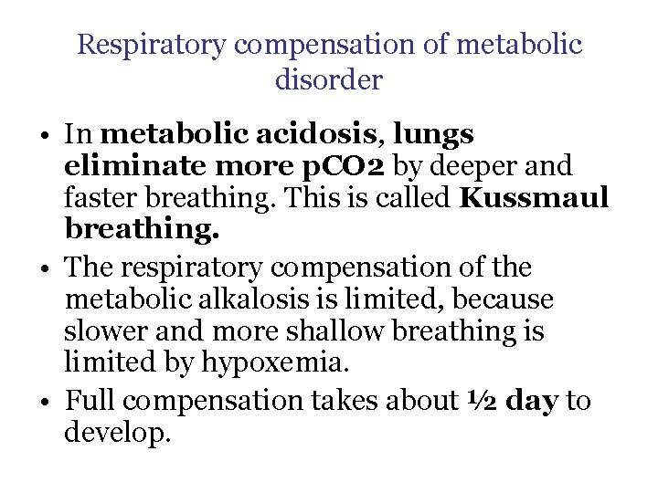
Respiratory compensation of metabolic disorder • In metabolic acidosis, lungs eliminate more p. CO 2 by deeper and faster breathing. This is called Kussmaul breathing. • The respiratory compensation of the metabolic alkalosis is limited, because slower and more shallow breathing is limited by hypoxemia. • Full compensation takes about ½ day to develop.
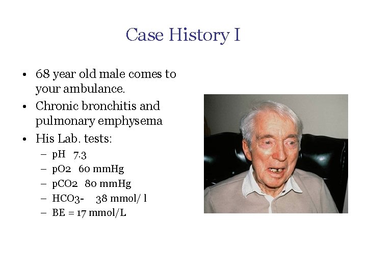
Case History I • 68 year old male comes to your ambulance. • Chronic bronchitis and pulmonary emphysema • His Lab. tests: – – – p. H 7. 3 p. O 2 60 mm. Hg p. CO 2 80 mm. Hg HCO 3 - 38 mmol/ l BE = 17 mmol/L
s 40 7 p. H esp = p. H , 5 7 H= dr ate 7, 6 = sis p. H mp ens alo sa pen 50 Acute metabolic acidosis 3 4 7, p Co cidosi 60 . ac =7 p. H resp. a 70 =7 , 3 , 2 =7 p. H 80 Acute =7, 1 90 p. H ted Alc. t me m Co Acute metabolic alkalosis s sis losi o cid lka a 30 c a li bo s. a e t r me d d e e t 20 sat sa n e n mp pe o C m o 10 C -25 -20 -15 -10 -5 Acute respiratory alkalosis PCO 2 torr ido sis Compensation of respiratory acidosis 0 5 10 15 20 25 30 Base Excess mmol/l
Case History II • • • 20 year old student is admitted to hospital for acute anxiety state Cannot concentrate, feeling of numbness or pins and needles in fingers She has split with her boyfriend recently Has not been seriously ill until now, no medication Physical examination – normal Lab. values: – – – p. H 7, 49 p. O 2 100 mm Hg p. CO 2 30 mm Hg HCO 3 - 22 mmol/l BE = -2 mmol/L
Causes of respiratory acidosis • Respiratory acidosis is part of Global (type II) respiratory insuficiency : • ↓ alveolar ventilation – Respiratory center depression • Drugs, medicaments • Respiratory centre hypoxia or damage – – Trauma Stroke Tumor Cerebral edema / increased intracranial pressure – Lung disease • Restrictive – ARDS – Fibroses • Trauma, pneumothorax, serial rib fractures • Obstructive – Astma – Tumor – Foreign body • Increase in dead space – Embolism – Emphysema – Nerve of muscle disease • Myasthenia gravis • Polyradiculoneuritis • Serious obesity • Breathing CO 2 in the inspired air (miners buried in a mine)
Causes of respiratory alkalosis • Hyperventilation – in mechanical ventilation – With hypoxemia • High altitude disease • Right-left shunting – In neurosis • Respiratory centre irritation – Trauma, salicylates, inflammation
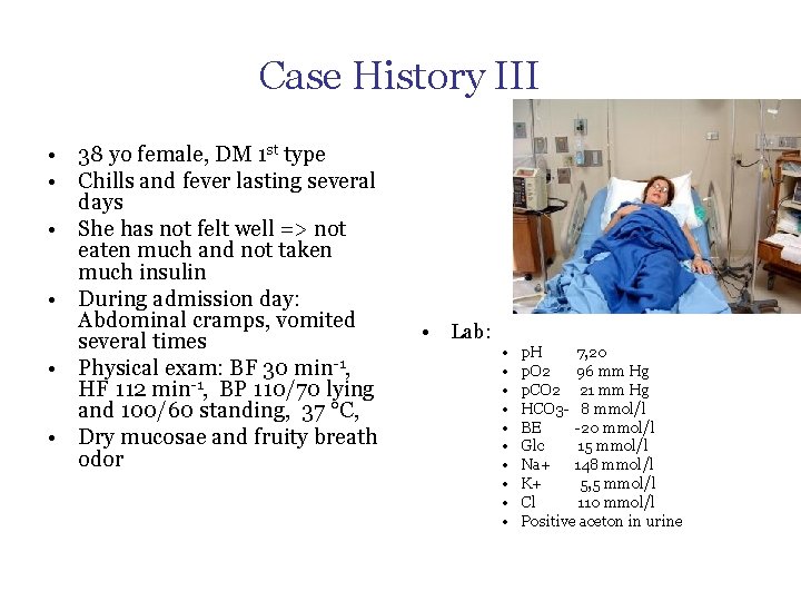
Case History III • 38 yo female, DM 1 st type • Chills and fever lasting several days • She has not felt well => not eaten much and not taken much insulin • During admission day: Abdominal cramps, vomited several times • Physical exam: BF 30 min-1, HF 112 min-1, BP 110/70 lying and 100/60 standing, 37 °C, • Dry mucosae and fruity breath odor • Lab: • • • p. H 7, 20 p. O 2 96 mm Hg p. CO 2 21 mm Hg HCO 3 - 8 mmol/l BE -20 mmol/l Glc 15 mmol/l Na+ 148 mmol/l K+ 5, 5 mmol/l Cl 110 mmol/l Positive aceton in urine
s 40 7 p. H esp = p. H , 5 7 H= dr ate 7, 6 = sis p. H mp ens alo sa pen 50 Acute metabolic acidosis 3 4 7, p Co cidosi 60 . ac =7 p. H resp. a 70 =7 , 3 , 2 =7 p. H 80 Acute =7, 1 90 p. H ted Alc. t me m Co Acute metabolic alkalosis s sis losi o cid lka a 30 c a li bo s. a e t r me d d e e t 20 sat sa n e n mp pe o C m o 10 C -25 -20 -15 -10 -5 Acute respiratory alkalosis PCO 2 torr ido sis Compensation of respiratory acidosis 0 5 10 15 20 25 30 Base Excess mmol/l
Case history IV • 23 yo male, admitted for suicide attempt • Has ingested large amount of aspirin • At admission somnolent, difficult to make contact with • BF 30 min-1 , HF 100 min-1, BP 142/88, t = 36. 8 °C • Lab: – – Toxic levels of salicylates, p. H 7. 25 p. CO 2 14 mm. Hg HCO 3 - 8 mmol/l
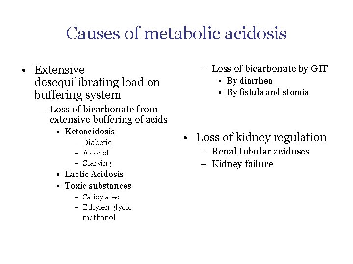
Causes of metabolic acidosis • Extensive desequilibrating load on buffering system – Loss of bicarbonate by GIT • By diarrhea • By fistula and stomia – Loss of bicarbonate from extensive buffering of acids • Ketoacidosis – Diabetic – Alcohol – Starving • Lactic Acidosis • Toxic substances – Salicylates – Ethylen glycol – methanol • Loss of kidney regulation – Renal tubular acidoses – Kidney failure
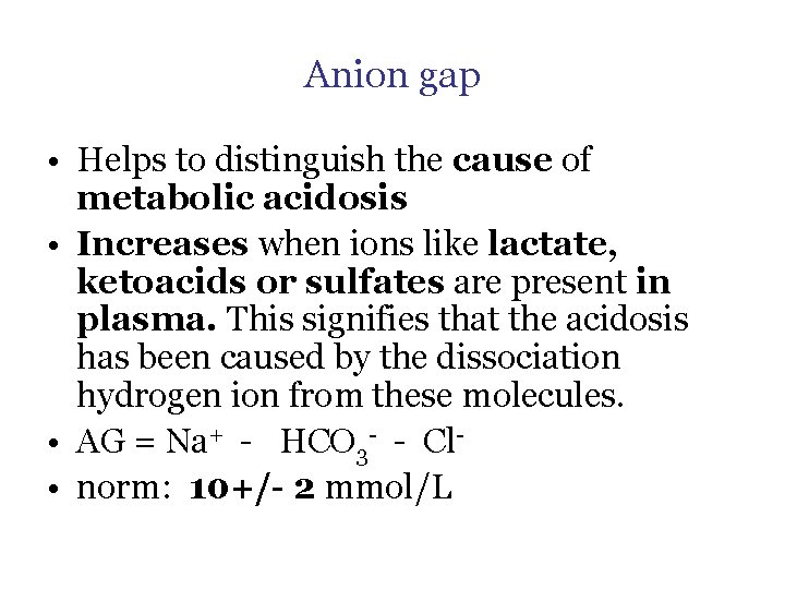
Anion gap • Helps to distinguish the cause of metabolic acidosis • Increases when ions like lactate, ketoacids or sulfates are present in plasma. This signifies that the acidosis has been caused by the dissociation hydrogen ion from these molecules. • AG = Na+ - HCO 3 - - Cl • norm: 10+/- 2 mmol/L
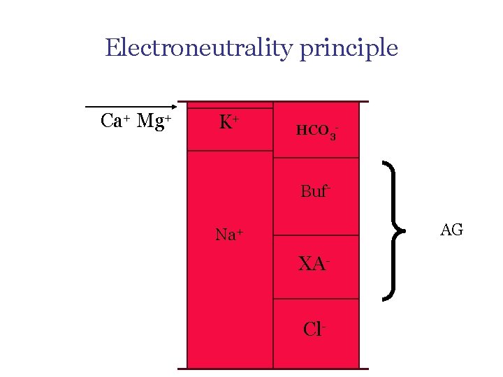
Electroneutrality principle Ca+ Mg+ K+ HCO 3 - Buf. AG Na+ XACl-
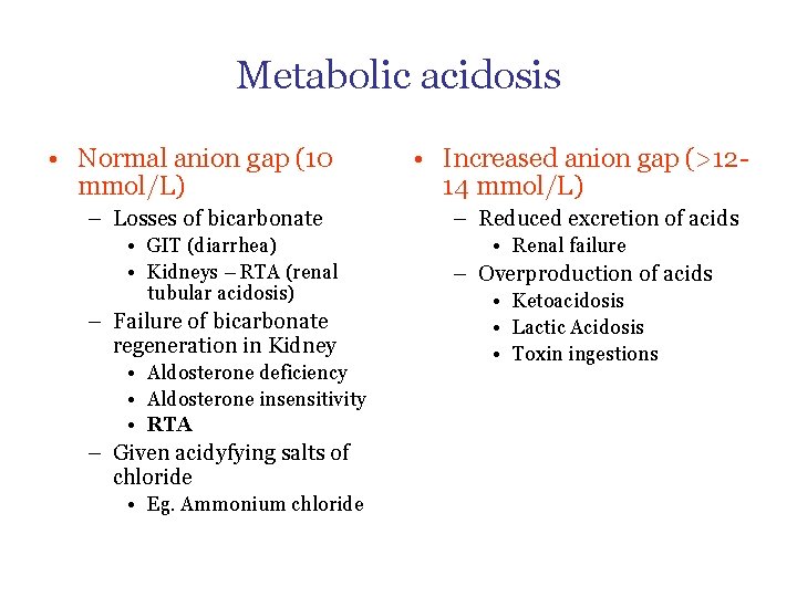
Metabolic acidosis • Normal anion gap (10 mmol/L) – Losses of bicarbonate • GIT (diarrhea) • Kidneys – RTA (renal tubular acidosis) – Failure of bicarbonate regeneration in Kidney • Aldosterone deficiency • Aldosterone insensitivity • RTA – Given acidyfying salts of chloride • Eg. Ammonium chloride • Increased anion gap (>1214 mmol/L) – Reduced excretion of acids • Renal failure – Overproduction of acids • Ketoacidosis • Lactic Acidosis • Toxin ingestions
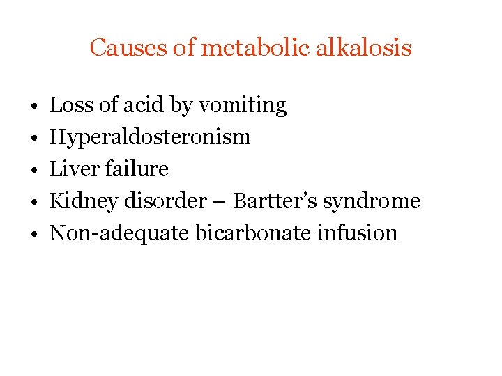
Causes of metabolic alkalosis • • • Loss of acid by vomiting Hyperaldosteronism Liver failure Kidney disorder – Bartter’s syndrome Non-adequate bicarbonate infusion
- Slides: 44