ACIDBASE DISORDERS Normal AcidBase Homeostasis Systemic arterial p

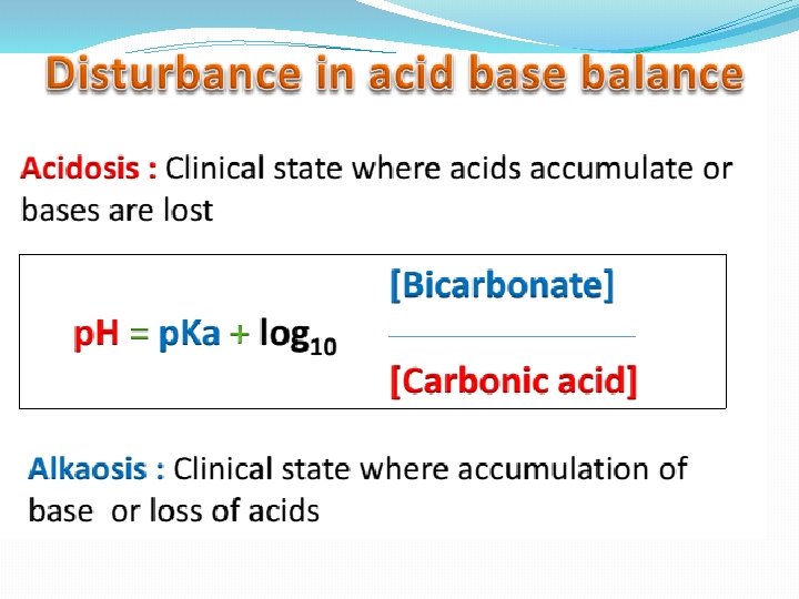
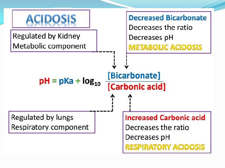
ACID–BASE DISORDERS

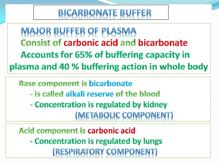
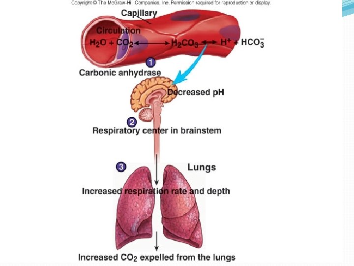
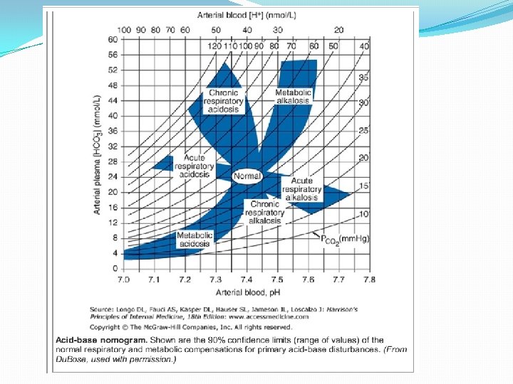
Normal Acid-Base Homeostasis Systemic arterial p. H is maintained between 7. 35 and 7. 45 by extracellular and intracellular chemical buffering together with respiratory and renal regulatory mechanisms. The control of arterial CO 2 tension (Paco 2) by the central nervous system (CNS) and respiratory systems and the control of the plasma bicarbonate by the kidneys stabilize the arterial p. H by excretion or retention of acid or alkali.

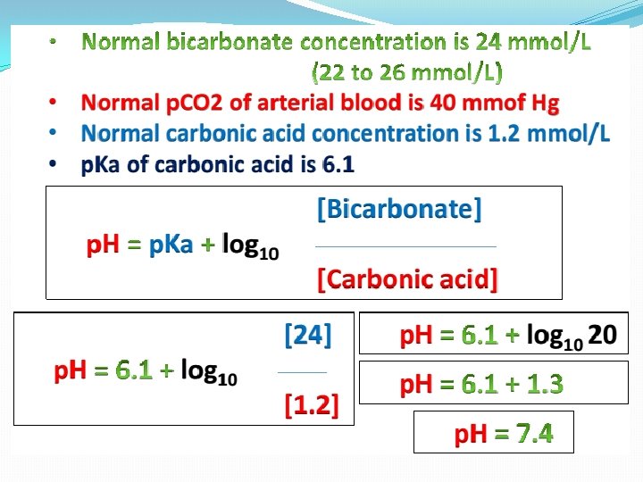
Normal Values Arterial Venous 7. 40 <7. 35 HCO 3 24 24 p. CO 2 40 >40 p. O 2 >70 <60 p. H

Effects of Metabolic acidosis Cardiovascular Impaired cardiac contractility Arteriolar dilation Venoconstriction Centralization of blood volume Increased pulmonary vascular resistance Decreased cardiac output Decreased systemic BP Decreased hepatorenal blood flow Decreased threshold for cardiac arrhythmias Attenuation of responsiveness to catecholamines

METABOLIC ACIDOSIS METABOLIC EFFECTS �Insulin resistance �Inhibition of anaerobic glycolysis �Reduction in ATP synthesis �Hyperkalemia �Protein degradation �Bone demineralization (chronic)

METABOLIC ACIDOSIS Neurologic Respiratory �Inhibition of metabolism and cell-volume regulation �Obtundation and coma �Compensatory hyperventilation with possible respiratory muscle fatigue

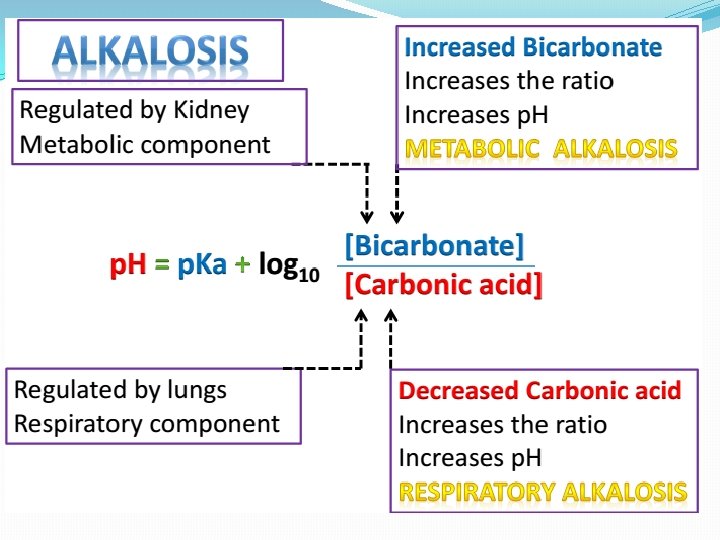
METABOLIC ALKALOSIS CARDIOVASCULAR METABOLIC �Stimulation of anaerobic �Arteriolar constriction glycolysis Reduced coronary blood flow Reduced anginal threshold �Formation of organic acids �Decreased oxyhemoglobin Decreased threshold for dissociation cardiac arrhythmias �Decreased ionized calcium �Hypokalemia �Hypomagnesemia �Hypophosphatemia

METABOLIC ALKALOSIS NEUROLOGIC RESPIRATORY �Tetany Seizures Lethargy Delirium Stupor �Compensatory hypoventilation with hypercapnia and hypoxemia

Acid–Base Balance Disturbances 1. Disorders: Circulating buffers � Respiratory performance � Renal function � 2. Cardiovascular conditions: Heart failure � Hypotension � 3. Conditions affecting the CNS: � Neural damage or disease that affects respiratory and cardiovascular reflexes

Overview of Acid-Base Physiology �Intracellular acid and base production �Intravascular transport of acids or bases �Elimination of acids and bases by kidneys and CO 2 by the lungs

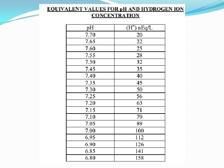
Normal Serum Values �Normal serum p. H: 7. 36 – 7. 44 �Normal serum [H+] : 40 m. Eq/L �Normal serum [HCO 3]: 24 m. Eq/L �Normal serum p. CO 2 : 40 mm. Hg

Acid–Base Balance
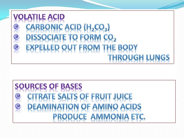

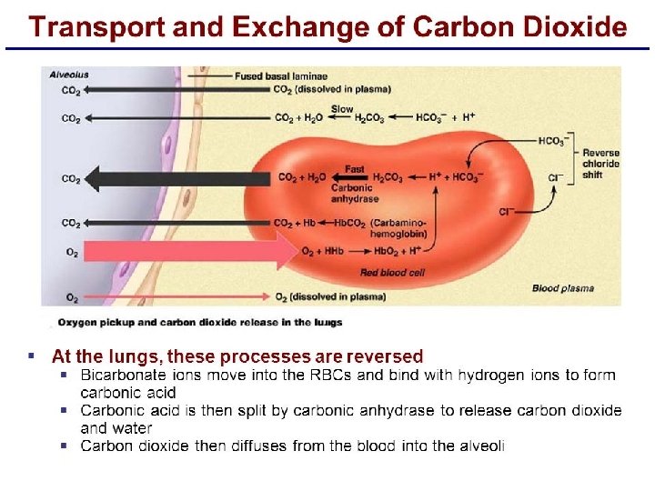
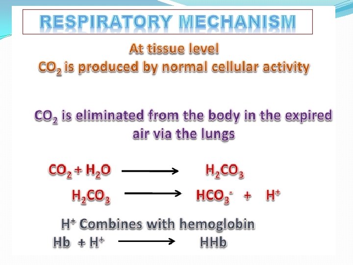
Sources of Hydrogen Ions �Most hydrogen ions originate from cellular metabolism �Breakdown of phosphorus-containing proteins releases phosphoric acid into the ECF �Anaerobic respiration of glucose produces lactic acid �Fat metabolism yields organic acids and ketone bodies �Transporting carbon dioxide as bicarbonate releases hydrogen ions
 �Are gained � At digestive tract � Through cellular")
Acid–Base Balance �Hydrogen Ions (H+) �Are gained � At digestive tract � Through cellular metabolic activities �Are eliminated � At kidneys and in urine � At lungs �Must be neutralized to avoid tissue damage �Acids produced in normal metabolic activity � Are temporarily neutralized by buffers in body fluids

Acid–Base Balance
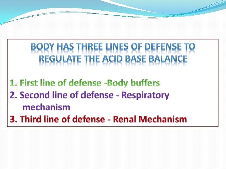

Figure 27. 7
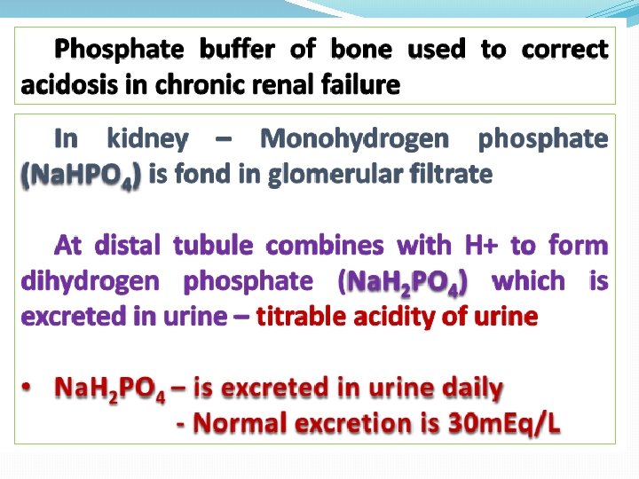
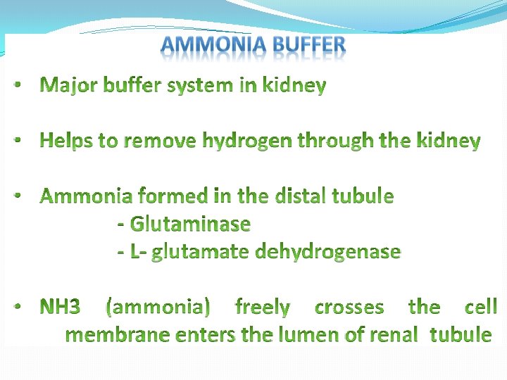
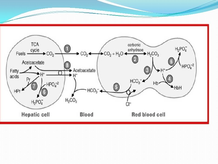
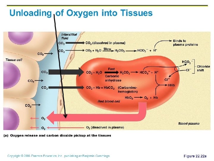
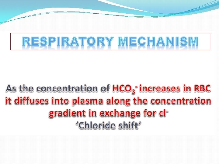

Acid–Base Balance �Major Buffers in Urine �Glomerular filtration provides components of �Carbonic acid–bicarbonate buffer system �Phosphate buffer system �Tubule cells of PCT �Generate ammonia

Carbonic acid– bicarbonate buffer system

Phosphate buffer system

Ammonium Ion Excretion Figure 26. 14
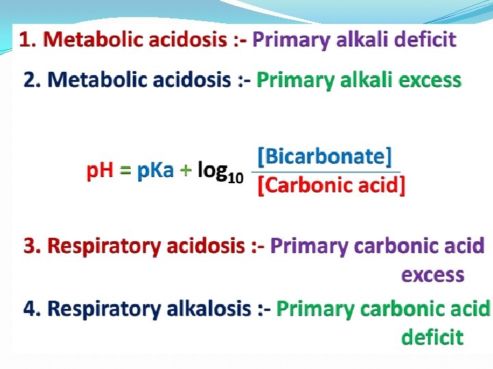

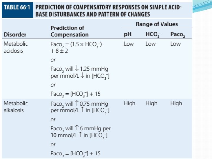
Primary and Compensatory Changes in Acid-Base Disorders Disorder Metabolic acidosis Metabolic alkalosis Respiratory acidosis Respiratory alkalosis Primary Process HCO 3 Compensation p. CO 2 HCO 3

MANAGEMENT

General Guidelines in Treatment of Metabolic Acidosis 1. Identifying and correcting the specific underlying cause of the metabolic acidosis. 2. some types of metabolic acidosis will require HCO 3 - therapy and some will not. 3. The decision to use HC 03 - replacement should be weighed carefully, depending upon the severity of the acidemia (blood p. H) and the type of acidosis. 4. If the p. H falls below 7. 10, emergency HC 03 - administration should be considered, regardless of the cause of the acidosis. This is especially important if there appears to be respiratory fatigue or developing hemodynamic instability

5. Never give HC 03 - without a determination of blood Ph 6. When the decision to give IV bicarbonate is made in the acute setting, calculate the amount of HCO 3 - required to increase the HC 03 - concentration to a specified value, often several m. Eq/L above the measured value. In general, assume that HC 03 - distributes in about 50% of Body weight (kg). HC 03 - deficit = 0. 5 X Body weight (kg) X ([HCO 3 -(desired)]- [HC 03(measured)]) 7. In severe acidosis (p. H in the 7. 10 range, HC 03 - <10 m. Eq/L), the amount of HCO 3 required to increase the HCO 3 - concentration to the range 10 -12 m. Eq/L is initially calculated. For example, in a 70 kg patient, if the HC 03 - is 6 m. Eq/L, and it is desired to bring the HC 03 - to 10 m. Eq/L: HC 03 - deficit =. 5 X 70 X (10 m. Eq/L - 6 m. Eq/L) = 140 m. Eqn Give this calculated amount slowly and remeasure p. H, HC 03 - and PCO~after the HCO 3 - is given to assess the effect of therapy on the acid-base status.

8. The relationship between amount of HCO 3 - given and the increase in HCO 3 - is not linear: At mild levels of acidemia, 2 m. Eq/kg will increase the HC 03 - by roughly 4 m. Eq. L. At severe levels of acidemia, 2 m. Eq/kg will only raise the HC 03 - by roughly 2 m. Eq/L. 9. In the case of an ongoing acidosis, repeated doses of HCO 3 - may be required until the underlying cause of the acidosis can be corrected. Treatment of L Lactic Acidosis Consider alkali therapy in cases of severe lactic acidosis when the p. H falls below 7. 10. When the underlying condition is corrected, however, lactate is converted to HC 03 -, and there may be an "overshoot alkalosis" during recovery.

Treatment of Diabetic Ketoacidosis Diabetic ketoacidosis generally responds well to therapy with insulin, saline, and potassium. Because the circulating ketoanions will be converted to HC 03 - by the liver once insulin and fluids reverse ketosis, they represent "potential" HC 03 -. The majority of patients should not receive HC 03 - replacement for this reason. Treatmentof. D-Lactic Acidosis Intravenous fluids and HC 03 -, but also requires oral antibiotics to eliminate the offending flora.

Treatment of Alcoholic Acidosis Treatment consists of the administrationof dextrose-containing saline to reverse ketogenesis and correct any ECFV depletion. D 5 0. 9% saline with supplemental KC 1 is usually appropriate for this purpose. HCO 3 - is not usually required because the ketones are converted to HC 03 -, once the ketosis is reversed and the ECFV normalized. In the case of alcoholic ketoacidosis with severe hypokalemia, the administration of Glucose should be postponed until potassium replacement is well underway
 RTA Determine and correct the cause, if possible, and")
Treatment of Distal (Type I) RTA Determine and correct the cause, if possible, and replace HC 03 and potassium. Distal RTA may require an amount of HC 03 - replacement that roughly equals the daily production of hydrogen ion (50 -100 m. Eq/ day). Some of the HC 03 - should be given as KHC 03 to correct potassium losses as long as there is no renal failure. Treatment of Type II (Proximal)RTA HC 03 - can be given as KHC 03 (often as K-citrate) as long as there is no significant degree of renal failure. Mild to moderate hypokalemia is common in PRTA and is worsened by alkali therapy.
 Vomiting/nasogastric suction Diuretic therapy Post")
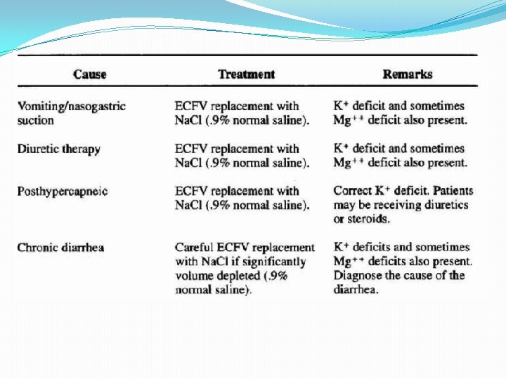
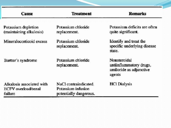
Causes of Metabolic Alkalosis ECFV depletion--chloride depletion syndrome (saline-responsive) Vomiting/nasogastric suction Diuretic therapy Post hypercapnea Chronic diarrheal laxative abuse Severe potassium depletion from any cause (saline-resistant) Mineralocorticoid excess syndromes (saline-resistant) Primary hyperaldosteronism Cushing's syndrome Ectopic ACTH Secondary hyperaldosteronism Renovascular disease Malignant hypertension Congestive heart failure (with diuretic therapy) Cirrhosis (with diuretic therapy) Gitelman's syndrome (saline-resistant) Bartter's syndrome (saline-resistant) Metabolic alkalosis maintained by renal failure (saline generally contraindicated)
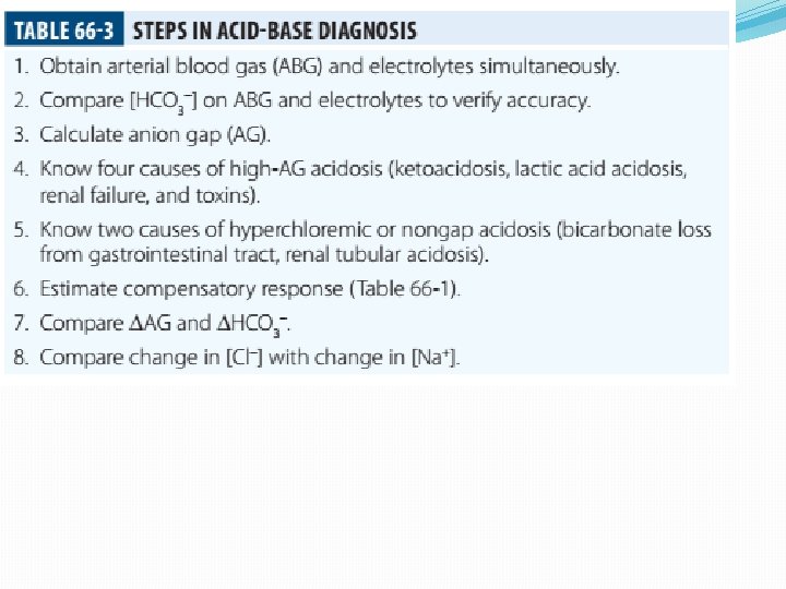

Interpreting ABG - Check p. H �Step 1 – evaluate p. H, narrow to 2 processes �If the p. H is < 7. 36 �Either metabolic acidosis or respiratory acidosis or both are present �If the p. H is >7. 44 �Either metabolic alkalosis or respiratory alkalosis or both are present

Check p. CO 2 �Step 2: evaluate p. CO 2 , narrow to 1 process �For a p. H < 7. 36 � If p. CO 2 < 40 - metabolic acidosis If p. CO 2 > 40 - respiratory acidosis � �For a p. H > 7. 44 � If p. CO 2 < 40 - respiratory alkalosis � If p. CO 2 > 40 - metabolic alkalosis

Use proper compensation formula �For metabolic acidosis: � p. CO 2 = 1. 5 [HCO 3] + 8 (WINTER’S) �For metabolic alkalosis: � p. CO 2 = 0. 9 [HCO 3] + 16

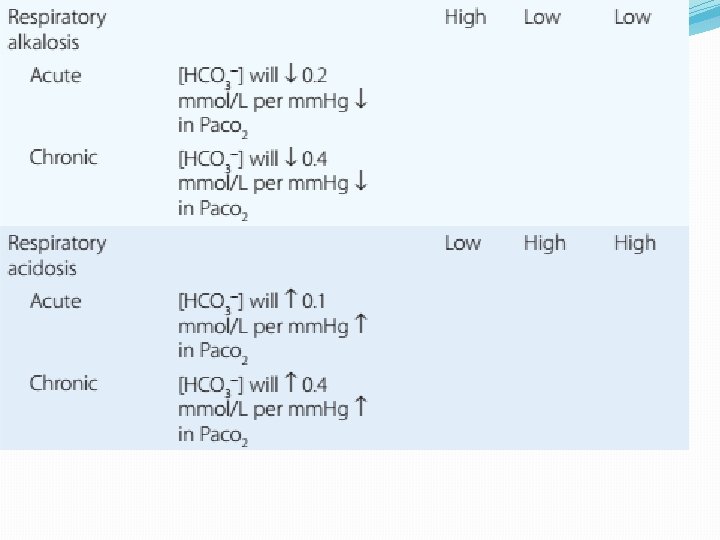
Use proper compensation formula �For respiratory acidosis: � For every increase of 10 in p. CO 2: � p. H decreases by : � 0. 08 (acute) / 0. 03 (chronic) � HCO 3 increases by: � 1 meq/l (acute) / 3 meq/l (chronic)

Use proper compensation formula �For respiratory alkalosis: � For every decrease of 10 in p. CO 2: � p. H increases by : � 0. 08 (acute) / 0. 03 (chronic) � HCO 3 decreases by: � 2 meq/l (acute) / 4 meq/l (chronic)

Use proper compensation formula �Simply choose the formula for the one disorder identified from step 1 and step 2. �The purpose of using the appropriate formula is to discover if any other acid-base process is present ( to a lesser / greater degree )
 value with the")
Identify other disorders �After applying chosen formula, compare the calculated (expected) value with the actual value �If the measured p. H, p. CO 2 , HCO 3 doesn’t coincide with calculated value, then another acid-base disturbance is present

Identify other disorders �Step 4: �For a metabolic acidosis: if actual p. CO 2 is � higher than calculated – respiratory acidosis � lower than calculated – respiratory alkalosis �For a metabolic alkolosis: if actual p. CO 2 is � higher by (>5 mm. Hg) – respiratory acidosis � lower by (>5 mm. Hg) – respiratory alkolosis

Identify other disorders �Step 4: �For a respiratory acidosis/ alkalosis: � if p. H or [HCO 3] is higher – metabolic alkalosis � if p. H or [HCO 3] is lower – metabolic acidosis �What if p. H is normal? [p. CO 2 & HCO 3] abnormal �An acidosis and alkalosis / to same degree

Identify other disorders �Possibilities: �Metabolic alkalosis and respiratory acidosis �Metabolic acidosis and respiratory alkalosis �Metabolic acidosis and metabolic alkalosis
")
Identify other disorders p. H in normal range ( 7. 36 – 7. 44) p. CO 2 < 36 mm. Hg [HCO 3 ] < 21 meq/l p. CO 2 >44 mm. Hg [HCO 3 ] > 27 meq/l Mixed respiratory alkalosis and Metabolic acidosis Mixed respiratory acidosis and Metabolic alkalosis
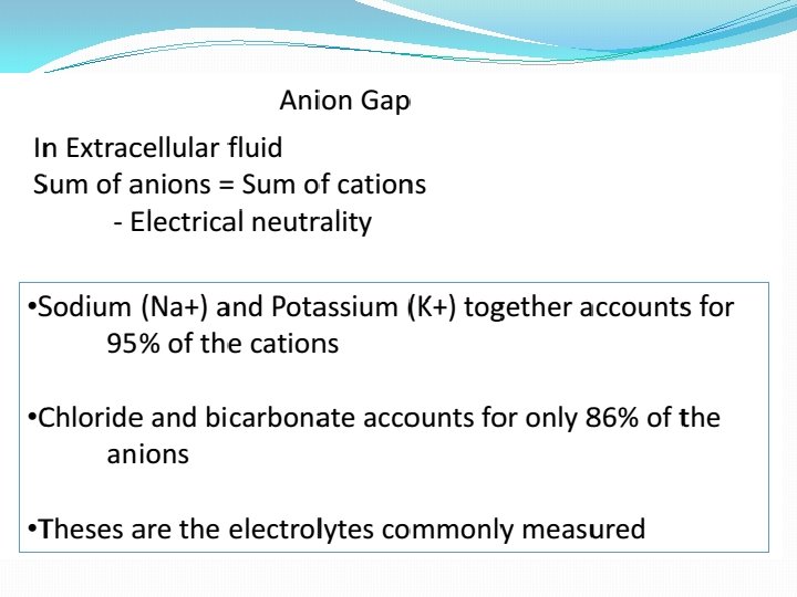
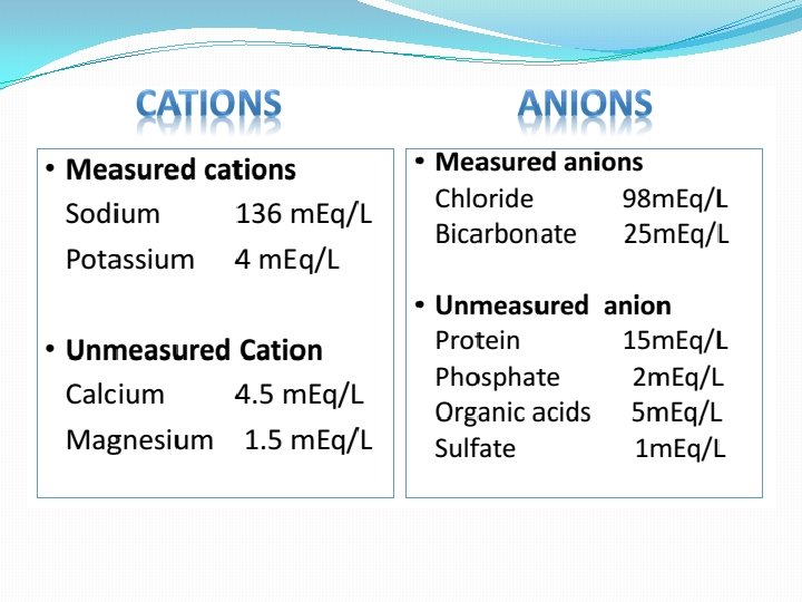
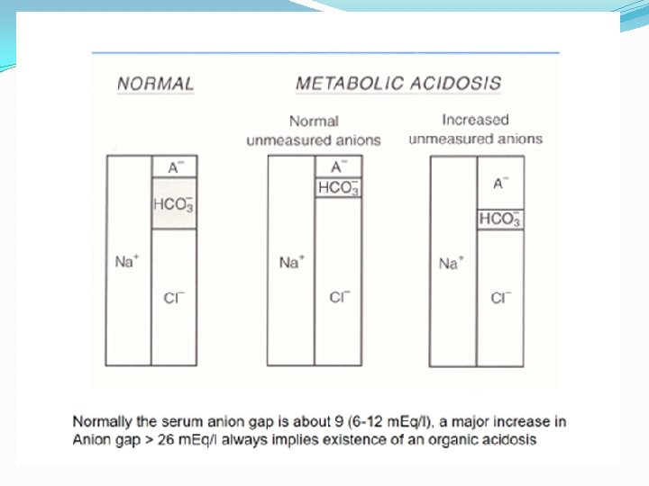
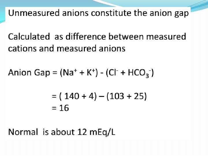
![Anion gap �Step 5: �Evaluate anion gap [ Na – (Cl + HCO 3)]](http://slidetodoc.com/presentation_image_h2/609686c2bb97a3d2724036d7ef9a459b/image-71.jpg "Anion gap �Step 5: �Evaluate anion gap [ Na – (Cl + HCO 3)]")
Anion gap �Step 5: �Evaluate anion gap [ Na – (Cl + HCO 3)] �If Elevated – indicate elevated gap metabolic acidosis

Check urine p. H �Step 6: �Urine is normally acidic unless the serum is alkalemic. �If urine is alkalotic (p. H > 6. 0) in face of an acidosis, a RTA or a UTI may be present

Generate differential diagnosis �Step 7: �Identifying the primary pathology from analyzing differential diagnosis of various acid-base disturbances.
 K – Ketosis (Diabetic, Alcoholic, ) U –")
Metabolic acidosis HIGH ANION GAP (KUSSMALE) K – Ketosis (Diabetic, Alcoholic, ) U – Uremia S – Salicylate poisoning S – Sepsis M – Methanol poisoning A – Alcohol (Ethanol Poisoning) L – Lactic acidosis E – Ethylene glycol NORMAL ANION-GAP RENAL RTA TYPE II RTA TYPE IV EARLY RENAL FAILURE NORMAL ANIONGAP EXTRARENAL DIARRHOEA GI-URETHRAL CONNECTIONS LOSS OF PANCREATIC AND BILIARY SECRETIONS CORRECTION PHASE OF DKA
 responsive Saline (chloride) unresponsive Diuretics Adenoma of colon Misc. (Bartter’s,")
Metabolic alkalosis Saline (chloride) responsive Saline (chloride) unresponsive Diuretics Adenoma of colon Misc. (Bartter’s, penicillin K+ defi. , bulimia) Posthypercapnia Emesis Nasogastric tube Alkali ingestion with decreased GFR 11 β hydroxylase deficiency Exogenous steroids Licorice ingestion Cushing’s syndrome Hyperaldosteronism

Respiratory acidosis Respiratory center depression: Sedative medications Brain stem lesions Central sleep apnoea, myxedema Neuromuscular failure: Myopathic and motor end plate dysfunction – polymyositis, hypokalemia, OPC Neuropathic – GBS, ALS, status epilepticus Decreased compliance Parenchymal – pulmonary fibrosis, ARDS Extra parenchymal – abdominal distension, severe kyphoscoliosis Increased airway resistance COPD, emphysema, severe asthma Obstructive sleep apnoea Increased dead space: Large pulmonary embolus Emphysema

Respiratory alkalosis �Hypoxia – pneumonia, pulmonary embolism, pulmonary edema, interstitial fibrosis �Hyperdynamic states – pain, fever, sepsis, pregnancy, hyperthyroidism, hepatic failure, anxiety, mechanical hyprventilation �CNS – CVA, tumor, infection, ICH, SAH �Drugs – salicylates, catecholamines, progestrone, nicotine

Pneumonics for pnuemonic lovers Metabolic Acidosis Anion Gap Metabolic Acidosis Non. Gap Acute Resp. Acidosis Metabolic Alkalosis Respiratory Alkalosis “CLEVERPD” “CHAMPS” “HARDUPS” “anything causing hypoventilation” “MUDPILERS” • Methanol • Hyperalimentatio • CNS • Contraction • CNS • Uremia n • Acetazolamide • Renal Tubular Acidosis • Diarrhea • Uretero-Pelvic shunt • Post-hypocapnia • Spironolactone depression • Licorice • Airway • Endocrine obstruction (Conn/Cushing/ • Pulmonary Bartters) edema • Vomiting • Pneumonia • Excess alkali • Hemo/Pneumo • Refeeding thorax • Post • Neuromuscular hypercapnia • Diuretics • DKA/Alcoholic ketoacidosis • Paraldehyde • Isoniazid • Lactic acidosis • Ethanol • Renal failure/Rhabdo • Salicylates disease • Hypocapnia • Anxiety • Mech. Ventilation • Progesterone • Salicylates • Sepsis
 A surgeon refers a 22 -year old man")
Interpretation – case 1 � 1) A surgeon refers a 22 -year old man with a hernia to you, with history of renal stones. �p. H - 7. 29 �p. CO 2 – 32 mm. Hg �[HCO 3] – 15 meq/l �Na+ - 138 meq/l �K+ - 3. 0 meq/l �Cl- - 110 meq/l �Urine p. H – 6. 0

Interpretation – case 1 �Step 1 – p. H < 7. 36 So, �Met. / resp. acidosis exist �Step 2 – p. CO 2 < 40 mm. Hg So, �At least a Metabolic acidosis exists �Step 3 – formula �Expected p. CO 2 = 1. 5[HCO 3] + 8; i. e. , = 30 �Actual p. CO 2 = 32 mm. Hg �p. H - 7. 29 �p. CO 2 – 32 mm. Hg �[HCO 3] – 15 meq/l

Interpretation – case 1 �Step 4 - any other process involved? �Actual and expected p. CO 2 values match closely �So, a metabolic acidosis, fully compensated exists �Step 5 – evaluate anion gap �Anion gap = 138 – (110 +15) = 13 �A normal gap METABOLIC ACIDOSIS exists

Case 2 �A 40 year old lady with obesity and newly detected hypertension �p. H - 7. 49 �p. CO 2 – 45 mm. Hg �[HCO 3] – 33 meq/l �Na+ - 142 meq/l �K+ - 4. 1 meq/l �Cl- - 98 meq/l �Urine p. H – 6. 5

Case 2 �Step 1 – p. H > 7. 44 So, �Met. / resp. alkalosis exist �Step 2 – p. CO 2 > 40 mm. Hg So, �At least a Metabolic alkalosis exists �Step 3 – formula �Expected p. CO 2 = 0. 9[HCO 3] + 16; � i. e. , = 46 �Actual p. CO 2 = 45 mm. Hg �p. H - 7. 49 �p. CO 2 – 45 mm. Hg �[HCO 3] – 33 meq/l

Case 2 �Step 4 - any other process involved? �Actual and expected p. CO 2 values appr. match closely �So, a metabolic alkalosis, fully compensated exists �Step 5 – evaluate anion gap �Anion gap = 142 – (98 +34) = 10 (normal) �Step 6 – check urine p. H = 6. 5 ( in alkalemia) �DD – cushings / hyperaldosteronism �So, a simple Metabolic alkalosis exists �p. H - 7. 49 �p. CO 2 – 45 mm. Hg �[HCO 3] – 33 meq/l

Case 3 �A 25 year old patient with fever, chest pain and breathlessness, was toxic and had a patch in his right middle lobe was treated for 3 days but respiratory rate was still high � p. H - 7. 47 �p. CO 2 – 21 mm. Hg �[HCO 3] – 15 meq/l �Na+ - 136 meq/l �Cl- - 110 meq/l �Urine p. H – 6. 5

Case 3 �Step 1 – p. H > 7. 44 So, �Met. / resp. alkalosis exist �Step 2 – p. CO 2 < 40 mm. Hg. So, �At least a respiratory alkalosis is present �Step 3 – apply formula for chronic resp. alk. �If we use p. H : �Expected p. H – 7. 40 + (2 x 0. 03) = 7. 46 �Actual p. H – 7. 47 �p. H - 7. 47 �p. CO 2 – 21 mm. Hg �[HCO 3] – 15 meq/l
![Case 3 �If we use [HCO 3]: �Expected [HCO 3] – 24 - (2](http://slidetodoc.com/presentation_image_h2/609686c2bb97a3d2724036d7ef9a459b/image-87.jpg "Case 3 �If we use [HCO 3]: �Expected [HCO 3] – 24 - (2")
Case 3 �If we use [HCO 3]: �Expected [HCO 3] – 24 - (2 x 4) = 16 meq/l �Actual [HCO 3] – 15 meq/l �Step 4 - any other process involved? �Actual and expected p. H &[HCO 3] values appr. match closely �So, a chronic respiratory alkalosis, fully compensated exists. �Step 5 – evaluate anion gap �Anion gap = 136 – (110 +15) = 11 �Step 6 – check urinep. H =6. 5(alkalemia) �A simple chronic (fully compensated) resp. alk. exists �p. H - 7. 47 �p. CO 2 – 21 mm. Hg �[HCO 3] – 15 meq/l

Case 4 �If in the same patient – history is not known: known � p. H - 7. 47 �p. CO 2 – 21 mm. Hg �[HCO 3] – 15 meq/l �Na+ - 136 meq/l �Cl- - 110 meq/l �Urine p. H – 6. 5 �With p. H and p. CO 2 – respiratory alkalosis exists

Case 4 �Step 3 – apply formula for acute resp. alk. �If we use p. H : �Expected p. H – 7. 40 + (2 x 0. 08) = 7. 56 �Actual p. H – 7. 47 �If we use [HCO 3]: �Expected [HCO 3] – 24 - (2 x 2) = 20 meq/l �Actual [HCO 3] – 15 meq/l �Step 4 - any other process involved? �Actual p. H &[HCO 3] values are both LOWER than the expected values. So, which could lower both? �Only a metabolic acidosis could lower both values �So, a mixed resp. alkalosis and metabolic acidosis exists. �p. H - 7. 47 �p. CO 2 – 21 mm. Hg �[HCO 3] – 15 meq/l

Case 5 �A 20 year old man is brought to the emergency room by his sister, who tells you he took a bottle of pills �p. H - 7. 35 �p. CO 2 – 15 mm. Hg �[HCO 3] – 8 meq/l �Na+ - 140 meq/l �K+ - 3. 5 meq/l �Cl- - 104 meq/l �Urine p. H – 5. 0

Case 5 �Step 1 – p. H < 7. 36 So, �Met. / resp. acidosis exist �Step 2 – p. CO 2 < 40 mm. Hg So, �At least a Metabolic acidosis exists �Step 3 – formula �Expected p. CO 2 = 1. 5[HCO 3] + 8; i. e. , = 20 �Actual p. CO 2 = 15 mm. Hg �p. H - 7. 35 �p. CO 2 – 15 mm. Hg �[HCO 3] – 8 meq/l

Case 5 �Step 4 - any other process involved? �Actual p. CO 2 value is less than expected �Only process that could decrease the p. CO 2 value beyond predicted is RESPIRATORY ALKALOSIS �So, a mixed metabolic acidosis & resp. alk. exists �Step 5 – evaluate anion gap �Anion gap = 140 - (104 + 8) = 28 (elevated) �Urine p. H = 5. 0 (fits to acidemia) �A mixed elevated gap Metabolic acidosis and Respiratory alkalosis exists �DD – only sepsis/salicylate overdose cause both �So, given the history salicyalate overdose is possibile

Case 6 �A 57 year old patient with long history of smoking presents without distress and with h/o BOE, ABG shows: �p. H - 7. 35 �p. CO 2 – 50 mm. Hg �[HCO 3] – 27 meq/l �Na+ - 143 meq/l �Cl- - 105 meq/l �Urine p. H – 5. 0

Case 6 �Step 1 – p. H < 7. 36 So, �p. H - 7. 35 �Met. / resp. acidosis present �p. CO 2 – 50 mm. Hg �Step 2 – p. CO 2 > 40 mm. Hg So, �At least a respiratory acidosis is present �[HCO 3] – 27 meq/l �Step 3 – apply formula for respiratory acidosis �If we use p. H : use chronic formula �Expected p. H – 7. 40 - (1 x 0. 03) = 7. 37 �Actual p. H – 7. 35
![Case 6 �p. H - 7. 35 �If we use [HCO 3]: �p. CO](http://slidetodoc.com/presentation_image_h2/609686c2bb97a3d2724036d7ef9a459b/image-95.jpg "Case 6 �p. H - 7. 35 �If we use [HCO 3]: �p. CO")
Case 6 �p. H - 7. 35 �If we use [HCO 3]: �p. CO 2 – 50 �Expected [HCO 3] – 24 + (1 x 3) = 27 meq/l mm. Hg �Actual [HCO 3] – 27 meq/l �[HCO 3] – 27 �Step 4 - any other process involved? meq/l �Actual and expected p. H &[HCO 3] values appr. match closely �So, a pure chronic respiratory acidosis, fully compensated exists. �Step 5 – evaluate anion gap �Anion gap = 143 – (105 +28) = 10(normal) �Step 6 – check urine p. H = 5. 0 ( fit for acidemia) �A simple chronic fully compensated resp. acid. exists

Case 6 �The same patient presents to emergency room 1 month later in respiratory distress. He has wheezing and his RR – 33 B/min �p. H - 7. 29 �p. CO 2 – 61 mm. Hg �[HCO 3] – 28 meq/l �Na+ - 142 meq/l �Cl- - 100 meq/l �Urine p. H – 5. 0

Case 6 �Step 1 – p. H < 7. 36 So, �Met. / resp. acidosis present �Step 2 – p. CO 2 > 40 mm. Hg So, �At least a respiratory acidosis is present �Step 3 – apply formula for respiratory acidosis, both acute and chronic �If we use p. H : chronic �Expected p. H – 7. 40 - (2 x 0. 03) = 7. 34 �Actual p. H – 7. 29 �p. H - 7. 29 �p. CO 2 – 61 mm. Hg �[HCO 3] – 28 meq/l
![Case 6 �If we use [HCO 3]: chronic �Expected [HCO 3] – 24 +](http://slidetodoc.com/presentation_image_h2/609686c2bb97a3d2724036d7ef9a459b/image-98.jpg "Case 6 �If we use [HCO 3]: chronic �Expected [HCO 3] – 24 +")
Case 6 �If we use [HCO 3]: chronic �Expected [HCO 3] – 24 + (2 x 3) = 30 meq/l �Actual [HCO 3] – 28 meq/l �Actual p. H &[HCO 3] values are lower than expected: �Possibilities: Possibilities � 1) Mixed resp. acid. / small met. acid. � 2) Resp. acid. – partially compensated �(acute – on –chronic / mixed acute & chronic resp. acid. ) �p. H - 7. 29 �p. CO 2 – 61 mm. Hg �[HCO 3] – 28 meq/l

Case 6 �If we use p. H : acute �Expected p. H – 7. 40 - (2 x 0. 08) = 7. 24 �Actual p. H – 7. 29 �If we use [HCO 3]: acute �Expected [HCO 3] – 24 + (2 x 1) = 26 meq/l �Actual [HCO 3] – 28 meq/l �Actual p. H &[HCO 3] values are higher than expected: �Possibilities: � 1) a mixed acute resp. acid. /small met. Alk. � 2) a resp. acid. that is partially compensated. �p. H - 7. 29 �p. CO 2 – 61 mm. Hg �[HCO 3] – 28 meq/l
A")
Case 6 �Step 4 – identify other processes �Summarising step 3 : � 1)A mixed chonic resp. acid. /small met. Acid. � 2)An acute resp. acid. / small met. Alk. � 3) A resp. acid. Not fully compensated �In our patient we consider the third possibility since patient is having acute exacerbation of COPD �So, a mixed acute and chronic resp. acidosis.

Case 7 �A 45 year old diabetic patient presents with obtundation �p. H - 7. 01 �p. CO 2 – 80 mm. Hg �[HCO 3] – 20 meq/l �Na+ - 140 meq/l �K+ - 5. 5 meq/l �Cl- - 97 meq/l �Urine p. H – 5. 0

Case 7 �Step 1 – p. H < 7. 36 So, �Met. / resp. acidosis exist �Step 2 – p. CO 2 > 40 mm. Hg So, �At least a respiratory acidosis exists �Step 3 – formula – acute resp. acid. �If we use p. H : acute �Expected p. H – 7. 40 - (4 x 0. 08) = 7. 08 �Actual p. H – 7. 01 �p. H - 7. 01 �p. CO 2 – 80 mm. Hg �[HCO 3] – 20 meq/l
![Case 7 �If we use [HCO 3]: acute �Expected [HCO 3] – 24 +](http://slidetodoc.com/presentation_image_h2/609686c2bb97a3d2724036d7ef9a459b/image-103.jpg "Case 7 �If we use [HCO 3]: acute �Expected [HCO 3] – 24 +")
Case 7 �If we use [HCO 3]: acute �Expected [HCO 3] – 24 + (4 x 1) = 28 meq/l �Actual [HCO 3] – 20 meq/l �Actual and expected p. H & [HCO 3] values are lower than expected: �Since [HCO 3] value is lower as against an increase a metabolic acidosis coexists �p. H - 7. 01 �p. CO 2 – 80 mm. Hg �[HCO 3] – 20 meq/l

Case 7 �Step 5 – evaluate anion gap �Anion gap = 140 – (97 +20) = 23(elevated) �Step 6 – check urine p. H = 5. 0 ( fit for acidemia) �DD – predominant respiratory acidosis and an elevated gap metabolic acidosis (DKA)

Case 8 �A 78 year old patient has been vomiting for several days, developed fever and breathlessnessover few hours. Her RR – 35 b/min and has consolidation over Rt. Base �p. H - 7. 69 �p. CO 2 – 20 mm. Hg �[HCO 3] – 25 meq/l �Na+ - 138 meq/l �Cl- - 97 meq/l �Urine p. H – 8. 0

Case 8 �Step 1 – p. H > 7. 44 So, �Met. / resp. alkalosis exist �Step 2 – p. CO 2 < 40 mm. Hg So, �At least a respiratory alkalosis exists �Step 3 – formula – acute resp. alkalosis �If we use p. H : �Expected p. H – 7. 40 + (2 x 0. 08) = 7. 56 �Actual p. H – 7. 69 �p. H - 7. 69 �p. CO 2 – 20 mm. Hg �[HCO 3] – 25 meq/l
![Case 8 �If we use [HCO 3]: �p. H - 7. 69 �Expected [HCO](http://slidetodoc.com/presentation_image_h2/609686c2bb97a3d2724036d7ef9a459b/image-107.jpg "Case 8 �If we use [HCO 3]: �p. H - 7. 69 �Expected [HCO")
Case 8 �If we use [HCO 3]: �p. H - 7. 69 �Expected [HCO 3] – 24 - (2 x 2) = 20 meq/l �p. CO 2 – 20 �Actual [HCO 3] – 25 meq/l mm. Hg �Step 4 - any other process involved? �[HCO 3] – 25 �Actual p. H &[HCO 3] values are both higher than the expected values. So, which could meq/l increase both? �Only a metabolic alkalosis could elevate both values �Step 5 – evaluate anion gap �Anion gap = 138 – (97 +28) = 13(normal) �Step 6 – check urine p. H = 8. 0 ( fit for alkalemia) �A mixed acute resp. alk. & met. Alk.

Case 9 �A 27 year old diabetic patient with 1 hr of shortness of breath. He was nauseated and has had increased urination over the past 2 days. So, he didn’t take insulin and was bedridden for past 2 days. He has family history of hypercoagulable disorder.

Case 9 �p. H - 7. 40 �p. CO 2 – 20 mm. Hg �[HCO 3] – 12 meq/l �Na+ - 136 meq/l �Cl- - 102 meq/l �Urine p. H – 5. 0, ketones + �Glucose – 650 mg/dl
 p. CO")
For p. H in normal range ( 7. 36 – 7. 44) p. CO 2 < 36 mm. Hg [HCO 3 ] < 21 meq/l p. CO 2 >44 mm. Hg [HCO 3 ] > 27 meq/l Mixed respiratory alkalosis and Metabolic acidosis Mixed respiratory acidosis and Metabolic alkalosis
 �p. CO 2")
Case 9 �Step 1 – p. H is 7. 40 (normal) �p. CO 2 and [HCO 3] are abnormal, So �A mixed acid-base disturbance is present �At least one acidosis and one alkalosis present �Alkalosis is exactly balancing acidosis �p. H - 7. 40 �p. CO 2 – 20 mm. Hg �[HCO 3] – 12 meq/l

Case 9 �Step 2 �p. H - 7. 40 �p. CO 2 < 40 mm. Hg and [HCO 3] < 25 meq/l �p. CO 2 – 20 �Respiratory alkalosis & metabolic acidosis mm. Hg is the process that can cause this �[HCO 3] – 12 �Step 3 – check with formula for both meq/l �Metabolic acidosis: �Expected p. CO 2 = 1. 5[HCO 3] + 8; i. e. , = 26 �Actual p. CO 2 = 20 mm. Hg �Actual is lower than expected value, So a respiratory alkalosis is lowering p. CO 2

Case 9 �For respiratory alkalosis: �If we use p. H : �Expected p. H – 7. 40 + (2 x 0. 08) = 7. 56 �Actual p. H – 7. 40 �If we use [HCO 3]: �Expected [HCO 3] – 24 - (2 x 2) = 20 meq/l �Actual [HCO 3] – 12 meq/l �Step 4 - any other process involved? �Actual p. H & [HCO 3] values are lower than expected. So, a metabolic acidosis exists. �p. H - 7. 40 �p. CO 2 – 20 mm. Hg �[HCO 3] – 12 meq/l
")
Case 9 �Step 5: 5 evaluate anion gap �Anion gap = 136 – (102+12) = 24 �An elevated gap metabolic acidosis is present �Step 6 : check the urine p. H �Urine p. H = 5. 0 (fit for acidemia) �DD – 1) DKA – elevated gap metabolic acidosis � 2) an acute respiratory alkalosis - ? PE �So, both equally balances to maintain p. H.

Thank you
- Slides: 118