
ACIDBASE BALANCE Dr zge Kner ACIDBASE BALANCE Y
ACID-BASE BALANCE Dr. Özge Köner ACID-BASE BALANCE Y. T. U. Hospital Anesthesiology Dept. Dr. Özge Köner YTÜ Hastanesi Anesteziyoloji A. D
Acid base imbalance: • Severe acid-base imbalance may lead to organ failure, such as brain edema, seizure activity, myocardial dysfunction and pulmonary vasoconstriction, systemic vasodilation. • Immune cell dysfunction
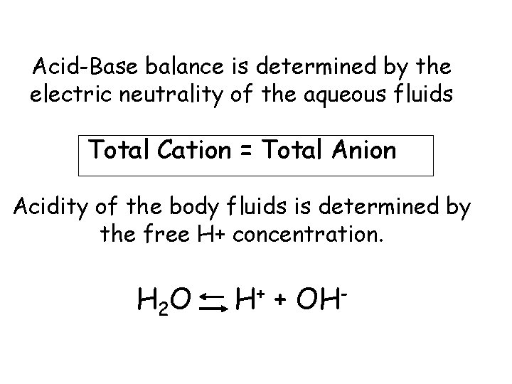
Acid-Base balance is determined by the electric neutrality of the aqueous fluids Total Cation = Total Anion Acidity of the body fluids is determined by the free H+ concentration. H 2 O H+ + OH-
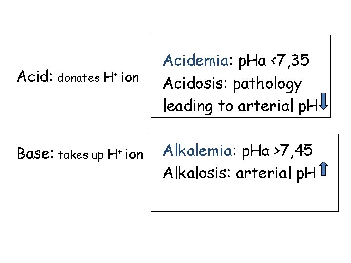
Acid: donates H+ ion Base: takes up H+ ion Acidemia: p. Ha <7, 35 Acidosis: pathology leading to arterial p. H Alkalemia: p. Ha >7, 45 Alkalosis: arterial p. H
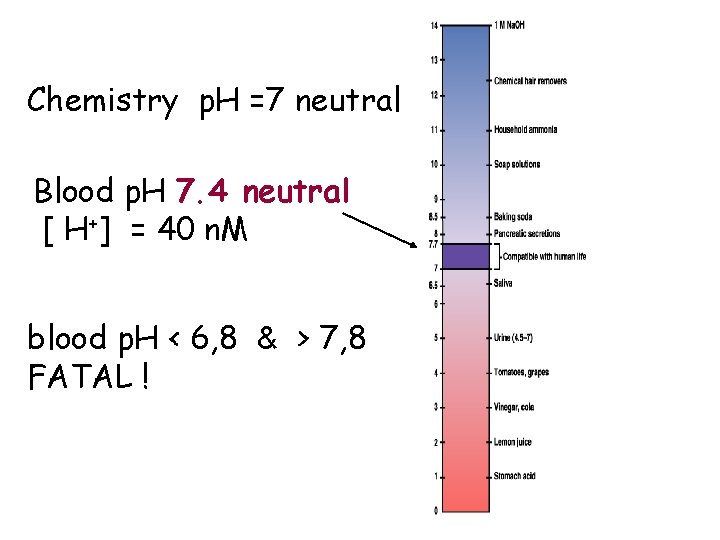
Chemistry p. H =7 neutral Blood p. H 7. 4 neutral [ H+] = 40 n. M blood p. H < 6, 8 & > 7, 8 FATAL !
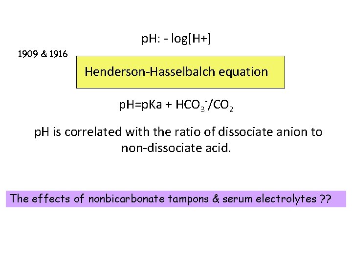
p. H: - log[H+] 1909 & 1916 Henderson-Hasselbalch equation p. H=p. Ka + HCO 3 -/CO 2 p. H is correlated with the ratio of dissociate anion to non-dissociate acid. The effects of nonbicarbonate tampons & serum electrolytes ? ?
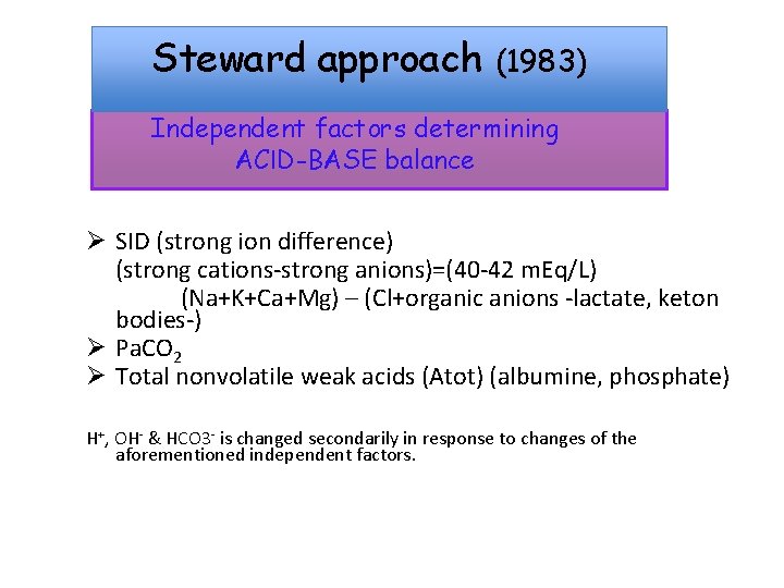
Steward approach (1983) Independent factors determining ACID-BASE balance Ø SID (strong ion difference) (strong cations-strong anions)=(40 -42 m. Eq/L) (Na+K+Ca+Mg) – (Cl+organic anions -lactate, keton bodies-) Ø Pa. CO 2 Ø Total nonvolatile weak acids (Atot) (albumine, phosphate) H+, OH- & HCO 3 - is changed secondarily in response to changes of the aforementioned independent factors.
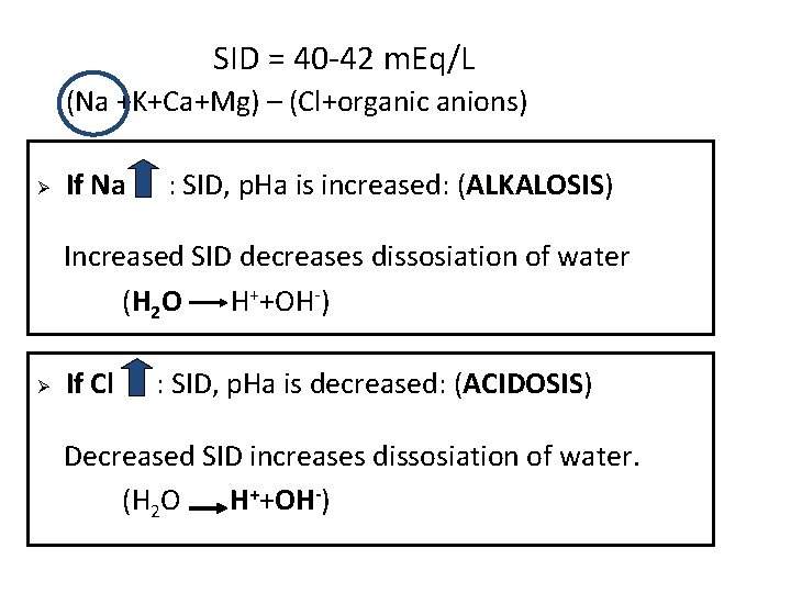
SID = 40 -42 m. Eq/L (Na +K+Ca+Mg) – (Cl+organic anions) Ø If Na : SID, p. Ha is increased: (ALKALOSIS) ALKALOSIS Increased SID decreases dissosiation of water (H 2 O H++OH-) Ø If Cl : SID, p. Ha is decreased: (ACIDOSIS) ACIDOSIS Decreased SID increases dissosiation of water. (H 2 O H++OH-)
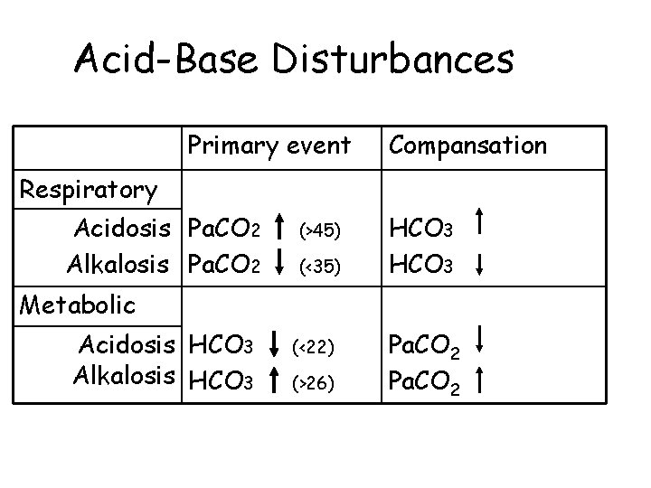
Acid-Base Disturbances Primary event Compansation Respiratory Acidosis Pa. CO 2 Alkalosis Pa. CO 2 (>45) (<35) HCO 3 Metabolic Acidosis HCO 3 Alkalosis HCO 3 (<22) (>26) Pa. CO 2
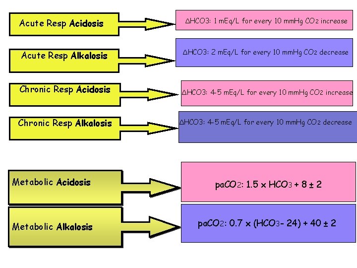
Acute Resp Acidosis ΔHCO 3: 1 m. Eq/L for every 10 mm. Hg CO 2 increase Acute Resp Alkalosis ΔHCO 3: 2 m. Eq/L for every 10 mm. Hg CO 2 decrease Chronic Resp Acidosis ΔHCO 3: 4 -5 m. Eq/L for every 10 mm. Hg CO 2 increase Chronic Resp Alkalosis ΔHCO 3: 4 -5 m. Eq/L for every 10 mm. Hg CO 2 decrease Metabolic Acidosis pa. CO 2: 1. 5 x HCO 3 + 8 ± 2 Metabolic Alkalosis pa. CO 2: 0. 7 x (HCO 3 - 24) + 40 ± 2
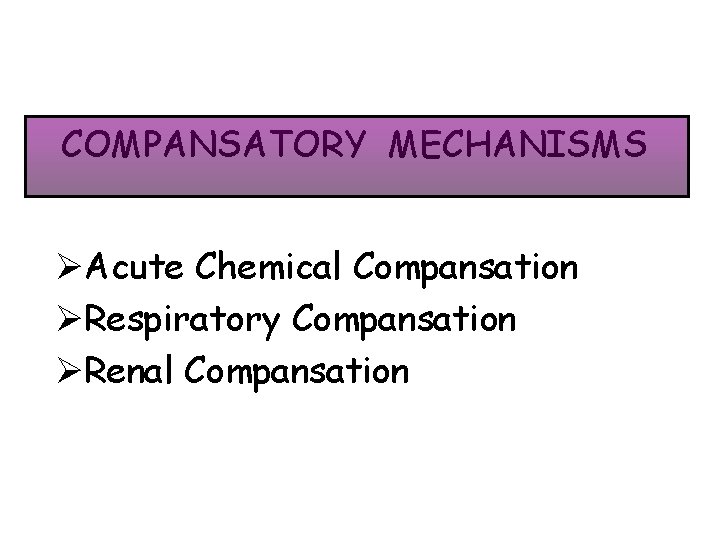
COMPANSATORY MECHANISMS ØAcute Chemical Compansation ØRespiratory Compansation ØRenal Compansation

1 -Chemical tampon system Ø HCO 3 -/H 2 CO 3* Ø Hb (Hb. H/Hb-) -extracellular-fast 15 -20 mins -extracellular, tampons volatile+nonvolatile acids Ø Proteins (Pr. H/Pr-) Ø HPO 4/H 2 PO 4 Ø NH 3/NH 4+ –intracellular-slow 2 -4 hrs –urinary, intracellular -urinary
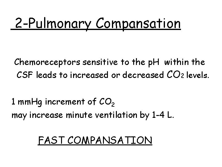
2 -Pulmonary Compansation Chemoreceptors sensitive to the p. H within the CSF leads to increased or decreased CO 2 levels. 1 mm. Hg increment of CO 2 may increase minute ventilation by 1 -4 L. FAST COMPANSATION
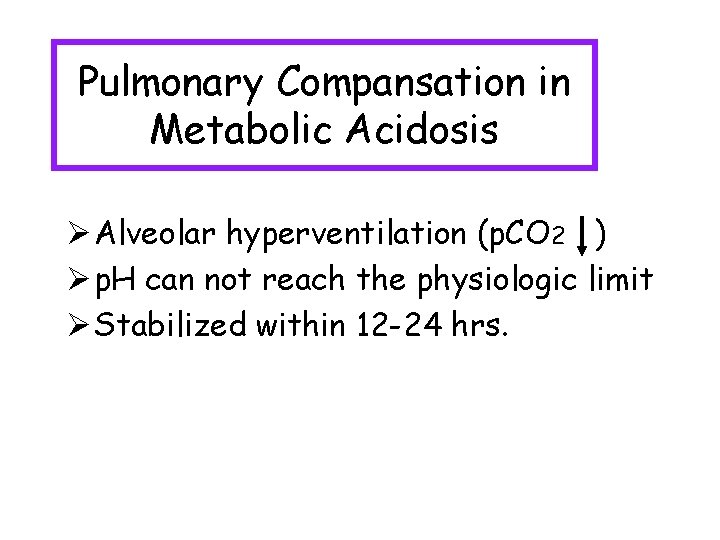
Pulmonary Compansation in Metabolic Acidosis Ø Alveolar hyperventilation (p. CO 2 ) Ø p. H can not reach the physiologic limit Ø Stabilized within 12 -24 hrs.
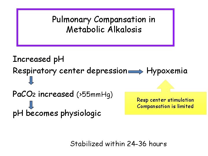
Pulmonary Compansation in Metabolic Alkalosis Increased p. H Respiratory center depression Pa. CO 2 increased (>55 mm. Hg) p. H becomes physiologic Hypoxemia Resp center stimulation Compansation is limited Stabilized within 24 -36 hours
3 - Renal Compansation
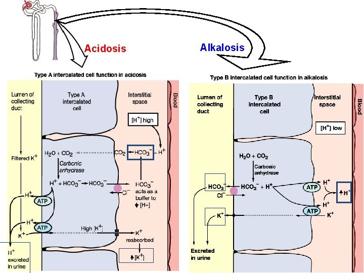
Acidosis Alkalosis
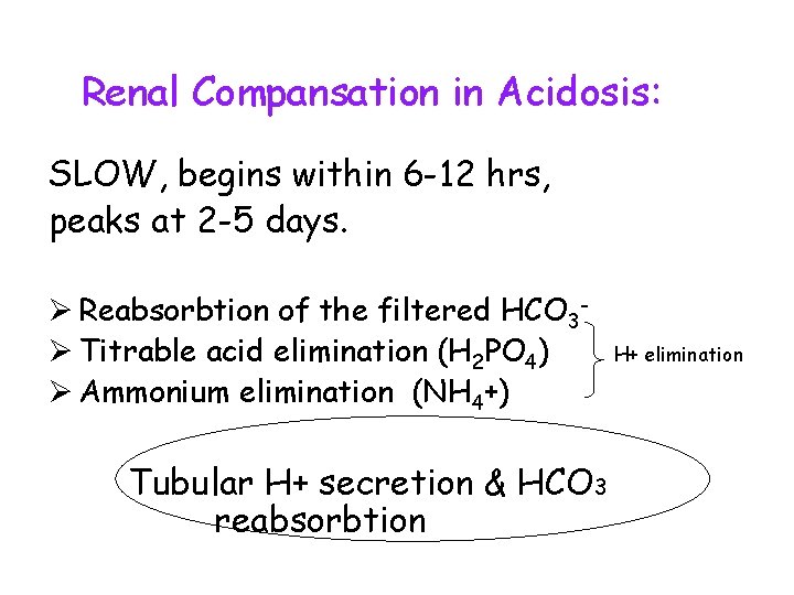
Renal Compansation in Acidosis: SLOW, begins within 6 -12 hrs, peaks at 2 -5 days. Ø Reabsorbtion of the filtered HCO 3Ø Titrable acid elimination (H 2 PO 4) Ø Ammonium elimination (NH 4+) Tubular H+ secretion & HCO 3 reabsorbtion H+ elimination
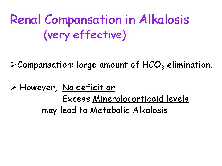
Renal Compansation in Alkalosis (very effective) ØCompansation: large amount of HCO 3 elimination. Ø However, Na deficit or Excess Mineralocorticoid levels may lead to Metabolic Alkalosis
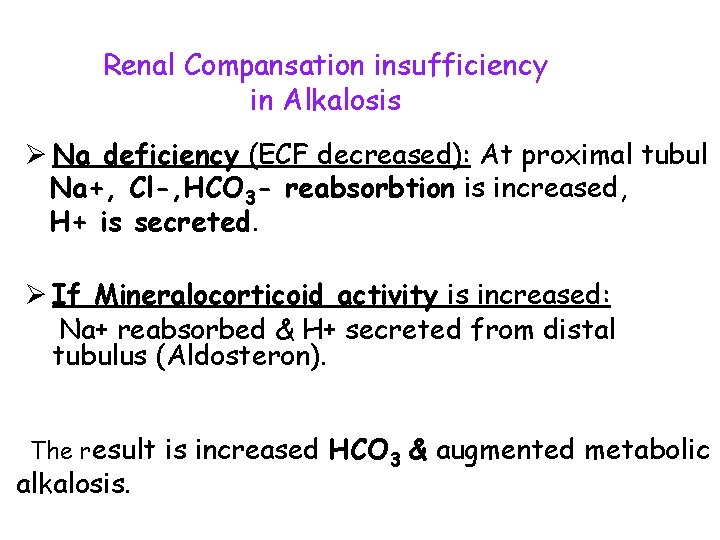
Renal Compansation insufficiency in Alkalosis Ø Na deficiency (ECF decreased): At proximal tubul Na+, Cl-, HCO 3 - reabsorbtion is increased, H+ is secreted. Ø If Mineralocorticoid activity is increased: Na+ reabsorbed & H+ secreted from distal tubulus (Aldosteron). The result is increased HCO 3 & augmented metabolic alkalosis.
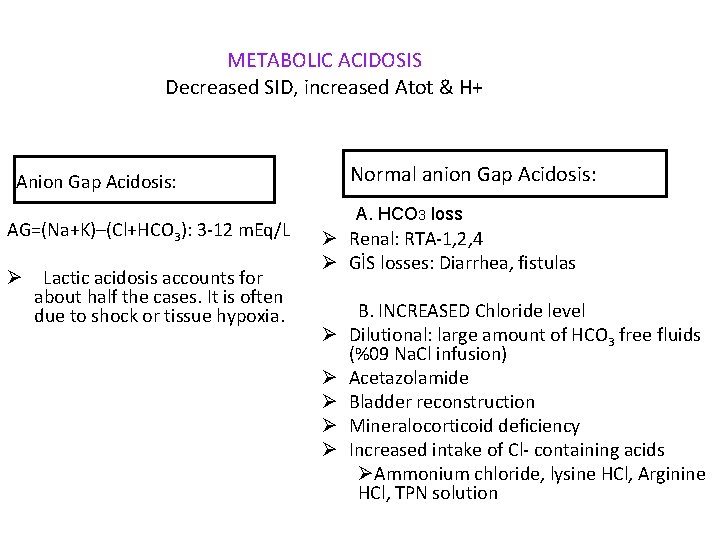
METABOLIC ACIDOSIS Decreased SID, increased Atot & H+ Normal anion Gap Acidosis: Anion Gap Acidosis: AG=(Na+K)–(Cl+HCO 3): 3 -12 m. Eq/L Ø Lactic acidosis accounts for about half the cases. It is often due to shock or tissue hypoxia. A. HCO 3 loss Ø Renal: RTA-1, 2, 4 Ø GİS losses: Diarrhea, fistulas Ø Ø Ø B. INCREASED Chloride level Dilutional: large amount of HCO 3 free fluids (%09 Na. Cl infusion) Acetazolamide Bladder reconstruction Mineralocorticoid deficiency Increased intake of Cl- containing acids ØAmmonium chloride, lysine HCl, Arginine HCl, TPN solution
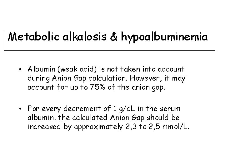
Metabolic alkalosis & hypoalbuminemia • Albumin (weak acid) is not taken into account during Anion Gap calculation. However, it may account for up to 75% of the anion gap. • For every decrement of 1 g/d. L in the serum albumin, the calculated Anion Gap should be increased by approximately 2, 3 to 2, 5 mmol/L.
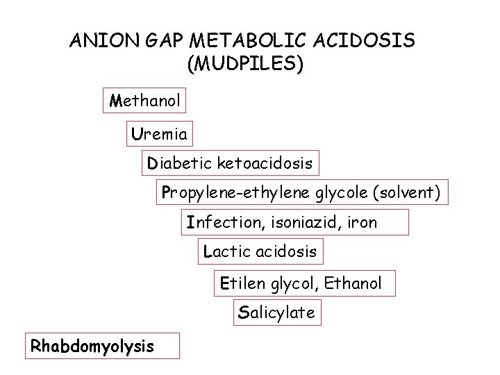
ANION GAP METABOLIC ACIDOSIS (MUDPILES) Methanol Uremia Diabetic ketoacidosis Propylene-ethylene glycole (solvent) Infection, isoniazid, iron Lactic acidosis Etilen glycol, Ethanol Salicylate Rhabdomyolysis
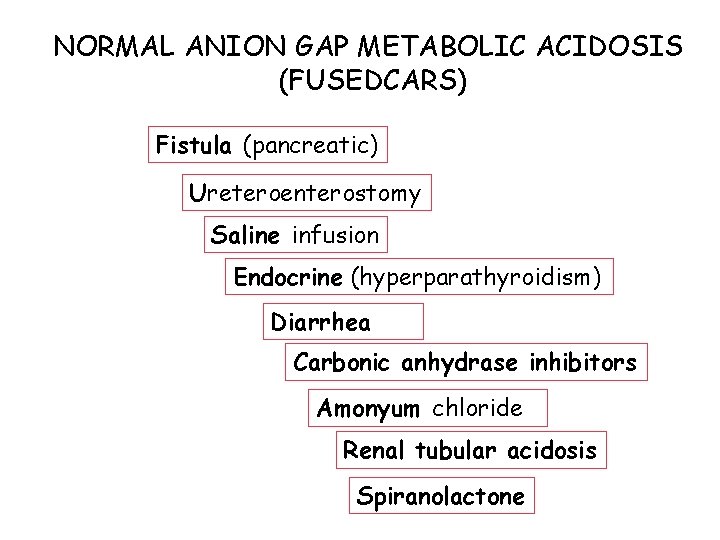
NORMAL ANION GAP METABOLIC ACIDOSIS (FUSEDCARS) Fistula (pancreatic) Ureteroenterostomy Saline infusion Endocrine (hyperparathyroidism) Diarrhea Carbonic anhydrase inhibitors Amonyum chloride Renal tubular acidosis Spiranolactone
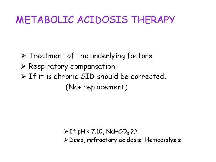
METABOLIC ACIDOSIS THERAPY Ø Treatment of the underlying factors Ø Respiratory compansation Ø If it is chronic SID should be corrected. (Na+ replacement) Ø If p. H < 7. 10, Na. HCO 3 ? ? Ø Deep, refractory acidosis: Hemodialysis
METABOLIC ACIDOSIS TREATMENT Na. HCO 3 disadvantages: • Hypertonisity • Intracellular acidosis (due to increased pa. CO 2).
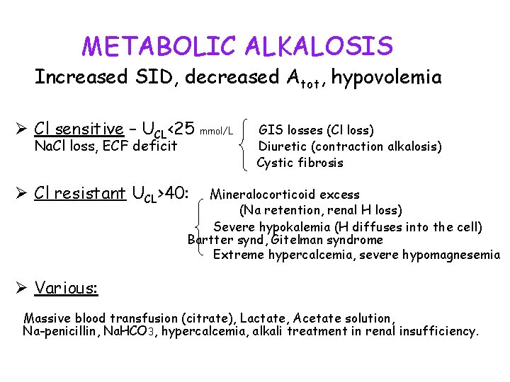
METABOLIC ALKALOSIS Increased SID, decreased Atot, hypovolemia Ø Cl sensitive – UCL<25 mmol/L Na. Cl loss, ECF deficit Ø Cl resistant UCL>40: GIS losses (Cl loss) Diuretic (contraction alkalosis) Cystic fibrosis Mineralocorticoid excess (Na retention, renal H loss) Severe hypokalemia (H diffuses into the cell) Bartter synd, Gitelman syndrome Extreme hypercalcemia, severe hypomagnesemia Ø Various: Massive blood transfusion (citrate), Lactate, Acetate solution, Na-penicillin, Na. HCO 3, hypercalcemia, alkali treatment in renal insufficiency.
METABOLIC ALKALOSIS TREATMENT Ø Cl sensitive: CL replacement Na. Cl (large amount), KCl, HCl Ø Cl resistant: Aldosteron antagonist Acetazolamide (edematous patients): 5 -10 mg/kg oral Ø p. H>7, 60: iv HCL (0, 1 N) or Hemodialysis In Metabolic Alkalosis, SID is increased due to : Cl loss or increased Na.
RESPIRATORY ACIDOSIS q CNS depression CNS diseases, drugs, sleep-apnea, obesity, q Neuromusculary disorders M. gravis, G. barre, myopathies, hipopotasemia q LUNG disease/Pleural abnormalities Asthma, ARDS, COPD, lung edema-fibrosis, pneumonia, pneumothorax q Chest wall abnormalities (cot fracture, flail chest) q Upper airway obstruction (croup, foreign body) q Mechanical hypoventilation q Increased CO 2 production: m. hyperthermia, intensive shivering, large caloric intake, prolonged seizure activity, thyroid storm, extensive thermal injury
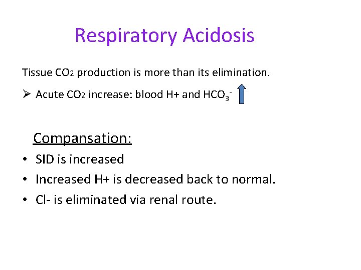
Respiratory Acidosis Tissue CO 2 production is more than its elimination. Ø Acute CO 2 increase: blood H+ and HCO 3 - Compansation: • SID is increased • Increased H+ is decreased back to normal. • Cl- is eliminated via renal route.
Respiratory Acidosis. Treatment Ø Mechanical ventilation Ø Nutritional carbonhydrate content should be decreased Ø Fever control & Shivering control
RESPIRATORY ALKALOSIS Ø CNS stimulation: Pain, anxiety, stroke, ischemia, tumor, infection, fever, drugs (salisilat, progesterone-pregnancy-, teophyillin, chatecolamine, analeptics-doxapram), liver dysfunction. Ø Peripheral stimulation: Ø Ø Hypoxemia High altitude Pulmonary disease (CHF, Noncardiogenic PE, Asthma, P. Embolism) Severe anemia Ø Unknown mechanism: Sepsis, Metabolic encephalopathy Ø Iatrogenic: Ventilator-induced
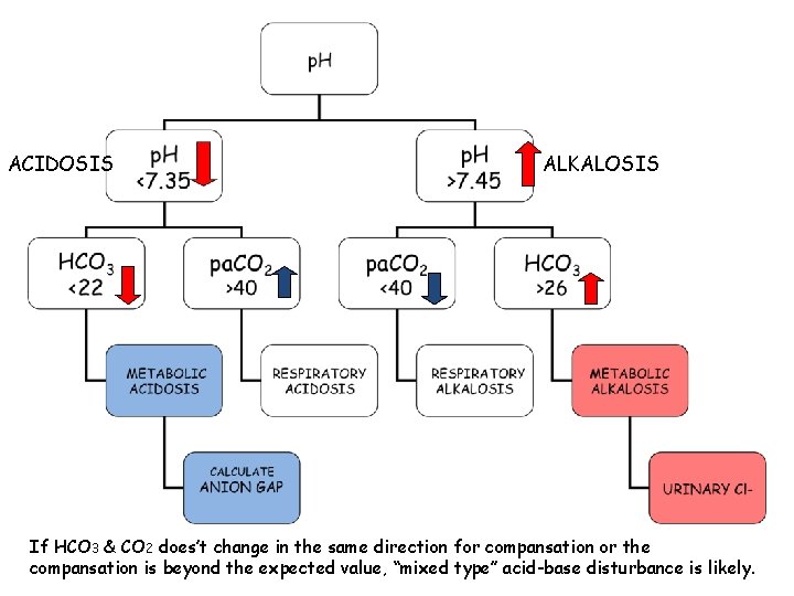
ACIDOSIS ALKALOSIS If HCO 3 & CO 2 does’t change in the same direction for compansation or the compansation is beyond the expected value, “mixed type” acid-base disturbance is likely.
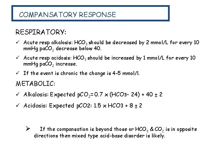
COMPANSATORY RESPONSE RESPIRATORY: ü Acute resp alkalosis: HCO 3 should be decreased by 2 mmol/L for every 10 mm. Hg pa. CO 2 decrease below 40. ü Acute resp acidosis: HCO 3 should be increased by 1 mmol/L for every 10 mm. Hg pa. CO 2 increase. ü If the event is chronic the change is 4 -5 mmol/l. METABOLIC: ü Alkalosis: Expected p. CO 2= 0. 7 x (HCO 3 - 24) + 40 ± 2 ü Acidosis: Expected p. CO 2: 1. 5 x HCO 3 + 8 ± 2 Ø If the compansation is beyond those or HCO 3 & CO 2 is in opposite directions then mixed type acid-base disorder is likely.
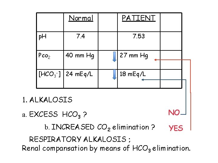
Normal PATIENT 7. 4 7. 53 p. H Pco 2 40 mm Hg [HCO 3 -] 24 m. Eq/L 27 mm Hg 18 m. Eq/L 1. ALKALOSIS a. EXCESS HCO 3 ? b. INCREASED CO 2 elimination ? NO YES RESPIRATORY ALKALOSIS ; Renal compansation by means of HCO 3 elimination.
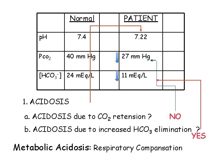
p. H Pco 2 Normal PATIENT 7. 4 7. 22 40 mm Hg [HCO 3 -] 24 m. Eq/L 27 mm Hg 11 m. Eq/L 1. ACIDOSIS a. ACIDOSIS due to CO 2 retension ? NO b. ACIDOSIS due to increased HCO 3 elimination ? YES Metabolic Acidosis: Respiratory Compansation
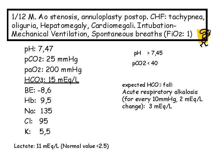
1/12 M. Ao stenosis, annuloplasty postop. CHF: tachypnea, oliguria, Hepatomegaly, Cardiomegali. Intubation. Mechanical Ventilation, Spontaneous breaths (Fi. O 2: 1) p. H: 7, 47 p. CO 2: 25 mm. Hg pa. O 2: 200 mm. Hg HCO 3: 15 m. Eq/L BE: -8, 6 Hb: 9, 5 Na: 135 Cl: 95 K: 5, 5 Lactate: 11 m. Eq/L (Normal value <2. 5) p. H > 7, 45 p. CO 2 < 40 expected HCO 3 fall: Acute respiratory alkalosis (for every 10 mm. Hg, 2 m. Eq/L change): 3 m. Eq/L
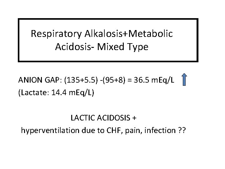
Respiratory Alkalosis+Metabolic Acidosis- Mixed Type ANION GAP: (135+5. 5) -(95+8) = 36. 5 m. Eq/L (Lactate: 14. 4 m. Eq/L) LACTIC ACIDOSIS + hyperventilation due to CHF, pain, infection ? ?
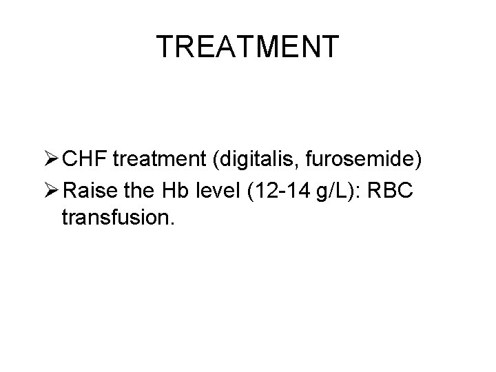
TREATMENT Ø CHF treatment (digitalis, furosemide) Ø Raise the Hb level (12 -14 g/L): RBC transfusion.
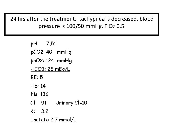
24 hrs after the treatment, tachypnea is decreased, blood pressure is 100/50 mm. Hg, Fi. O 2 0. 5. p. H: 7, 51 p. CO 2: 40 mm. Hg pa. O 2: 124 mm. Hg HCO 3: 28 m. Eq/L BE: 5 Hb: 14 Na: 136 Cl: 91 K: Urinary Cl=10 3. 2 Lactate 2. 7 mmol/L
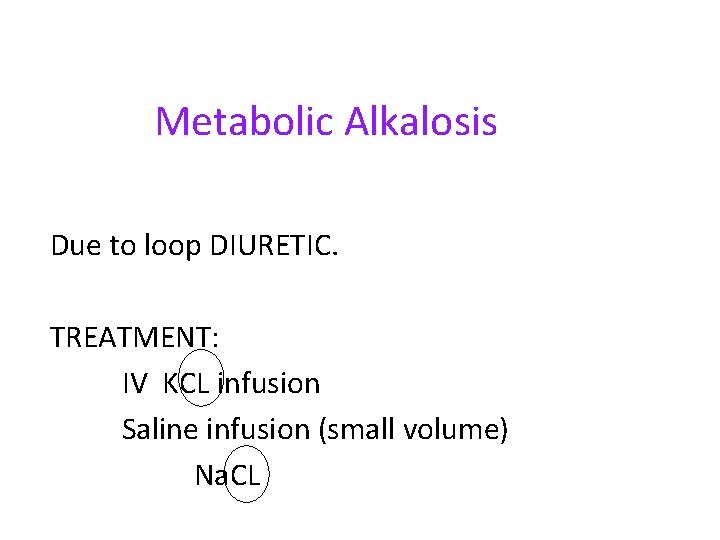
Metabolic Alkalosis Due to loop DIURETIC. TREATMENT: IV KCL infusion Saline infusion (small volume) Na. CL
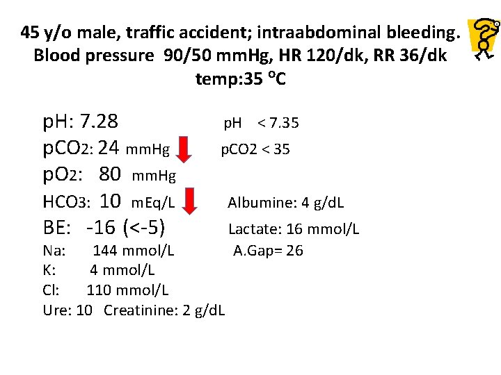
45 y/o male, traffic accident; intraabdominal bleeding. Blood pressure 90/50 mm. Hg, HR 120/dk, RR 36/dk temp: 35 o. C p. H: 7. 28 p. CO 2: 24 mm. Hg p. O 2: 80 mm. Hg HCO 3: 10 m. Eq/L BE: -16 (<-5) p. H < 7. 35 p. CO 2 < 35 Na: 144 mmol/L K: 4 mmol/L Cl: 110 mmol/L Ure: 10 Creatinine: 2 g/d. L Albumine: 4 g/d. L Lactate: 16 mmol/L A. Gap= 26
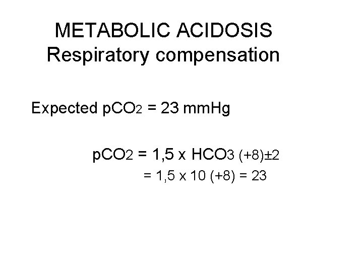
METABOLIC ACIDOSIS Respiratory compensation Expected p. CO 2 = 23 mm. Hg p. CO 2 = 1, 5 x HCO 3 (+8)± 2 = 1, 5 x 10 (+8) = 23
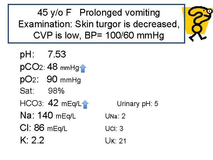
45 y/o F Prolonged vomiting Examination: Skin turgor is decreased, CVP is low, BP= 100/60 mm. Hg p. H: 7. 53 p. CO 2: 48 mm. Hg p. O 2: 90 mm. Hg Sat: 98% HCO 3: 42 m. Eq/L Na: 140 m. Eq/L Cl: 86 m. Eq/L K: 2. 2 Urinary p. H: 5 UNa: 2 UCl: 3 UK: 21
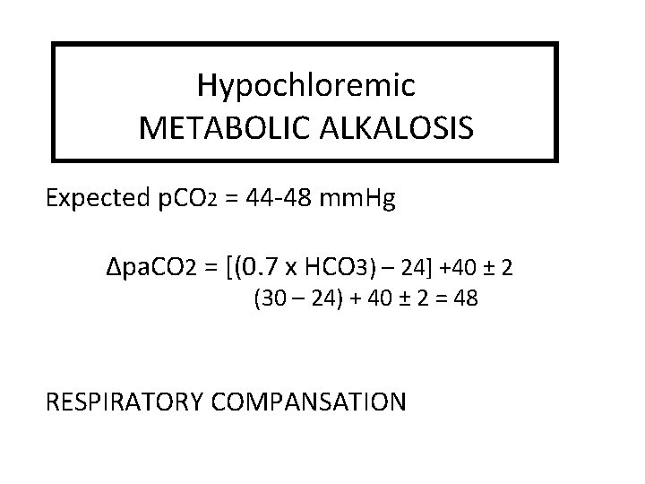
Hypochloremic METABOLIC ALKALOSIS Expected p. CO 2 = 44 -48 mm. Hg Δpa. CO 2 = [(0. 7 x HCO 3) – 24] +40 ± 2 (30 – 24) + 40 ± 2 = 48 RESPIRATORY COMPANSATION

4 y/o, F Severe diarrhea Exam: Skin turgor is decreased, BP is low, HR: 135, CVP=0 p. H: 7. 02 p. CO 2: 13 mm. Hg HCO 3: 3 m. Eq/L p. O 2: 70 mm. Hg Sat: 96% Na: 130 m. Eq/L Cl: 106 m. Eq/L K: 3
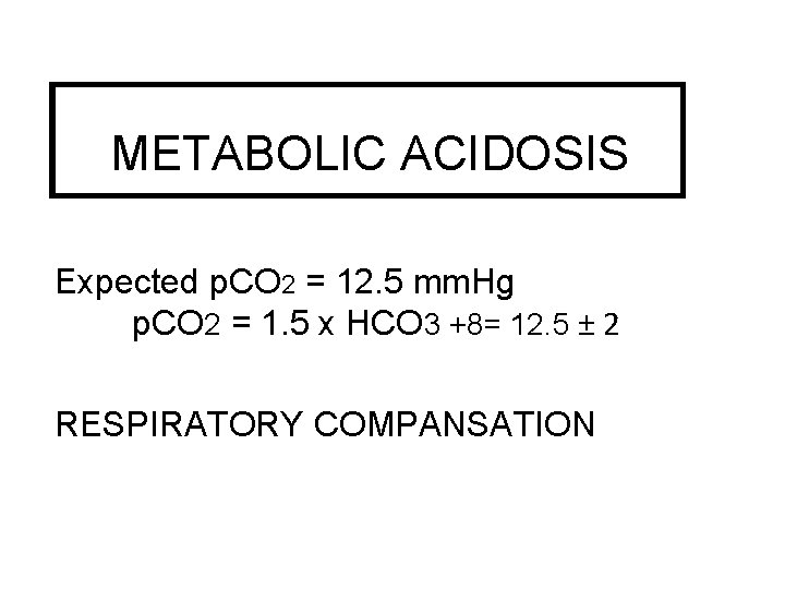
METABOLIC ACIDOSIS Expected p. CO 2 = 12. 5 mm. Hg p. CO 2 = 1. 5 x HCO 3 +8= 12. 5 ± 2 RESPIRATORY COMPANSATION

42 y/o male, IDDM, PONV for 2 days, stops insulin due to the insufficient oral intake. p. H: 7, 21 ACIDOSIS p. CO 2 25 METABOLIC HCO 3 10 ANION GAP ? = 35 Na 133 m. Eq/l Cl 88 m. Eq/l K 5 m. Eq/l DIABETIC KETOACIDOSIS Respiratory alkalosis (Compansation) Expected p. CO 2: 1. 5 x HCO 3+8 ± 2 = 21 -25 mm. Hg

70 y/o male, IDDM, chronic renal disease (creat 2. 0); ischemic heart disease, EF %25 -30; 8 kg weight gain within the last week, pretibial edema, crepitation with lung oscultation; mask O 2 -0. 6 Fi. O 2. BP: 130/65; HR: 90/min. p. H : 7. 17 p. CO 2 : 60 mm. Hg HCO 3: 18 m. Eq/L BE : -7. 5 pa. O 2 : 83 mm. Hg Sat : 94% Na : 148 m. Eq/L Cl : 110 m. Eq/L K: 4. 7 Hb: 10 g/d. L Blood glucose: 180 g/d. L p. H < 7, 40 p. CO 2 > 40
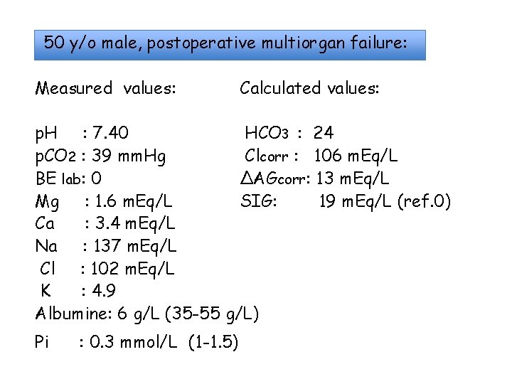
50 y/o male, postoperative multiorgan failure: Measured values: Calculated values: p. H : 7. 40 HCO 3 : 24 p. CO 2 : 39 mm. Hg Clcorr : 106 m. Eq/L BE lab: 0 ΔAGcorr: 13 m. Eq/L Mg : 1. 6 m. Eq/L SIG: 19 m. Eq/L (ref. 0) Ca : 3. 4 m. Eq/L Na : 137 m. Eq/L Cl : 102 m. Eq/L K : 4. 9 Albumine: 6 g/L (35 -55 g/L) Pi : 0. 3 mmol/L (1 -1. 5)
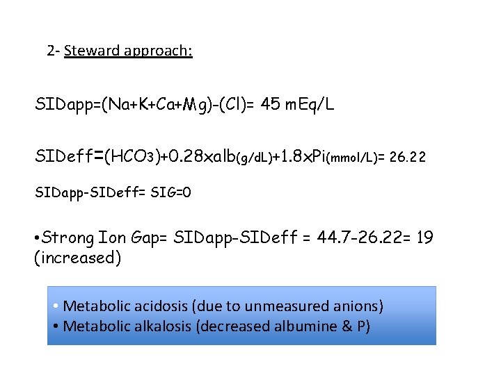
2 - Steward approach: SIDapp=(Na+K+Ca+Mg)-(Cl)= 45 m. Eq/L SIDeff=(HCO 3)+0. 28 xalb(g/d. L)+1. 8 x. Pi(mmol/L)= 26. 22 SIDapp-SIDeff= SIG=0 • Strong Ion Gap= SIDapp-SIDeff = 44. 7 -26. 22= 19 (increased) • Metabolic acidosis (due to unmeasured anions) • Metabolic alkalosis (decreased albumine & P)
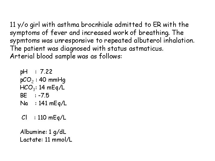
11 y/o girl with asthma brocnhiale admitted to ER with the symptoms of fever and increased work of breathing. The sypmtoms was unresponsive to repeated albuterol inhalation. The patient was diagnosed with status astmaticus. Arterial blood sample was as follows: p. H : 7. 22 p. CO 2 : 40 mm. Hg HCO 3: 14 m. Eq/L BE : -7. 5 Na : 141 m. Eq/L Cl : 110 m. Eq/L Albumine: 1 g/d. L Lactate: 11 mmol/L
- Slides: 52