ACID BASE BALANCE OR Homeostasis of Blood p

ACID BASE BALANCE OR Homeostasis of Blood p. H OR Regulation of Blood p. H Dr Anissa Atif Mirza

Synopsis Introduction Sources of Acids and Bases in body What is Acid Base Balance? Mechanisms Regulating Blood p. H. Significance of Maintaining Acid Base Balance • Acid Base Imbalance and their conditions. • Diagnostic Tests • • •

Introduction • Acid Base Balance is a physiological and biochemical mechanism associated to body/blood p. H.

What Is p. H? • p. H is a Hydrogen ion concentration. • p. H = - log [H+] • Different compartment of human body has specific p. H. • p. H has role in Enzyme activity.

Why blood p. H is Altered?

• Addition of various acids or alkalies by metabolic activities alters body/blood p. H.

Sources and Types of Acids and Alkalies Added During Metabolic Life Processes

+ H • Acids are donors. + • Bases are H acceptors, or give up OH in solution. 8
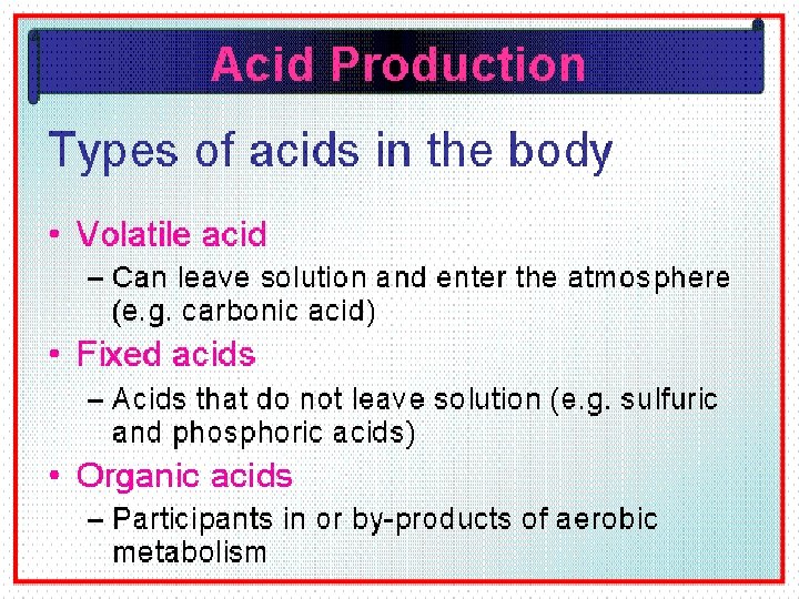

Acids and Bases can be strong or weak: • A strong acid or base is one that dissociates completely in a solution - HCl, Na. OH, and H 2 SO 4 • A weak acid or base is one that dissociates partially in a solution -H 2 CO 3, C 3 H 6 O 3, and CH 2 O, Lactate.
 – Phosphoric")
• Acidic Substances of body: – Carbonic acid(H 2 CO 3) – Phosphoric acid( H 3 PO 4) – Sulphuric acid (H 2 SO 4) • Organic Acids: – Lactate, Acetoactate, Pyruvate • Alkaline Substances of body: – Citrate – Bicarbonates.

What is Acid Base Balance?

Homeostatic Mechanisms That Regulate Blood/Body p. H

• Acid Base balance is a homeostatic mechanism • Carried out to regulate the altered p. H of blood and other body compartments to its normal constant range.

• Maintenance of Acid Base balance • Is a prime requisite to maintain normal healthy and active life.

Acid-Base Balance • It is the regulation of HYDROGEN ions. (The more Hydrogen ions, the more acidic the solution and the LOWER the p. H) – The acidity or alkalinity of a solution is measured as p. H

Acid Base Balance Regulates p. H Why it is Very Essential To Regulate p. H?

• p. H of blood and other body compartments are precisely regulated. • p. H is always tried to be maintained to its normal constant range.

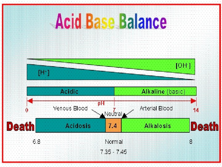
• Acid Base Balance maintains the blood p. H at normal constant narrow range of 7. 35 -7. 45.

• p. H of the medium directly affects the enzyme activities • Optimum p. H is an essential requisite for enzyme activities and normal metabolism.

• It is prerequisite for regulating blood/body p. H: • To maintain normal/optimal Enzyme activities • Normal metabolism • Normal Coordination • Normal Health

Factors Regulating Acid Base Balance 22

Acid Base Balance is Regulated By • First Line of Defense v. Blood Buffer System • Second Line of Defense –Respiratory Mechanism • Third Line of Defense ØRenal Mechanism
 Chemical Buffers • React very rapidly (less than a second) 2) Respiratory Regulation")
1) Chemical Buffers • React very rapidly (less than a second) 2) Respiratory Regulation • Reacts rapidly (seconds to minutes) 3) Renal Regulation • Reacts slowly (minutes to hours) 24

Role of Blood Buffer System • First line of defense in mechanism of Acid Base Balance. + • Acids (H ) added are neutralized by the salt part of buffer.

Extracellular Buffers • Bicarbonate Buffer –Na. HCO 3/H 2 CO 3 (20: 1 at 7. 4 p. H) • Phosphate Buffer –Na 2 HPO 4/Na. H 2 PO 4 (4: 1 at 7. 4 p. H) • Protein Buffer –Na-Protein/H-Protein

Intracellular Buffers • Bicarbonate Buffer –KHCO 3/H 2 CO 3 • Phosphate Buffer –K 2 HPO 4/KH 2 PO 4 • Protein Buffer –K-Hb/H-Protein

Mechanism Action of Buffer Systems • Buffers mixture of weak acids and its salts • Resist change in p. H of blood when small amount of acids or alkalis added to the medium.

• Buffers act quickly but not permanently

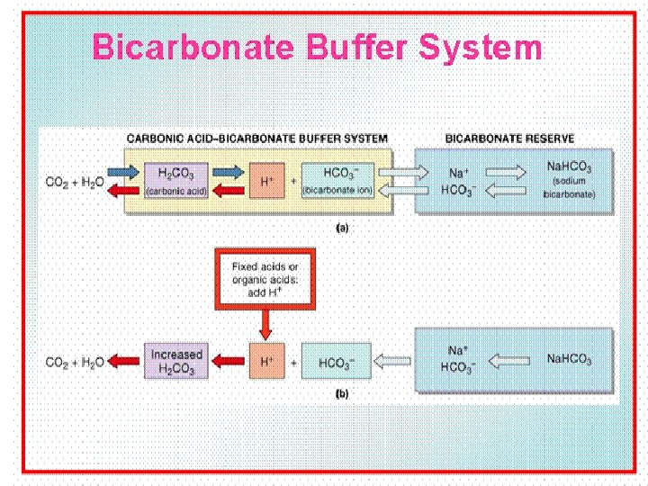
Bicarbonate Buffer System Respiratory Buffer System • Acid - Base balance is primarily concerned with Bicarbonate Buffer mechanism : – H 2 CO 3/ Hydrogen (H+) – Bicarbonate (HCO 3 - ) (Alkali Reserve) + H 30 HCO 3 -

Bicarbonate Buffer • Bicarbonate Buffer- Chief Buffer system of Blood. • Na. HCO 3 the salt part of buffer neutralizes the strong and non volatile acids added to blood. • It constitutes Alkali reserve(HCO 3 -)
 and carbonic acid (H 2 CO")
Bicarbonate Buffer • Sodium Bicarbonate (Na. HCO 3) and carbonic acid (H 2 CO 3) • Maintain a 20: 1 ratio : HCO 3 - : H 2 CO 3 HCl + Na. HCO 3 ↔ H 2 CO 3 + Na. Cl Na. OH + H 2 CO 3 ↔ Na. HCO 3 + H 2 O 33
 converts strong dissociable acid into weak")
• Action of Bicarbonate (Na. HCO 3) converts strong dissociable acid into weak non dissociable acid (H 2 CO 3) and a neutral salt without altering the p. H.

• Weak acid H 2 CO 3 formed during buffering action of Bicarbonate buffer is then expired out by Lungs. • Thus Bicarbonate buffer is connected to the respiratory system • Bicarbonate buffer is also termed as Respiratory buffer.

• Alkali reserve is represented by the concentration of Na. HCO 3 in the blood. • Alkali reserve concentration(HCO 3 -) determines the strength of buffering action towards added H+ ions by acids. • More the concentration of Alkali reserve , more is the buffering action and vice a versa.

• The blood buffers are effective as long as • The acid load added is not very high and • The alkali reserve (HCO 3 -) is not exhausted.

Phosphate Buffer/Urine Buffer Na 2 HPO 4/Na. H 2 PO 4 (4: 1 at 7. 4 p. H) • H+ + HPO 42 - ↔ H 2 PO 4 • OH- + H 2 PO 4 - ↔ H 2 O + H 2 PO 42 - 38

Phosphate Buffer Mechanism • When H+ ions added they are neutralized/fixed by Na 2 HPO 4 (Alkaline Phosphate) and converted to Na. H 2 PO 4 (Acid Phosphates). • These acid phosphates then excreted out through kidneys as acidic urine.

• Thus Phosphate Buffer is connected to Excretory system. • Phosphate Buffer also termed as Urine Buffer.

• When an alkali enters it is buffered by the acid phosphate Na. H 2 PO 4 which converted to Na 2 HPO 4 alkaline phosphate. • Excreted in urine making it alkaline urine.

Protein Buffers • Includes hemoglobin, work in blood. • Carboxyl group gives up H+ • Amino Group accepts H+ • The Imidazole group of Histidine present in Hb structure has buffering capacity. 42

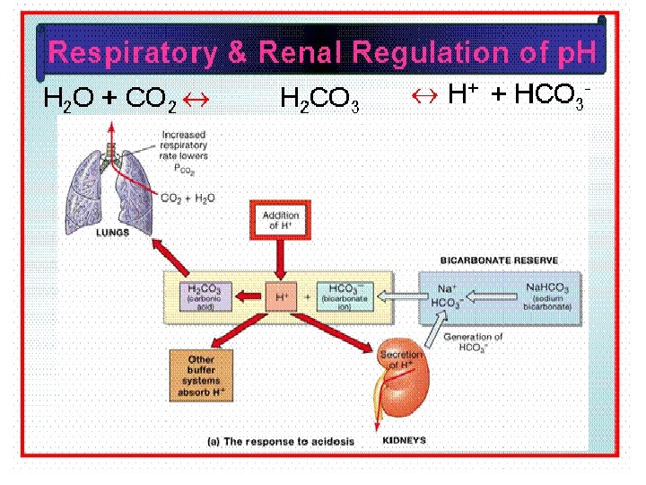
Role of Respiratory Mechanisms • Respiratory system plays second line of defense mechanism of Acid Base Balance. • Role of respiration in acid base balance is short term regulatory process.

• H 2 CO 3 formed from Bicarbonate Buffer, is exhaled out through respiratory system. • Increased H 2 CO 3 stimulates the respiratory centre in Medulla Oblongata. • This in turn stimulates hyperventilation which promptly removes H 2 CO 3 from blood by expiration.

• Exhalation of H 2 CO 3 is as carbon dioxide by activity of enzyme Carbonic Anhydrase of Lungs. • H+ + HCO 3 - ↔ H 2 CO 3 ↔ CO 2 + H 20

• Respiratory mechanism is powerful, but only works with volatile acids. • Doesn’t affect fixed acids like lactic acid.

• Blood p. H can be adjusted through respiratory mechanism • By changing rate and depth of breathing.

• Low H 2 CO 3 concentration in blood depresses respiratory centre , causes hypoventilation i. e slow and shallow respiration. • This retains H 2 CO 3 in blood.

• If Nervous centre / Respiratory system fails. • Acid Base Balance fails.

Generation of bicarbonate by RBC LACK OF AEROBIC ACTIVITY, DIFFUSION OF CARBONDIOXIDE, H+ BUFFERED BY HHb. 50

Events in lungs and tissue lung tissue HCO 3 - HCO 3 HHb O 2 H+ H 2 CO 3 H 2 O H+ Hb. O 2 CO 2 EXPIRED AIR H 2 CO 3 CO 2 H 2 O Isohydric transport of co 2 METABOLISM 51

Role of Renal Mechanism • Renal mechanism is the third line of defense mechanism. • Role of renal mechanism is long term regulatory process. 52

• The acid and alkaline phosphates formed during phosphate buffering mechanism are filtered from blood and excreted out through urine. • Thus the phosphate buffer system is directly connected to renal mechanism.
. • Renal")
• Renal mechanism conserve and produce Bicarbonate ions ( Alkali reserve). • Renal Mechanism is the most effective regulator of blood p. H. • If kidneys fail, p. H balance fails.

• Renal System maintains Acid Base Balance through: – Reabsorption of Bicarbonate (HCO 3 -) ions. – Excretion of H+ ions – Excretion of titrable acids(Acid Phosphates) – Excretion of Ammonium ions (Glutaminase activity)

REABSORPTION OF BICARBONATE ~Conservation of Bicarbonate ~Urine is free of HCO 3~Simultaneous excretion of H+ 56

EXCRETION OF TITRABLE ACIDS ~measure of acid excreated by kidney ~no. of millilitres of N/10 Na. OH required to titrate 1 litre of urine to p. H 7. 4 ~role of phosphate buffer 57

Excretion Of H+ ions ~Elimination of nonvolatile acid ~Excretion of H+ ~Occurs in PCT ~Regeneration of bicarbonate ~H+ combine with non carbonate base and excreated 58

EXCRETION OF AMMONIUM ION NH 3 is obtained from Deamination of Glutamine NH 4+ cant diffuse back 2/3 of body acid load liberated in the form of NH 4+ 59

Rates of correction • Buffers function almost instantaneously • Respiratory mechanisms take several minutes to hours • Renal mechanisms may take several hours to days 60

62

63

MECHANISM FOR REGULATION OF ACID BASE BALANCE • Buffer system: temporary solution • Respiratory mechanism provide short time regulation • Renal mechanism : permanent solution • Urine p. H < plasma p. H , 4. 5 -9. 5 • Eliminate nonvolatile acid, buffered by cation (principally Na+) • Maintain alkali reserve

Acid Base Imbalance OR Conditions Of Acid Base Disturbances

The Body and p. H • Homeostasis of blood p. H is tightly controlled by mechanisms of Acid Base Balance. • Extracellular fluid = 7. 4 • Blood p. H regulated to = 7. 35 – 7. 45 66

Occurrence of Acid Base Imbalance • When Factors involved in homeostatic mechanisms to regulate Acid Base Balance fails to work efficiently. • Does not maintain the altered p. H of blood to normal constant range. • Results into Acid Base Imbalance.

ACIDOSIS / ALKALOSIS • Two major disturbances in Acid-Base balance –Acidosis –Alkalosis
")
Conditions Of Acid Base Imbalance • Acidosis /Acidemia ( Decreased p. H/Increased H+ ions) • Alkalosis/Alkalemia (Increased p. H/Decreased H+ ions)
 below 7. 35 • Alkalosis (Alkalemia) above 7. 45 •")
• Acidosis (Acidemia) below 7. 35 • Alkalosis (Alkalemia) above 7. 45 • Blood p. H < 6. 8 or > 8. 0 death occurs

ACIDOSIS / ALKALOSIS • Acidosis – A condition in which the blood has too much acid (or too little base), frequently resulting in a decrease in blood p. H. • Alkalosis – A condition in which the blood has too much base (or too little acid), occasionally resulting in an increase in blood p. H. 71

72

74

Effect of Altered p. H • Altered p. H may seriously disturbs the vital processes. • Might lead to fatality.

• Most enzymes function only with narrow p. H ranges. • Extremes of p. H affects the enzymatic action by protonation or deprotonation at the active sites of Enzymes. • Makes Enzymes inactive.

• Inactivated Enzymes affect metabolic reactions and metabolic pathways. • Metabolism gets deranged. • Leads to metabolic syndromes.

p. H also affect excitability of Nerve and Muscle cells 78 p. H Excitability

ACID-BASE REGULATION • Enzymes, Hormones and ion distribution are all affected by Hydrogen ion concentrations 79

ACIDOSIS / ALKALOSIS • p. H changes have dramatic effects on normal cell function 1) Changes in excitability of nerve and muscle cells 2) Influences Enzyme activity 3) Influences K+ levels/Retention of K+ 80
 depresses the central nervous")
CHANGES IN CELL EXCITABILITY • p. H decrease (more acidic) depresses the central nervous system – Can lead to loss of consciousness • p. H increase (more basic)causes over excitability of nervous system. – Tingling sensations, nervousness, muscle twitches

INFLUENCES ON ENZYME ACTIVITY • p. H increases or decreases can alter the shape of the enzyme rendering it non-functional • Changes in enzyme structure can result in accelerated or depressed metabolic actions within the cell 82
 than H+ is")
INFLUENCES ON K+ LEVELS • If H+ concentrations are high (acidosis) than H+ is secreted in greater amounts • This leaves less K+ than usual excreted. • The resultant K+ retention can affect cardiac function and other systems K+ H+ 83 Na+

Small changes in p. H can produce major disturbances • Acid-base balance can also + + affect Electrolytes (Na , K , Cl ) • Can also affect Hormones 84
 • Bicarbonate")
ACID-BASE IMBALANCE • Derangements of • Hydrogen/Carbonic acid (H+/H 2 CO 3) • Bicarbonate (HCO 3 -) concentrations In body fluids are common in conditions of Acid Base Imbalance

Acid-Base Imbalances • p. H< 7. 35 Acidosis • p. H > 7. 45 Alkalosis

4 Types of Primary Acid-Base Disorders Acid Base Imbalances Biochemical Change Respiratory Acidosis Increased H 2 CO 3 Respiratory Alkalosis Decreased H 2 CO 3 Metabolic Acidosis Metabolic Alkalosis 87
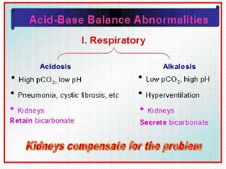

RESPIRATORY ACIDOSIS 90

Respiratory Acidosis Primary Carbonic acid excess Increased H 2 CO 3/Increased p. CO 2 Defect in respiratory centre of brain Defect in respiratory organ system Decreased elimination of H 2 CO 3 by the lungs. • Hypoventilation • • •

• Increased blood levels of CO 2 above 45 mm Hg. • Hypercapnia – high levels of p. CO 2 in blood 92

RESPIRATORY ACIDOSIS • Respiratory acidosis develops when the lungs don't expel CO 2 adequately. • This can happen in diseases that severely affect the lungs. 93

• Chronic conditions: – Depression of respiratory center in brain that controls breathing rate – drugs or head trauma – Paralysis of respiratory or chest muscles – Emphysema – Asthma – Pneumonia – Pulmonary edema – Obstruction of respiratory tract – Congestive Cardiac Failure

HYPOVENTILATION Causes Respiratory Acidosis • Hypo = “Under” Elimination of CO 2 + H p. H

RESPIRATORY ACIDOSIS CO 2 - HCO 3 CO 2 H 2 CO 3 2 : 20 -breathing is suppressed holding CO 2 in body -p. H = 7. 1 96 CO 2
 Obstruction of air passages – Vomit, Anaphylaxis, Tracheal Cancer 97")
RESPIRATORY ACIDOSIS • 1) Obstruction of air passages – Vomit, Anaphylaxis, Tracheal Cancer 97
 Decreased Respiration – Shallow, slow breathing – Depression of the")
RESPIRATORY ACIDOSIS • 2) Decreased Respiration – Shallow, slow breathing – Depression of the respiratory centers in the brain which control breathing rates • Drug overdose 98
 Collapse of lung – Compression injury, open thoracic wound Left")
RESPIRATORY ACIDOSIS • 4) Collapse of lung – Compression injury, open thoracic wound Left lung collapsed 99

Respiratory Acidosis • Acute conditions: –Adult Respiratory Distress Syndrome –Pulmonary edema –Pneumothorax 100

Compensation for Respiratory Acidosis • Kidneys eliminate hydrogen ion and retain bicarbonate ions. 101

Signs and Symptoms of Respiratory Acidosis Breathlessness Restlessness Lethargy and disorientation Tremors, convulsions, coma Respiratory rate rapid, then gradually depressed • Skin warm and flushed due to vasodilation caused by excess CO 2 • • • 102

Treatment of Respiratory Acidosis • Restore ventilation • IV lactate solution • Treat underlying dysfunction or disease 103

RESPIRATORY ALKALOSIS 104

Respiratory Alkalosis • • • Primary Carbonic acid deficit Decreased H 2 CO 3 p. CO 2 less than 35 mm Hg (hypocapnea) Most common acid-base imbalance Primary cause is hyperventilation Washes out excessive quantity of H 2 CO 3 through expiration process of lungs. 105

• Stimulation of respiratory centre in brain • Hyperventilation

Respiratory Alkalosis • Conditions that stimulate respiratory center: – Oxygen deficiency at high altitudes – Pulmonary disease and Congestive heart failure – caused by hypoxia – Respiratory center lesions – Acute anxiety – Fever, anemia – Early salicylate intoxication – Cirrhosis – Gram-negative sepsis/Meningitis 107

RESPIRATORY ALKALOSIS • Anxiety is an emotional disturbance • The most common cause of hyperventilation, and thus respiratory alkalosis, is noted in anxiety 108

RESPIRATORY ALKALOSIS • Respiratory center lesions – Damage to brain centers responsible for monitoring breathing rates • Tumors • Strokes 109

RESPIRATORY ALKALOSIS • High Altitude – Low concentrations of O 2 in the arterial blood reflexly stimulates ventilation in an attempt to obtain more O 2 – Too much CO 2 is “blown off” in the process 110

RESPIRATORY ALKALOSIS • Fever – Rapid shallow breathing blows off too much CO 2 111
 – Ventilation is stimulated without regard to")
RESPIRATORY ALKALOSIS • Salicylate poisoning (Aspirin overdose) – Ventilation is stimulated without regard to the status of O 2, CO 2 or H+ in the body fluids 112

RESPIRATORY ALKALOSIS • Kidneys compensate by: – Retaining hydrogen ions – Increasing bicarbonate excretion HCO 3 H+ HCO 3 - HCO 3 H+ H+ HCO 3 H+ HCO 3+ H + HCO 3 - H H+ 113 H+

HYPERVENTILATION Causes Respiratory Alkalosis • Hyper = “Over” Elimination of CO 2 + H 114 p. H

Compensation of Respiratory Alkalosis • If kidneys are functioning normal • The conditions of respiratory acidosis or alkalosis are compensated. • Kidneys conserve hydrogen ion • Excrete bicarbonate ion 115

Treatment of Respiratory Alkalosis • Treat underlying cause • Breathe into a paper bag • IV Chloride containing solution Cl ions replace lost bicarbonate ions 116

METABOLIC ACIDOSIS 117

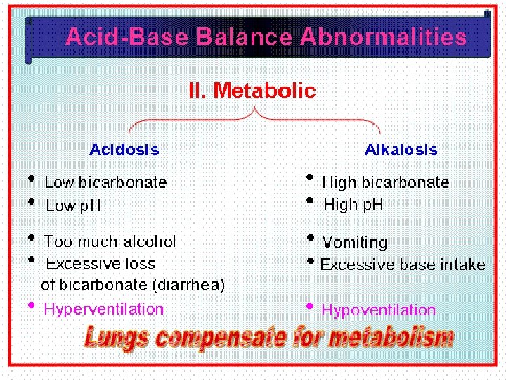
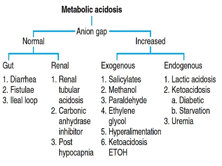
Metabolic Acidosis • Primary Alkali deficit • Bicarbonate deficit - blood concentrations of bicarbonate drop below 22 m. Eq/L • Causes: – Loss of bicarbonate through diarrhea or renal dysfunction. – Overproduction of acids (lactic acid or ketones) – Failure of kidneys to excrete H+ 118

METABOLIC ACIDOSIS • Occurs when there is a decrease in the normal 20: 1 ratio – Decrease in blood p. H and bicarbonate level • Excessive H+ or decreased HCO 3 - - O 3 3 HCHCO H 2 CO 3 119 1 : 10 20 == 7. 4

METABOLIC ACIDOSIS • Any acid-base imbalance not attributable to CO 2 is classified as metabolic – Metabolic production of Acids – Or loss of Bases 120

METABOLIC ACIDOSIS • The causes of metabolic acidosis can be grouped into five major categories – 1) Ingesting an acid or a substance that is metabolized to acid – 2) Abnormal Metabolism – 3) Kidney Insufficiencies – 4) Strenuous Exercise – 5) Severe Diarrhea 121
 Ingesting An Acid – Most substances that cause acidosis when")
METABOLIC ACIDOSIS • 1) Ingesting An Acid – Most substances that cause acidosis when ingested are considered poisonous – Examples include wood alcohol (methanol) and antifreeze (ethylene glycol) – However, even an overdose of aspirin (acetylsalicylic acid) can cause metabolic acidosis 122
 Abnormal Metabolism – The body can produce excess acid as")
METABOLIC ACIDOSIS • 2) Abnormal Metabolism – The body can produce excess acid as a result of several diseases • Ketoacidosis • Type I Diabetes Mellitus • Uncontrolled Diabetes mellitus • Prolonged Starvation • Lacticacidosis • Shock • Haemorrhage • Violent Exercise-

METABOLIC ACIDOSIS • Unregulated diabetes mellitus causes ketoacidosis – Body metabolizes fat rather than glucose – Accumulations of metabolic acids (Keto Acids) cause an increase in plasma H+ 124
 Kidney Insufficiencies – This type of kidney malfunction is called")
METABOLIC ACIDOSIS • 3) Kidney Insufficiencies – This type of kidney malfunction is called renal tubular acidosis or uremic acidosis and may occur in people with kidney failure or with abnormalities that affect the kidneys' ability to excrete acid
 Kidney Insufficiencies – Kidneys may be unable to rid the")
METABOLIC ACIDOSIS • 3) Kidney Insufficiencies – Kidneys may be unable to rid the plasma of even the normal amounts of H+ generated from metabolic acids – Kidneys may be also unable to conserve an adequate amount of HCO 3 - to buffer the normal acid load 126
 Strenuous Exercise – Muscles resort to anaerobic glycolysis during strenuous")
METABOLIC ACIDOSIS • 4) Strenuous Exercise – Muscles resort to anaerobic glycolysis during strenuous exercise – Anaerobic respiration leads to the production of large amounts of lactic acid C 6 H 12 O 6 127 Enzymes 2 C 3 H 6 O 3 + ATP (energy) Lactic Acid
 Severe Diarrhea – Fluids rich in HCO 3 - are")
METABOLIC ACIDOSIS • 5) Severe Diarrhea – Fluids rich in HCO 3 - are released and reabsorbed during the digestive process – During diarrhea this HCO 3 - is lost from the body rather than reabsorbed
 Severe Diarrhea – The loss of HCO 3 - without")
METABOLIC ACIDOSIS • 5) Severe Diarrhea – The loss of HCO 3 - without a corresponding loss of H+ lowers the p. H – Less HCO 3 - is available for buffering H+ – Prolonged deep (from duodenum) vomiting can result in the same situation

Symptoms of Metabolic Acidosis • Headache, lethargy • Nausea, vomiting, diarrhea • Coma • Death 130

Compensation for Metabolic Acidosis • Increased ventilation. • Renal excretion of hydrogen ions if possible. • K+ exchanges with excess H+ in ECF. • H+ into cells, K+ out of cells. 131

Treatment of Metabolic Acidosis • IV lactate solution 132

METABOLIC ALKALOSIS 133

Metabolic Alkalosis • Bicarbonate Excess - concentration in blood is greater than 26 m. Eq/L • Causes: – Excess vomiting = loss of stomach acid – Excessive use of alkaline drugs – Certain diuretics – Endocrine disorders – Heavy ingestion of antacids – Severe dehydration – Cushings Syndrome – Prolonged exposure to x rays and UV rays 134

METABOLIC ALKALOSIS • Elevation of p. H due to an increased 20: 1 ratio – May be caused by: • An increase of bicarbonate • A decrease in hydrogen ions – Imbalance again cannot be due to CO 2 – Increase in p. H which has a non-respiratory origin 135 7. 4
 Ingestion of Alkaline Substances")
METABOLIC ALKALOSIS • Can be the result of: – 1) Ingestion of Alkaline Substances – 2) Vomiting ( loss of HCl ) 136
 often used as a remedy for")
METABOLIC ALKALOSIS • Baking soda (Na. HCO 3) often used as a remedy for gastric hyperacidity – Na. HCO 3 dissociates to Na+ and HCO 3 - 137

Compensation for Metabolic Alkalosis • Alkalosis most commonly occurs with renal dysfunction, so can’t count on kidneys. • Respiratory compensation difficult – hypoventilation limited by hypoxia. 138

Symptoms of Metabolic Alkalosis • Respiration slow and shallow • Hyperactive reflexes ; tetany • Often related to depletion of electrolytes • Atrial tachycardia • Dysrhythmias 139

Treatment of Metabolic Alkalosis • Electrolytes to replace those lost • IV chloride containing solution • Treat underlying disorder 140

Acidosis • Principal effect of acidosis is depression of the CNS through ↓ in synaptic transmission. • Generalized weakness • Deranged CNS function the greatest threat • Severe acidosis causes – Disorientation – Coma – Death 141

Alkalosis • Alkalosis causes over excitability of the central and peripheral nervous systems. • Numbness • Light headedness • Severe Alkalosis causes : – Nervousness – muscle spasms or Tetany – Convulsions – Loss of consciousness – Death 142

Compensation Of Acid Base Imbalance • The body response to acid-base imbalance is called compensation • May be complete compensation if altered p. H brought back within normal limits • Partial compensation if p. H range is still outside norms. • Uncompensated if p. H range is very out from norms.

• If underlying problem is respiratory, renal mechanisms can bring about metabolic compensation. • If underlying problem is metabolic, hyperventilation or hypoventilation can help : respiratory compensation.

ACIDOSIS decreased removal of CO 2 from lungs accumulation of CO 2 in blood respiratory acidosis 145 failure of kidneys to excrete acids metabolic acid production of keto acids absorption of metabolic acids from GI tract accumulation of acid in blood increase in plasma H+ concentration depression of nervous system prolonged diarrhea excessive loss of Na. HCO 3 from blood metabolic acidosis deep vomiting from GI tract kidney disease (uremia)

ALKALOSIS anxiety overdose of certain drugs hyperventilation loss of CO 2 and H 2 CO 2 from blood high altitudes prolonged vomiting loss of acid decrease in plasma H+ concentration overexcitability of nervous system excess aldosterone accumulation of base metabolic alkalosis respiratory alkalosis 146 ingestion of excessive alkaline drugs
 and alkalosis")
Organ dysfunction And Acid Base Imbalance • CNS – respiratory acidosis (suppression) and alkalosis (stimulation) • Pulmonary – respiratory acidosis (COPD) and • • alkalosis (hypoxia, pulmonary embolism) Cardiac – respiratory alkalosis, respiratory acidosis, metabolic acidosis (pulmonary edema) GIT – metabolic alkalosis (vomiting) and acidosis (diarrhea) Liver – respiratory alkalosis, metabolic acidosis (liver failure) Kidney – metabolic acidosis (RTA) and alkalosis (1 st Aldosterone)

Organ Dysfunction • Endocrine – Diabetes mellitus – metabolic acidosis – Addisons Disease/Adrenal insufficiency – metabolic acidosis. (Decreased H+ ions excretion) – Cushing’s Syndrome – metabolic alkalosis (Increased Cortisol- Increased H+ ions excretion) – Primary aldosteronism – metabolic alkalosis • Drugs/toxins – Toxic alcohols – metabolic acidosis – ASA/Aspirin – metabolic acidosis and respiratory alkalosis( Causes Hyperventilation) – Theophylline overdose – respiratory alkalosis

ACID – BASE DISORDERS Clinical State Acid-Base Disorder Pulmonary Embolus Respiratory Alkalosis Cirrhosis Respiratory Alkalosis Pregnancy Respiratory Alkalosis Diuretic Use Metabolic Alkalosis Vomiting Metabolic Alkalosis Chronic Obstructive Pulmonary Disease Respiratory Acidosis Shock Metabolic Acidosis Severe Diarrhea Metabolic Acidosis Renal Failure Metabolic Acidosis Sepsis (Bloodstream Infection) 149 Respiratory Alkalosis, Metabolic Acidosis

150

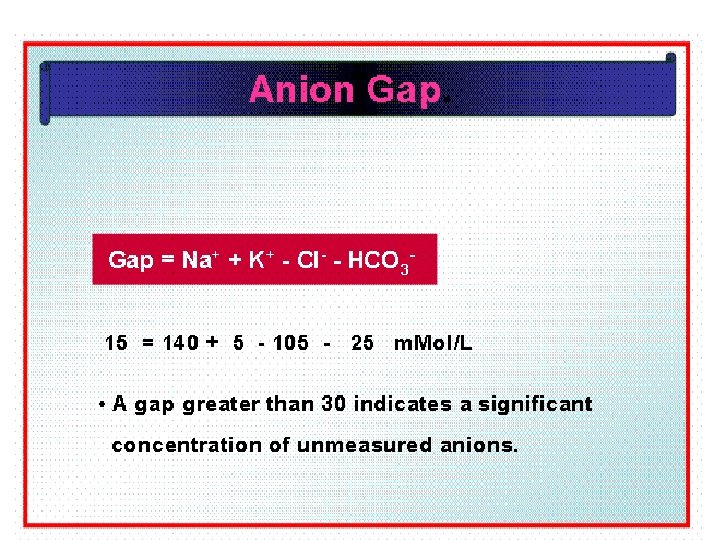
Anion Gap

• Sum of anion and cations is always equal • Sodium and Potassium accounts for 95% of cations • Chloride and bicarbonate accounts for 68% of anions • There is difference between measured anion and cation • The unmeasured anions constitute the ANION GAP.
")
• They are protein anions , sulphates , phosphates and organic acid(Unmeasured Anions) • AG can be calculated as (Na+ + K+)—(HCO 3 - + Cl-) • High anion gap acidosis: renal failure, DM • Normal anion gap acidosis: diarrhea • Hyperchloremic acidosis

Calculation Of Anion Gap • Na ++ K+ = Cl- + HCO 3 - + A • 136+ 4 = 100 + 25 • A- = 15 m. Eq/L

• Normal AG is typically 12 ± 4 m. Eq/L. • If AG is calculated using K+, the normal AG is 16 ± 4 m. Eq/L

Significance of Anion Gap Calculation • Calculation of Anion gap and its values help in diagnosing conditions of Acid Base Balance and Imbalance.

• The anion gap is increased in conditions such as metabolic acidosis: • That result from elevated levels of metabolic acids (metabolic acidosis) –Lactic acidosis –Diabetic Ketoacidosis –Renal Failure

• A low anion gap occurs in conditions that cause a fall in unmeasured anions • (primarily albumin) OR a rise in unmeasured cations

Calculate the Anion Gap • 1. Calculate the anion gap as described. • 2. An anion gap , over 25 suggests a severe metabolic acidosis. • 3. Causes of an high anion gap: ethylene glycol, lactic acid, methanol, paraldehyde, aspirin, renal failure, ketoacidosis (diabetic or ethanol).

Anion Gap Acidosis: • Anion gap >12 mmol/L; caused by a decrease in [HCO 3 -] • Balanced by an increase in an unmeasured acid ion from either endogenous production or exogenous ingestion (normochloremic acidosis).

Metabolic Acidosis and the Anion gap 1. Normal gap 1. Renal “HCO 3” 2. GI “HCO 3” losses Proximal RTA Distal RTA Diarrhea 2. Increased gap 1. Acid prod Lactate DKA Ketosis Toxins Alcohols Salicylates Iron 2. Acid elimination Renal disease
![Henderson Hasselbalch Equation • p. H= pka +log [HCO 3 -]/[H 2 CO 3]](http://slidetodoc.com/presentation_image_h/8d759d485107a2865cc553f3c06df23e/image-164.jpg "Henderson Hasselbalch Equation • p. H= pka +log [HCO 3 -]/[H 2 CO 3]")
Henderson Hasselbalch Equation • p. H= pka +log [HCO 3 -]/[H 2 CO 3] • At p. H 7. 4 the ratio of HCO 3 -/H 2 CO 3 is 1: 20. • A buffer is most effective when p. H=p. Ka • When concentration of salt and acid are equal.

Significance of Henderson Hasselbalch Equation • The equation helps in calculating p. H of Buffers. • The equation helps in assessing status of Acid Base balance.

Stepwise Approaches • History & physical examination • Arterial blood gas for p. H, p. CO 2, (HCO 3) – Use the HCO 3 from ABG to determine compensation • Serum Na, K, Cl, CO 2 content – Use CO 2 content to calculate anion gap • Calculate anion gap – Anion gap = {Na - (Cl + CO 2 content)} • Determine appropriate compensation • Determine the primary cause

DIAGNOSTIC LAB VALUES & INTERPRETATION
Analyzer determines Acid Base Balance and Imbalance.")
• Arterial Blood Gas(ABG )Analyzer determines Acid Base Balance and Imbalance.
 or")
Diagnosis of Acid-Base Imbalances 1. Note whether the p. H is low (acidosis) or high (alkalosis) 2. Decide which value, p. CO 2 or HCO 3 - , is outside the normal range 3. If the cause is a change in p. CO 2, /H 2 CO 3 the problem is respiratory. 4. If the change is in HCO 3 - the problem is metabolic. 169
 Lab Values: • • Arterial p. H: 7. 35")
Normal Arterial Blood Gas (ABG) Lab Values: • • Arterial p. H: 7. 35 – 7. 45 HCO 3 -: 22 – 26 m. Eq/L PCO 2: 35 – 45 mm. Hg TCO 2: 23 – 27 mmol/L PO 2: 80 – 100 mm. Hg Base Excess: -2 to +2 Anion Gap: 12 – 14 m. Eq/L

Example • A patient is in intensive care because he suffered a severe myocardial infarction 3 days ago. The lab reports the following values from an arterial blood sample: – p. H 7. 3 – HCO 3 - = 20 m. Eq / L ( 22 - 26) – p. CO 2 = 32 mm Hg (35 - 45) 171

Diagnosis • Metabolic acidosis • With compensation 172

Questions • Long Essays. • What is acid-base balance? Describe the homeostatic mechanism by which the blood p. H is regulated. • Short Notes • • Blood Buffer System. Role of Kidney in acid-base balance. Hb as Buffer system. Acid-Base imbalance. Metabolic Acidosis. Difference between acidosis & alkalosis. Anion Gap.

END ACID - BASE BALANCE THANKS 174

THANK YOU
- Slides: 175