Acid base balance Mohammed AlGhonaim MD FFRCPC FACP

Acid base balance Mohammed Al-Ghonaim MD, FFRCPC, FACP

Objectives At the end of this tutorial participant will be able to: – State the normal value for PH, PCO 2, HCO 3 – Understand the basic mechanism of acid base disturbance – Interpret basic acid base disturbance – List common differential diagnosis for different acid base disorder

Normal Value v. Normal arterial blood p. H = 7. 35 – 7. 45 v Pa. CO 2 = 35 -45 v Serum HCO 3 -. = 22 -26 v. Anion gap = 8 -12

primary disturbance Primary Disorder Problem Metabolic acidosis gain of H+ or loss of HCO 3 Metabolic alkalosis gain of HCO 3 or loss of H+ Respiratory acidosis hypoventilation Respiratory alkalosis hyperventilation p. H HCO 3 Pa. CO 2 ↓ ↓ ↓ ↑ ↑ ↑ ↓ ↓

Respiratory acidosis

Respiratory acidosis • Primary mechanism: Hypoventilation • CNS • Peripheral nerve • Neuro muscular junction • Chest wall • Bronchial tree

Acute respiratory acidosis Causes: – Respiratory pathophysiology - airway obstruction, severe pneumonia, chest trauma/pneumothorax – Acute drug intoxication (narcotics, sedatives) – Residual neuromuscular blockade – CNS disease (head trauma)

Chronic Respiratory Acidosis • pa. CO 2 is elevated with a p. H in the acceptable range • Renal mechanisms increase the excretion of H+ within 24 hours and may correct the resulting acidosis caused by chronic retention of CO 2 to a certain extent
 – Neuromuscular disease –")
Chronic Respiratory Acidosis Causes – Chronic lung disease ( COPD) – Neuromuscular disease – Extreme obesity – Chest wall deformity

Respiratory alkalosis

Respiratory Alkalosis – Pain – Drugs – Sepsis – Fever – Thyrotoxicosis – Pregnancy – Overaggressive mechanical ventilation – Hepatic failure – Anxiety – Hypoxemia

Metabolic acidosis v. Increase acid production v. Decrease acid excretion v. Loss of bicarbonate

![Metabolic acidosis • Anion gap = [Sodium] - ([Chloride] + [Bicarbonate]) Or AG =](http://slidetodoc.com/presentation_image_h2/78ca8eaff3f122a05756c1cd70f69c2c/image-14.jpg "Metabolic acidosis • Anion gap = [Sodium] - ([Chloride] + [Bicarbonate]) Or AG =")
Metabolic acidosis • Anion gap = [Sodium] - ([Chloride] + [Bicarbonate]) Or AG = [Na+] - ([Cl-] + [HCO 3 -]). • OR Anion gap = ([Na+] + [K+]) - ([Cl-] + [HCO 3 -]) Anion gap = cations - anions

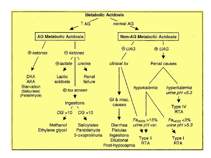
Increased anion gap metabolic acidosis • Methanol other alcohols, and ethylene glycol intoxication Uremia (renal failure) Lactic acidosis Ethanol Paraldehyde and other drugs Aspirin Ketones (starvation, alcoholic and diabetic ketoacidosis)

Anion Gap High AG

Anion Gap Normal AG

Etiologies of AG Metabolic Acidosis Ketoacidosis Lactic acidosis Renal failure Ingestions Diabetes mellitus, alcoholism, starvation Type A: impairment in tissue oxygenation, eg. Circulatory or Respiratory failure, sepsis, ischemic bowel, carbon monoxide Type B: no impairment in tissue oxygenation, eg. Malignancy, alcoholism, meds (metformin, NRTIs, salicylates) D-lactic acidosis: short bowel syndrome glc metab by colonic bacteria To D-lactate, which is absorbed; not detected by standard lactate assay Accumulation of organic anions such as phosphates, sulfates, etc. Methanol: manifestations include blurred vision Ethylene glycol: manifestations include ∆MS, cardiopulmonary failure, calcium oxalate crystals and renal failure Paraldehyde Salicylates: metabolic acidosis (from lactate, ketones) + respiratory alkalosis due to stimulation of CNS respiratory center Acetaminophen: glutathione depletion accumulation of the Endogenous organic acid 5 -oxoproline in susceptible host

Etiologies of Non-AG Metabolic Acidosis GI losses of HCO 3 Diarrhea, intestinal or pancreatic fistulas or drainage RTAs See section on renal tubular acidoses below Early renal failure Impaired generation of ammonia Ingestions Acetazolamide, sevelamer, cholestyramine, toluene Dilutional Due to rapid infusion of bicarbonate-free intravenous fluids Respiratory alkalosis renal wasting of HCO 3 rapid correction Of resp. alk. Transient acidosis until HCO 3; regenerated Posthypocapnia Ureteral diversion Colonic CI- /HCO 3 - exchange, ammonium reabsorption

Metabolic alkalosis

Etiologies of Metabolic Alkalosis Saline – responsiv e GI loss of H+ : vomiting, NGT drainage, villous adenoma Diuretic use posthypercapina Saline – resistant Hypertensive (mineralocorticoid excess) 10 hyperaldosteronism (eg. Conn's) 20 hyperaldosteronism (eg, renovascular dis. Renninsecreting tumor) Non-aldo (eg. Cushing's, Liddle's, exogenousmineralocorticoids) Normotensive Severe hypokalemia Exogenous alkali load Bartter's syndrome, Gitelman's syndrome

primary disturbance Primary Disorder Problem Metabolic acidosis gain of H+ or loss of HCO 3 Metabolic alkalosis gain of HCO 3 or loss of H+ Respiratory acidosis hypoventilation Respiratory alkalosis hyperventilation p. H HCO 3 Pa. CO 2 ↓ ↓ ↓ ↑ ↑ ↑ ↓ ↓

Steps in Acid-Base Analysis • Step 1: Acidemic or Alkalemic? • Step 2: Is the primary disturbance respiratory or metabolic? • Step 3: Is the respiratory disturbance acute or chronic? • Step 4: For a metabolic acidosis, is there an increased anion gap? • Step 5: Are there other metabolic processes present in a patient with an increased anion gap metabolic acidosis? • Step 6: Is the respiratory system compensating adequately for a metabolic disturbance

Step 1: Acidemic or Alkalemic? • The p. H of the arterial blood gas measurement identifies the disorder as alkalemic or acidemic. • Normal arterial blood p. H = 7. 35 – 7. 45 • Acidemic: p. H < 7. 35 • Alkalemic: p. H > 7. 45

Step 2: Is the primary disturbance respiratory or metabolic? • To determine whether the disturbance affects primarily – The arterial Pa. CO 2 or – The serum HCO 3 -. • Respiratory disturbances alter the arterial Pa. CO 2 (normal value 35 -45) • Metabolic disturbances alter the serum HCO 3 - (normal value 22 -26)

Quiz? p. H p. CO 2 HCO 3 1 7. 41 40 24 2 7. 5 42 35 3 6. 72 40 5 4 7. 26 63 25 5 7. 52 18 25 Interpretation

Quiz? p. H p. CO 2 HCO 3 Interpretation 1 7. 41 40 24 normal 2 7. 5 42 35 metabolic alkalosis 3 6. 72 40 5 metabolic acidosis 4 7. 26 63 25 respiratory acidosis 5 7. 52 18 25 respiratory alkalosis

Step 3: Is the respiratory disturbance acute or chronic? • Acute respiratory acidosis: – HCO 3 - increase by 1 m. Eq/l for every 10 mm. Hg increase in Pa. CO 2 • Chronic respiratory acidosis: – HCO 3 - increase by 3 -3. 5 m. Eq/l for every 10 mm. Hg increase in Pa. CO 2 • Acute respiratory alkalosis: – HCO 3 - decrease by 2 m. Eq/l for every 10 mm. Hg decrease in Pa. CO 2 • Chronic respiratory alkalosis: – HCO 3 - decrease by 4 -5 m. Eq/l for every 10 mm. Hg decrease in Pa. CO 2

Case study -1 • p. H =7. 2, p. CO 2 = 60, HCO 2 = 24. • What it is the primary problem? Compensation? • Differential diagnosis? • Treatment ?

Respiratory acidosis • • • Is it acute or chronic? Note that the PH is abnormal Note the HCO 2 is with in normal Remember: Acute respiratory acidosis: – HCO 3 - increase by 1 m. Eq/l for every 10 mm. Hg increase in Pa. CO 2 • Chronic respiratory acidosis: – HCO 3 - increase by 3 -3. 5 m. Eq/l for every 10 mm. Hg increase in. Pa. CO 2

Case 2 • What do you expect the ABG in the following patients to be: • 24 years old male with acute SOB, and wheezes for 2 days. • Past hx: Bronchial asthma • 67 years old women, HTN, DMII, COPD presenting with cough and SOB
![Case 3 • p. H: 7. 25 • [HCO 3 -]: 20 m. Eq/L](http://slidetodoc.com/presentation_image_h2/78ca8eaff3f122a05756c1cd70f69c2c/image-32.jpg "Case 3 • p. H: 7. 25 • [HCO 3 -]: 20 m. Eq/L")
Case 3 • p. H: 7. 25 • [HCO 3 -]: 20 m. Eq/L • Pa. CO 2: 52 mm. Hg • What it is the primary problem? Compensation? • Differential diagnosis?
![Case 4 • p. H: 7. 32 • [HCO 3 -]: 19 m. Eq/L](http://slidetodoc.com/presentation_image_h2/78ca8eaff3f122a05756c1cd70f69c2c/image-33.jpg "Case 4 • p. H: 7. 32 • [HCO 3 -]: 19 m. Eq/L")
Case 4 • p. H: 7. 32 • [HCO 3 -]: 19 m. Eq/L • Pa. CO 2: 55 mm. Hg • What it is the primary problem? Compensation? • Differential diagnosis? • What other investigation you want to do?

Step 4: For a metabolic acidosis, is there an increased anion gap? • Anion gap = [Sodium] - ([Chloride] + [Bicarbonate]) Or AG = [Na+] - ([Cl-] + [HCO 3 -]). • Normal AG 8 -16 • Serum Osmolality = (2 x (Na + K)) + (BUN ) + (glucose )

Step 5: Are there other metabolic processes present in a patient with an increased anion gap metabolic acidosis?

Step 6: Is the respiratory system compensating adequately for a metabolic disturbance • Metabolic acidosis: – PCO 2 decreases by 1 mm. Hg for every 1 m. Eq/l decrease in HCO 3 • Metabolic alkalosis: • PCO 2 increases by 0. 6 mm. Hg for every 1 m. Eq/l increases in HCO 3

Case 5 • 56 yo M with Hx of COPD is admitted with 1 wk Hx of dyspnea, productive cough and diarrhea (Na) 125, (Cl) 103 , (BUN) 42, (Glucose) 100, (K) 3. 5, (HCO 3 -) 10, (Creat) 1. 4 • ABG 7. 14 p. CO 2 30 p. O 2 50 • What is the predominant acid base disorder ?

Case 5 continue • What p. CO 2 is expected with normal respiratory compensation ? 40 – (1. 2 * (2410)) = 23. 2, this is not full compensation b/c p. CO 2 is 30 – indicates an underlying primary respiratory acidosis, suggested by the Hx of COPD, dyspnea, and productive cough (lungs not able to appropriately compensate) • What is the Anion Gap ? 125 – (103+10) = 12 – normal AG etiology is either diarrhea or RTA – most likely diarrhea b/c of the history

Case 6 • 32 y/o male present w/ 2 d Hx of intractable vomiting. ; p. H 7. 51, p. CO 2 41 Na 132, Cl 90 32 K 3. 4 HCo 2= 33 creatinine 1. 6 • What is the predominant acid-base disorder? Alkalosis (Metabolic )

Case 6 continue • What p. CO 2 is expected w/ normal respiratory compensation? = 40 + (32 – 24) * (~ 0. 6 0. 7) = 44. 8 45. 6 mm. Hg; since the measured p. CO 2 < 44. 8 45. 6, there is also a primary respiratory alkalosis (inappropriate hyperventilation) • Tx: Isotonic saline to correct for volume depletion –

Case 7 • A 58 - year old man presents to the Emergency Department with abdominal pain and hypotension. Investigation reveal the following: • Na 140 K 4 Cl 90 HCO 3 = 5 PH 6. 8 PCO 2 36 PO 2 7 • Analyze the acid-base disorder(s) seen in the patient. •

Summary • First, does the patient have an acidosis or an alkalosis – Look at the p. H • Second, what is the primary problem – metabolic or respiratory – Look at the p. CO 2 – If the p. CO 2 change is in the opposite direction of the p. H change, the primary problem is respiratory

Summary • Third, is there any compensation by the patient - do the calculations – For a primary respiratory problem, is the p. H change completely accounted for by the change in p. CO 2 • if yes, then there is no metabolic compensation • if not, then there is either partial compensation or concomitant metabolic problem
- Slides: 44