Achalasia of the Esophagus Dr Saleh M Aldaqal


 or secondary. �Primary achalasia : absence of inhibitory")


 and multichannel intraluminal impedance (MII) studies Normal Achalasia")


 �Anterior fundus is laid across the")
, �The anterior fundus is brought to either side")


 have been shown to respond less")


- Slides: 35
Achalasia of the Esophagus Dr. Saleh M. Aldaqal MBBS, FRCSI, SBGS Assistant Professor and Consultant General And laparoscopic Surgery(France), Department of Surgery, Faculty of Medicine, King Abdulaziz University. www. dr-aldaqal. com
Introduction �Primary esophageal motility disorder of unclear etiology. �Uncommon, but not rare. affecting approximately 1 in 100, 000/ year in America, 5 in 100, ooo/year in Europe. �Equally in men and women. �Usually diagnosed between 20 and 50 years of age
History �In 1672, Sir Thomas William first described the disease as “cardiospasm” and treated the problem with dilation using a whale sponge attached to a whale bone. � 1927 A F Hurst named the disease achalasia, a Greek term meaning failure to relax.
Etiology �Achalasia can be primary (idiopathic) or secondary. �Primary achalasia : absence of inhibitory ganglion cells in the myenteric (Auerbach's) plexus of the esophagus. �? Infectious agent, such as a virus, and its subsequent immune response.
Etiology �The most common secondary cause of achalasia is Chagas' disease a systemic disease caused by infestation by the protozoan Trypanosoma cruzi. Chagas is transmitted to humans by the reduviid or “kissing bug”. �worldwide is considered to be the most common cause of achalasia.
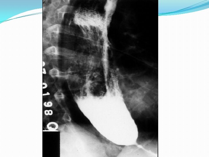
Diagnosis �The gold standard for diagnosis of achalasia is esophageal manometry. �Manometry is especially helpful in early disease state when other diagnostic evaluations appear normal.
Diagnosis �Residual LES relaxation pressure to be the most accurate diagnostic manometric criteria. �A residual pressure of >12 mm. Hg had 92% sensitivity. � Aperistalsis, a residual pressure of >10 mm. Hg had 100% sensitivity and 100% positive predictive value.
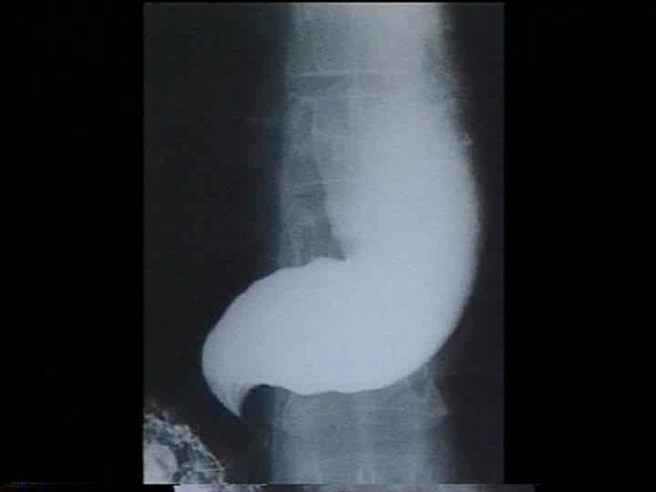
Diagnosis �High-Resolution manometry (HRM) and multichannel intraluminal impedance (MII) studies Normal Achalasia
Treatment �Achalasia is an incurable disease and treatment is focused on relief of symptoms. �The goal of both surgical and nonsurgical treatment is to eliminate the outflow obstruction afforded by a nonrelaxing sphincter, relieve dysphagia, and maintain a barrier against gastroesophageal reflux.
Pharmacologic treatment �Oral nitrates and calcium channel blockers, act to inhibit intramural neurons. �Poor outcomes and potentially harmful systemic effects, these agents are largely unreliable and unrealistic modalities for longterm symptom relief.
Endoscopic injection of Botox �Injection of Botox into the LES aims to block release of acetylcholine from cholinergic neurons in an effort to lower both basal and residual LES pressures. �Short-lived, rarely results in substantial reduction in LES pressure, and, although dysphagia can be improved, often requires repeat injections for continued relief.
Endoscopic injection of Botox �In a prospective randomized controlled trial comparing Botox injection (n = 40) with laparoscopic myotomy and partial fundoplication (n = 40), �Zaninotto and colleagues � 6 months after treatment dysphagia and regurgitation recurred in nearly half (45%) of those treated with Botox. � symptom-free at 2 years was considerably higher after myotomy (87. 5%) than after Botox injection (34%).
Endoscopic injection of Botox �It is now recognized that Botox injection creates an inflammatory reaction in the distal esophagus with consequent submucosal fibrosis, which can make subsequent surgical myotomy more difficult. �Botox should be reserved for those who are not surgical candidates.
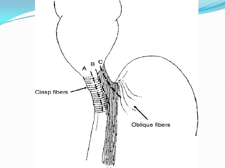
Pneumatic balloon dilation �Decreases esophageal outflow resistance by forceful “tearing” of the LES muscle fibers. �The most common balloon dilators in current use are the Rigiflex and Witzel polyethylene balloons. �Rigiflex is a balloon on a catheter, which is placed using fluoroscopy, and the Witzel balloon is inserted while attached to the endoscope. Risk of perforation using the Rigiflex is about 3% and that of the Witzel is 6%.
Pneumatic balloon dilation �Graded balloon dilation is begun using a 30 -mm balloon as the initial dilator. �Repeat balloon dilation can be performed using progressively larger dilator sizes to 35 mm and 40 mm
Pneumatic balloon dilation �The only prospective randomized trial comparing pneumatic dilation to surgical myotomy was reported by Csendes and colleagues from Chile. �They found that 100% of patients treated with myotomy (n = 19) had only mild or no dysphagia at a mean of 3. 5 years, as compared with only 61% of those treated with balloon dilation (n = 18).
Pneumatic balloon dilation �Two more recent evaluations by. West and colleagues �Retrospectively evaluated longterm outcomes of 125 achalasia patients followed prospectively for >5 years after pneumatic dilation. �In this experience, only 50% of patients had no or occasional (less than once per week) dysphagia at 5 years.
�Laparoscopic myotomy and partial fundoplication was superior to pneumatic dilation in newly diagnosed patients with achalasia.
Surgical treatment �Esophageal myotomy for achalasia was first described by Ernest Heller in 1913. �In this operation, both the anterior and posterior lower esophageal sphincter muscle fibers were disrupted. �A modified version of this procedure, referred to today as the Heller myotomy, consists of a single anterior longitudinal myotomy and has become the standard operative technique. performed through either a thoracotomy or laparotomy.
Thoracoscopic or laparoscopic. �Recent review of studies evaluating thoracoscopic myotomy from 1993 to 2005 (n = 10 studies; 204 patients) and laparoscopic myotomy from 1995 to 2005 (n = 15 studies; 499 patients) by Abir and colleagues. �Symptom relief to occur in 76% versus 94% of patients, respectively. �Development of gastroesophageal reflux disease occurred in 35% of patients after thoracoscopic myotomy and 13% of patients after laparoscopic myotomy.
Fundoplication ? �An anterior partial fundoplication (Dor ) �Anterior fundus is laid across the myotomy site and sewn to the cut edge of the esophageal myotomy with three or four interrupted sutures of 2 -0 silk. �Highest stitch is taken through the crural pillar as an anchor to prevent torsion of the fundus.
Fundoplication ? �Posterior partial fundoplication (Toupet), �The anterior fundus is brought to either side of the myotomy, and two columns of sutures are placed on either side, leaving the myotomy site bare and open
Fundoplication ? �A recent prospective randomized study has put this issue to rest. �Richards and colleagues reported on 43 achalasia patients randomized to laparoscopic Heller myotomy with and without Dor fundoplication. �Gastroesophageal reflux, defined by 24 -hour distal esophageal acid exposure time >4. 2%, � was present in 47. 5% of patients undergoing Heller myotomy alone compared with 9. 1% of patients undergoing Heller myotomy with partial fundoplication.
Results of laparoscopic Heller myotomy with fundoplication �Complications average 10% to 15%, and commonly include pneumothorax, wound infection, and esophageal leak. � Mortality is uncommon, zero in most large published series. �Injury of the esophageal mucosa occurs in 0 to 14% of procedures and, when recognized and repaired, is rarely of clinical consequence.
Results of laparoscopic Heller myotomy with fundoplication �Most published results report similar results. �More than 91% were symptom-free at a median of 2 years followup. � 90% probability of remaining asymptomatic at 5 years. �symptoms were found in 7% of patients <10 years, 23% of those 10 to 20 years, and 35% of patients >30 years postmyotomy.
Outcomes predictors �Young patients (younger than 40 years) have been shown to respond less well to pneumatic dilation and are good candidates for primary myotomy. �Lowering resting LES pressure to near 10 mm. Hg post operative has been reported by several authors to substantially affect longterm success of surgery or pneumatic dilation.
Extent of myotomy �Better symptomatic improvement and a lower incidence of recurrent dysphagia by increasing the distal extent of myotomy onto the proximal stomach from 1. 5 cm to 3 cm �Postoperative 24 -hour p. H data showed that extension of the distal aspect of the myotomy did not result in a higher prevalence of gastroesophageal reflux.
Persistent or recurrent symptoms �Mild to moderate symptoms of dysphagia, regurgitation, heartburn, and chest pain recur with time in as many as 15% to 20% of patients. � Lifestyle and behavioral changes or proton-pump inhibitor therapy
Persistent or recurrent symptoms �Early failures are usually caused by failure to extend the myotomy far enough onto the stomach. �Late failures occur secondary to development of complicated gastroesophageal reflux with stricturing or Barrett's changes and disease progression
Persistent or recurrent symptoms �Pneumatic dilation should be considered as the firstline treatment for patients with persistent dysphagia and that reoperation should be reserved for those who do not respond.
THANK YOU