Accessory Respiratory Organs in Teleosts Core Course No


 and hill streams")


 the highly vascularised buccopharyngeal epithelium helps in absorbing")


")


 – Ganguly, Sinha, Adhikari")
- Slides: 28
Accessory Respiratory Organs in Teleosts Core Course No. ZOOA P 2 T Group A, Unit: 1, Topic No. 6
What is ARO? • Generally adult fishes depended chiefly on pharyngeal gills for aquatic respiration. However, other devices also occur to supplement or replace gill respiration. • All such additional respiratory organs, other than gills, are known as accessory respiratory organs. The development of accessory respiratory organs is found mostly in freshwater fishes of tropical region and very rarely in marine fishes.
Sometimes the fishes of the tropical freshwaters (Barbs, Dianos etc. ) and hill streams develop accessory respiratory organs to meet extra demand for oxygen, because depletion of oxygen occurs during summers as the water level falls to a considerable degree. Hillstream Loach Barbs Dianos
The ARO is primarily important because… • Accessory respiratory organs enable the fishes to live in oxygen deficient water, to aestivate over prolonged droughts in dry summer, to take excursions on land or simply to meet extra demand for oxygen.
and also To overcome these adverse situations, accessory respiratory organs functionable in aquatic and/or aerial environment have been developed in fishes. So the development of such structures is essentially adaptive in native. Some accessory organs sub serve aquatic respiration, while others aerial respiration
Types
1. Skin or Integument In the eel, Anguilla anguilla, Amphipnous cuchia and in Periophthalmus and Boleophthalmus, the skin is highly vascular and serves for exchange of gases as in frog, when the fish is out of water. These fishes habitually leave the water and migrate from one place to another through damp vegetation. During this period, the moist skin serves as an important organ in respiration. They can respire cutaneously both in air and in water. Anguilla
Besides, the highly vascular opercular folds of Sturgeons and many cat fishes serve as accessory respiratory structures. Acipenser Huso huso (European Sturgeon )
2. Bucco-Pharyngeal Epithelium In most of the fishes, the epithelial lining of buccal cavity and pharynx is usually highly vascular and permeable to gases in water. It may remain simple or may develop folds, pleats or tongues projecting into the buccal cavity and pharynx to make it an efficient respiratory organ.
But in mudskippers (Periophthalmus and Boleophthalmus) the highly vascularised buccopharyngeal epithelium helps in absorbing oxygen directly from the atmosphere. These tropical fishes leave water and spend most of the time skipping or walking about through dampy areas particularly round the roots of the mangroove trees. The old idea that the mudskippers use the vascular tail as the respiratory organ is not supported by recent ichthyologists. Periophthalmus Boleophthalmus
3. Gut Epithelium In several fishes epithelial lining of certain parts of alimentary canal becomes vascular and modified to serve as a respiratory organ. It may be just behind stomach (Misgurus fossilis) or intestine (Lepidocephalus guntea), Gobitus (giant loach of Europe) or rectum (Callichthyes, Hypostomus and Doras). Misgurus fossilis Lepidocephalus guntea Callichthyes Hypostomus
Fresh air is drawn through mouth or anus and after gaseous exchange the gas is voided through the anus. In these fishes the wall of the gut is modified to perform the respiratory function. The walls of the gut in these areas become thin due to the reduction of muscular layers.
4. Outgrowths of Pelvic Fins • In American lung fish, Lepidosiren, during breeding time, the pelvic fins of male become enlarged and grow filamentous vascular outgrowths which provide fresh oxygen to the guarded eggs.
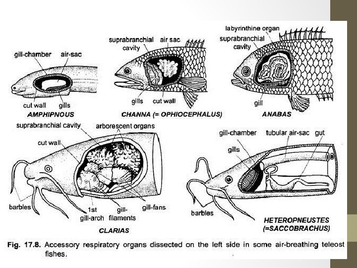
5. Pharyngeal Diverticula: Pharyngeal diverticula are a pair of simple sac like outgrowths of pharynx, lined by thickened vascular epithelium and extending above the gills. In Channa (= Ophiocephalus), the accessory respiratory organs are relatively simpler and consist of a pair of air chambers
5. Pharyngeal Diverticula • These are developed from the pharynx and not from the branchial chambers as seen in others. The air chambers are lined by thickened epithelium which is highly vascularised. The air chambers are simple sac like structures and do not contain any structure. • These chambers function as the lung like reservoirs. In Channa striatus, the vascular epithelium lining the chambers becomes folded to form some alveoli. The gill filaments are greatly reduced in size.
• In cuchia eel, Amphipnous cuchia, the accessory respiratory organs consist of a pair of vascular sac like diverticula from the pharynx above the gills. These diverticula open anteriorly into first gill slit. • These diverticula function physiologically as the lungs. The gills are greatly reduced and a few rudimentary gill filaments are present on the second of the three remaining gill arches. The third gill arch is found to bear fleshy vascular (respiratory) epithelium.
In Periophthalmus also, a small, shallow pharyngeal diverticulum lined with respiratory epithelium (vascular epithelium) is present on each side of the roof of the pharynx.
6. Opercular Chamber Modified for Aerial Respiration • In some species, the inhaled air is passed through the gill slits into the opercular chamber where it is stored for some time. The opercular chamber becomes bulged out in the form of two little balloons in the hinder region of the head and after sometimes its walls collapse and the air is passed out through the small external branchial opening. The membrane lining the opercular chamber becomes thin and highly vascular to allow exchange of gases. This is seen in Periophthalmus and Boleophthalmus. Opercular series in bony fish: operculum (yellow), preoperculum (red), interoperculum (green) and suboperculum (pink)
7. Branchial Diverticula • The outgrowths from gill chambers form more complicated aerial accessory respiratory organs than the simpler pharyngeal outgrowths in other fishes. Such air breathing organs are present in Heteropneustes, Clarias, Anabas, Trichogaster, Macropodus, Betta, etc.
• Anabas Testudineus - The Indian climbing perch has two, spacious, suprabranchial cavities as dorsal outgrowths of the two gill chambers. Each cavity contains a special labyrinthine organ formed of much folded, concentric bony plates developed from the first epibranchial bone and covered with thin vascular mucous membrane. Margins of these plates are wavy and the plates are covered with vascular gill like epithelium. • Each branchial outgrowth communicates freely not only with the opercular cavity but also with buccopharyngeal cavity. Air is drawn through mouth into suprabranchial cavities and expelled through opercular opening. The fish is so dependent on atmospheric oxygen that it will drown if denied access to surface to gulp air.
• Trichogaster fasciatus The accessory respiratory organs in this species consist of a suprabranchial chamber, a labyrinthine organ and the respiratory membrane. The suprabranchial chamber is situated above the gills on either side as in Anabas, communicates with the pharynx by means of inhalent aperture and with the exterior through the opercular chamber by means of an exhalent aperture. • The labyrinthine organ develops from the epibranchial of the first gill arch and is simpler in structure than that of Anabas. It is in the form of a spiral organ possessing two leaf like expansions and is composed of loose connective tissue covered by a vascular epithelium. • The respiratory membrane lining the air chamber and covering the labyrinthine organs consist of vascular and non vascular areas, of which the former possesses a large number of ‘islets’ containing parallel blood capillaries. The islets are believed to be derived from the secondary lamellae of a typical gill filament. Trichogaster fasciatus
8. Pneumatic Sacs • In Heteropneustes fos silis, a pair of tubular pneumatic sacs, one on each side of the body, act as the accessory respiratory organs. • These long tubular sacs arise as the outgrowths from the branchial chamber and extend almost up to the tail between the body musculature near the verte bral column. In Saccobranchus, similar tubular lung like outgrowths of the branchial chamber extend back into the body musculature.
Functions of Accessory Respiratory Organs • The accessory respiratory organs contain a higher percentage of oxygen. The fishes possessing such respiratory organs are capable of living in water where oxygen concentration is very low. Under this condition these fishes come to the surface of water to gulp in air for transmission to the accessory respiratory organs. If these fishes are prevented from coming to the surface, they will die due to asphyxiation for want of oxygen. So the acquisition of accessory respiratory organs in fishes is an adaptive feature. • Further it has been observed that the rate of absorption of oxygen in such organs is much higher than the rate of elimination of carbon dioxide. Hence, it is natural that the gills excrete most of the carbon dioxide. Absorption of oxygen appears to be the primary function of the accessory respiratory organs.
Origin and Significance of Accessory Respiratory Organs • During development, the fifth gill arch does not develop gill lamellae, and its embryonic gill material forms rudiments of the gill arch, and aggregates to form a structure called the ‘gill mass’. The air breathing organs or accessory respiratory organs develop from gill mass. In some species, the gill arches other than the fifth arch, also take part in the formation of accessory respiratory organs. • The gill lamellae which normally develop on gill arches for aquatic respiration, become modified to form the respiratory epithelium of the suprabranchial chamber, dendritic organs and the air sacs for aerial respiration. According to Singh (1993), the air sacs have evolved from the same basic material which has given rise to the gill in teleosts fishes.
Origin and Significance of Accessory Respiratory Organs • Most of the fishes possessing air breathing organs or accessory respiratory organs are capable of living in highly deoxygenated water of the swamps and muddy ponds infested with weeds. They have been observed to gulp in air from the surface and to pass it to the accessory respiratory organs. If prevented from reaching the surface, these fishes die due to asphyxiation. • This shows that the accessory respiratory organs are capable of maintaining life of the fish in oxygen deficient water. It has been shown that the absorption of oxygen in the accessory respiratory organs is much greater than the excretion of carbon dioxide. Hence, most of the carbon dioxide is excreted in the gills and the chief function of the accessory respiratory organs is the absorption of oxygen required for sustenance of life.
Origin and Significance of Accessory Respiratory Organs • The accessory respiratory organs of Heteropneustes and Clarias, are modifications of the gills. In these species, swim bladder is either absent or greatly reduced. During the Tertiary and Quaternary period of the Cenozoic era, the oxygen level of the atmosphere and of water was reduced. Due to depletion of oxygen in rivers and swamps, the gills were unable to cope with the requirements of the body. Hence, several teleostean species developed accessory respiratory organs to absorb oxygen from air.
• Ref: Biology of Animals (Vol 2) – Ganguly, Sinha, Adhikari