Accessory Glands of Digestive System The liver The


- Slides: 24

Accessory Glands of Digestive System

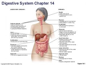
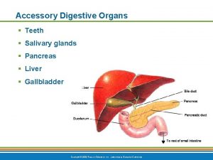
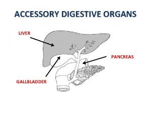
The liver • The liver is soft and pliable and occupies the upper part of the abdominal cavity just beneath the diaphragm. • The greater part of the liver is situated under cover of the right costal margin, and the right hemidiaphragm. • The liver extends to the left to reach the left hemidiaphragm. • Convex upper surface of the liver is molded to the undersurface of the domes of the diaphragm.

The liver • The visceral surface, is molded to adjacent viscera and is therefore irregular in shape; it lies in contact with: • • • The abdominal part of the esophagus. The stomach. The duodenum. The right colic flexure. The right kidney and suprarenal gland. And the gallbladder

The liver

The liver • The liver may be divided into a large right lobe and a small left lobe by the attachment of the peritoneum of the falciform ligament. • The right lobe is divided into a quadrate lobe and a caudate lobe by the presence of the gallbladder, the fissure for the ligamentum teres, the inferior vena cava, and the fissure for the ligamentum venosum.

The liver • The porta hepatis, or hilum of the liver, is found on the posteroinferior surface and lies between the caudate and quadrate lobes. • The upper part of the free edge of the lesser omentum is attached to its margins. • In it lie the right and left hepatic ducts, the right and left branches of the hepatic artery, the portal vein, and sympathetic and parasympathetic nerve fibers. A few hepatic lymph nodes lie here; they drain the liver and gallbladder and send their efferent vessels to the celiac lymph nodes. • The liver is completely surrounded by a fibrous capsule but only partially covered by peritoneum.

Porta Hepatis

Important Relations • Anteriorly: • • • Diaphragm. Right and left costal margins. Right and left pleura and lower margins of both lungs. Xiphoid process. And anterior abdominal wall in the subcostal angle. • Posteriorly: • • Diaphragm. Right kidney. Hepatic flexure of the colon. Duodenum. Gallbladder. Inferior vena cava. and esophagus and fundus of the stomach.

The liver

Peritoneal Ligaments of the Liver • The falciform ligament, which is a two-layered fold of the peritoneum, ascends from the umbilicus to the liver. • It has a sickle-shaped free margin that contains the ligamentum teres, the remains of the umbilical vein. • The falciform ligament passes on to the anterior and then the superior surfaces of the liver and then splits into two layers. The right layer forms the upper layer of the coronary ligament; the left layer forms the upper layer of the left triangular ligament. • The right extremity of the coronary ligament is known as the right triangular ligament of the liver. • It should be noted that the peritoneal layers forming the coronary ligament are widely separated, leaving an area of liver devoid of peritoneum ( bare area of the liver ).

Peritoneal Ligaments of the Liver • The ligamentum teres passes into a fissure on the visceral surface of the liver and joins the left branch of the portal vein in the porta hepatis. • The ligamentum venosum, a fibrous band that is the remains of the ductus venosus, is attached to the left branch of the portal vein and ascends in a fissure on the visceral surface of the liver to be attached above to the inferior vena cava. • The lesser omentum arises from the edges of the porta hepatis and the fissure for the ligamentum venosum and passes down to the lesser curvature of the stomach.

Peritoneal Ligaments of the Liver

Hepatic Ducts and Bile Duct

Gallbladder • The gallbladder is a pear-shaped sac lying on the undersurface of the liver. • gallbladder is divided into the fundus, body, and neck. • The fundus is rounded and projects below the inferior margin of the liver, where it comes in contact with the anterior abdominal wall at the level of the tip of the 9 th right costal cartilage. • The body lies in contact with the visceral surface of the liver. • The neck becomes continuous with the cystic duct, which turns into the lesser omentum to join the common hepatic duct, to form the bile duct. • The peritoneum completely surrounds the fundus of the gallbladder and binds the body and neck to the visceral surface of the liver.

Relations • Anteriorly: • The anterior abdominal wall • And the inferior surface of the liver. • Posteriorly: • The transverse colon. • And the first and second parts of the duodenum.

Gallbladder

Three common variations of terminations of the bile and main pancreatic ducts

Gallbladder

Pancreas • Consists of exocrine portion and endocrine portion, the pancreatic islets (islets of Langerhans). • The pancreas is an elongated structure that lies in the epigastrium and the left upper quadrant. • It is soft and lobulated and situated on the posterior abdominal wall behind the peritoneum. • It crosses the transpyloric plane. • The pancreas is divided into a head, neck, body, and tail.

Pancreas • The head of the pancreas is disc shaped and lies within the concavity of the duodenum. • A part of the head extends to the left behind the superior mesenteric vessels and is called the uncinate process. • The body runs upward and to the left across the midline. It is somewhat triangular in cross section. • The tail passes forward in the splenicorenal ligament and comes in contact with the hilum of the spleen.

Relations • Anteriorly: From right to left: • The transverse colon and the attachment of the transverse mesocolon. • The lesser sac. • And the stomach. • • • Posteriorly: From right to left: The bile duct. The portal and splenic veins. The inferior vena cava. The aorta. The origin of the superior mesenteric artery. The left psoas muscle. The left suprarenal gland, the left kidney. And the hilum of the spleen.

Pancreas

Pancreas

Thank you
 Male accessory glands
 Mouth function in digestive system
 Accessory organs of the digestive system
 Accessory organs
 Where is bile stored
 Which is an accessory organ of the digestive system
 Transitional epithelium
 Accessory digestive organs
 Accessory digestive organs
 Sinusoids
 Respiratory system circulatory system digestive system
 Ovarian ligament.
 Glands in integumentary system
 Endocrine glands
 Nervous system and digestive system
 Accessory structure of the skin
 Tongue epithelium
 Expiratory accessory muscles
 What is accessory pigment
 Accessory plates in optical mineralogy
 Nails integumentary system
 Accessory fruits
 Accessory fruit
 Pukpos
 Accessory structures of the eye

