Abortion INTRODUCTIONDEFINITION Termination or loss of pregnancy before

Abortion

INTRODUCTION/DEFINITION • Termination or loss of pregnancy before the age of viability( 28, 24 , 22 wks or <500 g) • WHO-24 wks or 500 g • In our environment- Officially still 28 wks • UK- 24 wks • USA-22 wks

�Abortion is a significant public health problem and an important cause of maternal mortality in the developing world � An estimated 70, 000 women die from complications of induced abortion annually in the world � A large number of these deaths (over 99%) are due to unsafe procedures carried out in developing countries � Preventing maternal deaths is an important Millennium Development Goal

DEFINITION Abortion is the expulsion or extraction of an embryo or fetus weighing 500 g or less from its mother when it is not capable of independent survival (i. e. before the period of viability)

Incidence • 10– 20% of all clinical pregnancies • 75% abortions occur before the 16 th week • Rates vary with maternal age; also high in women with past miscarriages

TYPES Spontaneous Isolated Threatened Recurrent Inevitable Complete Induced MTP Incomplete Illegal Missed Septic

Etiology • Fetal Factors • Maternal Factors

Fetal Factors • Genetic • 50% of early miscarriage is due to chromosomal abnormalities • Numerical defects like Trisomy, Polyploidy, Monosomy • Structural defects like translocation, deletion, inversion • Multiple Pregnancies • Degeneration of villi
: • Luteal Phase Defect •")
Maternal Factors • ENDOCRINE AND METABOLIC FACTORS (10– 15%): • Luteal Phase Defect • Thyroid abnormalities • Diabetes mellitus • Anatomical abnormalities (10– 15%) Cervicouterine factors • • Cervical incompetence & insufficiency Congenital malformation of the uterus Uterine Fibroid Intrauterine adhesions
 • Viral: rubella, cytomegalo, HIV, . . • Parasitic: toxoplasma,")
• Infections (5%) • Viral: rubella, cytomegalo, HIV, . . • Parasitic: toxoplasma, malaria, . . • Bacterial: ureaplasma, chlamydia, . . • IMMUNOLOGICAL DISORDERS (5– 10%)— • • Autoimmune disease • • Alloimmune disease • • Antifetal antibodies

• Environmental Factors • Cigarette smoking • Alcohol consumption • Contraceptive agents • Maternal medical illness • Cyanotic heart disease • Hemoglobinopathies • Unexplained (40 -60%) – In majority, the exact cause is not known.

Threatened Abortion • Condition in which miscarriage has started but has not progressed to a state from which recovery is impossible
 Slight bleeding per vaginam")
CLINICAL FEATURES: • The patient, having amenorrhea, complains of: (1) Slight bleeding per vaginam (2) Pain: Usually painless; there may be mild backache or dull pain in lower abdomen

CONTD • The uterus and cervix feel soft. • Digital examination reveals closed external os • Differential diagnosis includes • • cervical ectopy polyps or carcinoma ectopic pregnancy molar pregnancy • Ultrasound is diagnostic; Pelvic examination is avoided when USG is available

Management & Prognosis • Rest: Patient should be in bed for few days until bleeding stops • Relief of pain: Diazepam 5 mg BD • 80% of pregnancies with threatened abortions go on until term • If a live fetus is seen on USG, pregnancy is likely to continue in over 95% cases. • If pregnancy continues, there is increased frequency of preterm labor, placenta praevia & IUGR

Inevitable Abortion It is the clinical type of abortion where the changes have progressed to a state from where continuation of pregnancy is impossible.
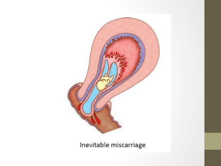

CLINICAL FEATURES: • The patient, having the features of threatened miscarriage, presents with • vaginal bleeding • Aggravation of colicky pain in the lower abdomen • Sometimes, the features may develop quickly without prior clinical evidence of threatened miscarriage • Internal examination reveals dilated internal os through which the products of conception are felt

Management • Management is aimed: • To accelerate the process of expulsion • To maintain strict asepsis • If pregnancy < 12 weeks, suction evacuation is done • If pregnancy > 12 weeks, expulsion by oxytocin infusion • General measures: • Excessive bleeding is controlled by administering methergin 0. 2 mg • Blood loss is corrected by IV fluid therapy and blood transfusion

Incomplete abortion The process of abortion has already taken place, but the entire products of conception are not expelled & a part of it is left inside the uterine cavity
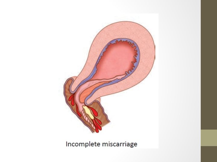

Clinical features: • History of expulsion of a fleshy mass per vaginam; • Continuation of pain in lower abdomen • Persistence of vaginal bleeding • Internal examination reveals • uterus smaller than the period of amenorrhea • Open internal os • varying amount of bleeding • On examination, the expelled mass is found incomplete Complications: • The retained products may cause: (a) bleeding (b) sepsis or (c) placental polyp.
 • Early abortion: Dilatation")
MANAGEMENT: • Evacuation of the retained products of conception (ERCP) • Early abortion: Dilatation and evacuation under analgesia or general anesthesia is to be done. • Late abortion: Uterus is evacuated under general anesthesia and the products are removed by ovum forceps or by blunt curette. In late cases, D&C is to be done to remove the bits of tissues left behind. • Prophylactic antibiotics are given; removed materials are subjected to a histological examination. • Medical management - Tab. Misoprostol 200 μg is used vaginally every 4 hours

Complete Abortion • When the products of conception are completely expelled from the uterus, it is called complete miscarriage.

Clinical features • There is history of expulsion of a fleshy mass per vaginam followed by • Subsidence of abdominal pain • Vaginal bleeding becomes trace or absent • Internal examination reveals: • Uterus smaller than the period of amenorrhea • Cervical os is closed • Bleeding is trace. • Transvaginal sonography confirms that uterus is empty

Missed Abortion • The fetus is dead and retained passively inside the uterus for a variable period • It is diagnosed when there is a fetus with a crown rump length of 5 mm without a fetal heart.

CLINICAL FEATURES: The patient usually presents with features of threatened miscarriage followed by: • Subsidence of pregnancy symptoms • Uterus becomes smaller in size • Cervix feels firm with closed internal os • Nonaudibility of the fetal heart sound even with Doppler ultrasound • Immunological test for pregnancy becomes negative

Complications • Retaining the products for long time can lead to sepsis • DIC [Disseminated Intravascular Coagulation] – (very rare) in gestations exceeding 16 weeks
 800 mg")
Management Uterus is less than 12 weeks: • Prostaglandin E 1 (Misoprostol) 800 mg is given vaginally and repeated after 24 hours if needed. Expulsion usually occurs within 48 hours • Suction evacuation is done when the medical method fails Uterus more than 12 weeks • 6 th or 12 th hourly misoprostol tablets given vaginally • If this fails, extraamniotic instillation of ethacridine lactate is used • Antibiotics are given

Septic Abortion • Any abortion associated with clinical evidences of infection of the uterus and its contents • Most common cause – Attempt at induced abortion by an untrained person without the use of aseptic precautions

Clinical Grading: • Grade–I: The infection is localized in the uterus. • Grade–II: The infection spreads beyond the uterus to the parametrium, tubes and ovaries or pelvic peritoneum. • Grade–III: Generalized peritonitis and/or endotoxic shock or jaundice or acute renal failure. Grade-I is the commonest and is usually associated with spontaneous abortion

Clinical Features • Fever, abdominal pain and vomiting or diarrhoea • A rising pulse rate of 100– 120/min or more is a significant finding than even pyrexia. It indicates spread of infection beyond the uterus. • Examination shows abdominal tenderness, guarding, rigidity • Internal examination reveals: • offensive purulent vaginal discharge • tender uterus usually with patulous os or a boggy feel • Soft cervix with open internal os

Investigations • CBC • Serum urea, creatinine, electrolytes • High vaginal swab • Blood culture in suspected septicaemia • Pelvic USG to detect retained products of conception • X-ray abdomen in suspected bowel injury • X-ray chest if there is difficulty in respiration

Complications Immediate: • Hemorrhage • Injury may to uterus & adjacent structures • Spread of infection leads to: • • • Generalized peritonitis Endotoxic shock—mostly due to E. Coli DIC Acute renal failure Thrombophlebitis • All these lead to increased maternal deaths

Management • Mild cases • Broad spectrum antibiotics started • Uterus is evacuated • Severe Cases • Vigorous IV infusion with crystalloid • Oxygen given by nasal catheter • Broad spectrum antibiotics – combination of ampicillin, gentamycin, metronidazole is started • Uterus is evacuated in 4 -6 hrs of commencing therapy.

Recurrent Miscarriage/ Pregnancy loss

Recurrent Abortion • Recurrent miscarriage is defined as a sequence of three or more consecutive spontaneous abortion • Seen in ~ 1% of all women • Risk increases with each successive abortion • No underlying cause is found for 50% of recurrent pregnancy loss
: § Parental chromosomal abnormalities §")
Etiology FIRST TRIMESTER ABORTION: • Genetic factors (3– 5%): § Parental chromosomal abnormalities § The most common abnormality is a balanced translocation. § This leads to unbalanced translocation in the fetus, causing early miscarriage or a live birth with congenital malformations § Risk of miscarriage in couples with a balanced translocation is > 25%. § This is the most common cause for 1 st trimester loss

• Endocrine and Metabolic: • Poorly controlled diabetic patients • Presence of thyroid autoantibodies • Luteal phase defect • Hypersecretion of luteinizing hormone (e. g. in PCOS). • Infection: • Infection in the genital tract - (Transplacental fetal infection) • Syphilis • Inherited thrombophilia • Protein C deficiency, Protein S deficiency, factor V Leiden mutation, prothrombin gene mutation
. • Antiphospholipid antibodies present in")
• Immunological cause: Autoimmunity – Antiphospholipid antibody syndrome(15%). • Antiphospholipid antibodies present in mother produce adverse fetal outcome • Diagnosis by presence of lupus anticoagulant/Ig. G/Ig. M anticardiolipin antibodies Alloimmune factors • Immune response against paternal antigens in the fetus • This is a result of lack of production of blocking antibodies by the mother due to failure of recognition of TLX (trophoblast-lymphocyte crossreactive antigens)

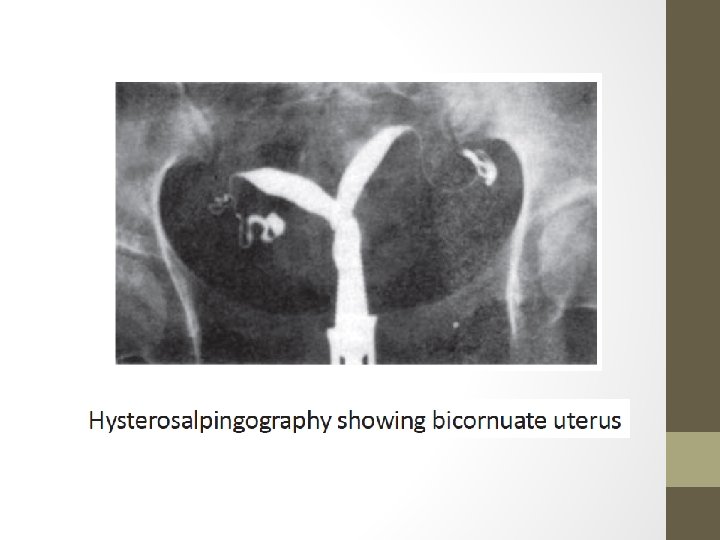
SECOND TRIMESTER MISCARRIAGE: • Anatomic abnormalities - responsible for 10– 15% of recurrent abortion. • Causes may be (a) Congenital - defects in the mullerian duct fusion (e. g. unicornuate, bicornuate, septate or double uterus) (b) Acquired - intrauterine adhesions, uterine fibroids and endometriosis, cervical incompetence

Uterine Causes • Defects of mullerian fusion • Double uterus, septate or bicornuate uterus • About 12% cases of recurrent abortion. • Implantation on the septum leads to defective placentation • Asherman syndrome – Intrauterine adhesions due to previous curettage – can lead to early miscarriage • Transvaginal ultrasound is used for diagnosis; • Hysteroscopic resection for septum or division of adhesions in Asherman’s syndrome. • Submucous fibroids - managed by myomectomy

Septate Uterus Double Uterus
 • Painless cervical dilatation with ballooning of amniotic sac into vagina,")
Cervical Insufficiency (Incompetence) • Painless cervical dilatation with ballooning of amniotic sac into vagina, followed by rupture of membrane and expulsion of fetus • Usually at 16 – 24 weeks
—common,")
Etiology • Congenital • Developmental weakness of cervix • Uterine anomalies • Acquired (iatrogenic)—common, following: (i) (iii) (iv) D&C operation Induced abortion by D and E vaginal operative delivery through an undilated cervix amputation of the cervix or cone biopsy. • Multiple gestations, prior preterm birth.

Diagnosis • History - Repeated mid trimester painless cervical dilatation and escape of liquor amnii followed by painless expulsion of the products of conception • Internal examination: Interconceptual period: • Passage of no. 6– 8 Hegar dilator beyond the internal os without any resistance or pain • Funnelling of internal os seen in hysterosalpingography

During pregnancy • Clinical digital – Painless cervical shortening and dilatation • Sonography: Trans vaginal ultrasound is performed. Short cervix < 25 mm; Funnelling of the internal Os > 1 cm.

Management • Surgical management – Cervical circlage • Usually at 12 -14 weeks • The procedure reinforces the weak cervix by a non-absorbable tape, placed around the cervix at the level of internal os.
 cervix Incompetent cervix with herniation of the membranes")
Normal (Competent) cervix Incompetent cervix with herniation of the membranes

Competency restored after encirclage operation

• Contraindications • Intrauterine infection • Ruptured membranes • History of vaginal bleeding • Severe uterine irritability • Cervical dilatation > 4 cm. • 2 main methods – Mc. Donald and Modified Shirodkar • Success rates - 80 – 90%

Types of circlage • History Indicated • Definite history of 3 previous second trimester losses/ preterm births • Ultrasound indicated • Short ended cervix or early funnelling in ultrasound in a woman with 1 or 2 spontaneous losses • Examination indicated / Rescue circlage • Performed after the cervix is found dilated • Also called emergency circlage
 is placed as")
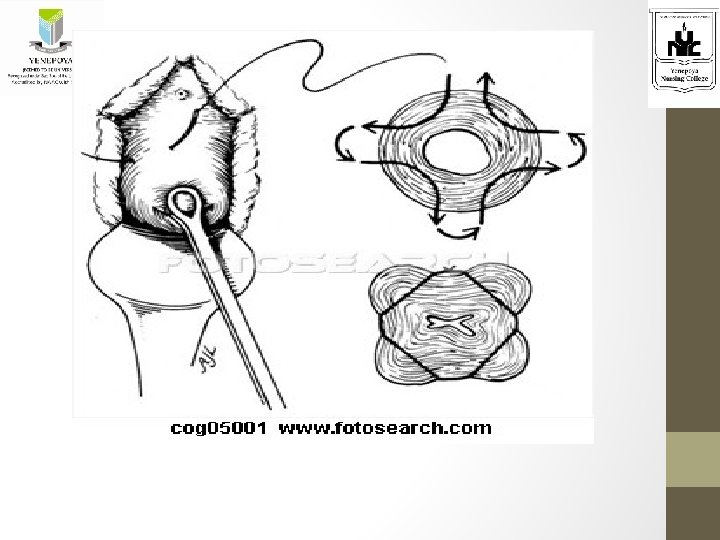
Methods I. Mc. DONALD’S OPERATION • The non-absorbable suture material (Mersilene) is placed as a purse string suture, as high as possible (level of internal os) • The suture starts at the anterior wall of the cervix. Taking successive deep bites (4– 5 sites) it is carried around the lateral and posterior walls back to the anterior wall again where the two ends of the suture are tied. • Commonly performed method nowadays.

Patient is in lithotomy position and cervix is exposed with Sim’s speculum. The cervical lips are held with sponge holding forceps and a purse string suture with a non absorbable material like black silk is taken all around the cervix. Disadvantage –suture may be below internal os.

Mc. Donald’s cerclage
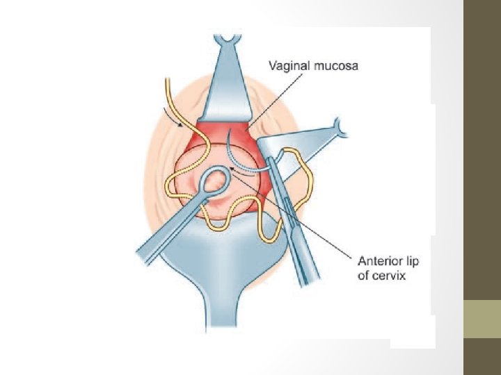

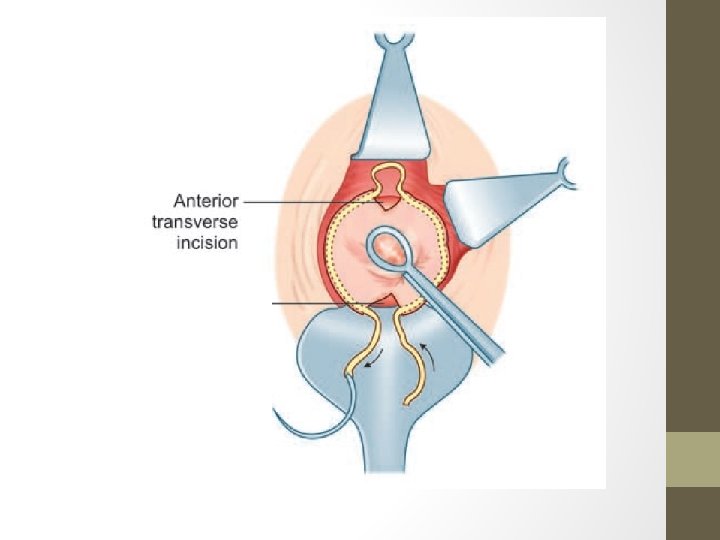
II. Modified Shirokdar Circlage • A transverse incision is made on the vaginal wall and the bladder is pushed up to expose the level of the internal os. • The non-absorbable suture material—Mersilene tape is passed submucously with the help of any curved round bodied needle so as to bring the suture ends to the posterior. • The ends of the tapes are tied up posteriorly by a knot. • The anterior incision is repaired using chromic catgut.

Shirodkar’s cerclage

III. Transabdominal Cerclage • Rarely done in cases of repeated failure of vaginal approach • Cerclage is placed at the level of isthmus • Delivery by CS

• Postoperative care: • The patient should be in bed for at least 2– 3 days • Progesterone supplementation - Weekly injections of 17 α hydroxy progesterone caproate 500 mg IM • Patient is asked to avoid sexual inercourse • Antibiotic cover • Cerclage is removed at 37 weeks or at the onset of labour, if not it can result in rupture uterus. • Removal of stitch: • The stitch should be removed at 37 th week, or earlier if labor pain starts or features of abortion appear. • If the stitch is not cut in time, uterine rupture or cervical tear may occur.

• Complications: • • Slipping or cutting through the suture Chorioamnionitis Rupture of the membranes Cervical scarring and dystocia requiring cesarean delivery.

Prognosis of recurrent miscarriage • The overall risk of recurrent miscarriage is about 25– 30% irrespective of the number of previous spontaneous miscarriage. • The overall prognosis is good even without therapy. • The chance of successful pregnancy is about 70– 80% with an effective therapy.

Other cases of recurrent miscarriage Chromosomal abnormalities-karyotyping of both parents and prenatal diagnosis in the next pregnancy. Uterine factors-hysteroscopic resection in case of a septum or division of the adhesion in Asherman’s syndrome. Myomectomy in case of fibroid. APLA Syndrome-Combination of low dose aspirin and low MW heparin as soon as pregnancy is confirmed. preconceptionally. Inherited thrombophilia-Low dose aspirin and heparin. Aspirin

- Slides: 67