Abnormalities of presentation position and lie Presented by

Abnormalities of presentation position and lie Presented by: Haneen Omar Abuhani

Outlines • Malpresentation • Malposition • Abnormal lies • Shoulder dystocia

Definitions • Presentation : the part of the fetus that is lowermost in the pelvis Ø Malpresentation: any presentation other than vertex • Position: the relationship of the part of the fetus that presents in the pelvis to the four quadrants of the maternal pelvis ØMalpositions: Abnormal positions of vertex relative to maternal pelvis o Any position other than occcipto-anterior • Lie: the relationship between the longitudinal axis of the fetus and the longitudinal axis of the mother

Associations : Maternal morbidity • Anaesthetic risks • Surgical risks “Obstructed labour” Fetal morbidity • Intrapartum : hypoxia (prolonged labour or cord prolapse) • Morbidity related to CS • Morbidity related to instrumental vaginal deliveries

Presetation • Portion of the fetus overlying the pelvic inlet. • The most common presentation is cephalic. • This is 96% of fetuses at term. • Malpresentations • Breech • Face • Brow • Cord presentation and prolapse • Shoulder / Compound

Breech • Incidence 28 % at 20 weeks, 15 % at 28 weeks, 3 % at term • Predisposing factors for breech presentation o Maternal Fibroids. Congenital uterine abnormalities (? ? ). Uterine surgery. ? o Fetal/placental Multiple gestation. Prematurity. Placenta praevia. Abnormality (e. g. anencephaly or hydrocephalus). Fetal neuromuscular condition. Oligohydramnios or Polyhydramnios.
 • Footling Breech (25%) • Complete Breech")
Types of breech • Frank Breech (65%) • Footling Breech (25%) • Complete Breech (10%)

 • Vaginal breech delivery")
Antenatal management of breech presentation • External cephalic version (ECV) • Vaginal breech delivery • Elective caesarean section
 • Reduces incidence of non-cephalic")
Antenatal management of breech presentation External cephalic version (ECV) • Reduces incidence of non-cephalic presentation at delivery • Reduces incidence of CS rate • Recommended to all women with an uncomplicated breech presentation from 36 weeks • Should not be offered routinely before term, as it has not been shown to improve outcomes if performed before term

ECV • Success rates 40 - 80 % • Following successful ECV, 97% remain cephalic Factors that may increase the likelihood of success o Multiparity o Adequate liquor volume o Breech above the pelvic brim o Fetal head easy to feel o Placenta not anterior o ? Low BMI
 •")
ECV: contraindications Relative • Previous lower segment CS • Maternal disease (hypertension, diabetes) • IUGR or oligohydramnios • Maternal high BMI Absolute • Multiple pregnancy • Antepartum haemorrhage (within the last 7 days) • CS indicated for other reasons • Ruptured membranes • Fetal abnormality

Risks of ECV • Placental abruption. • Premature rupture of the membranes. • Cord accident. • Transplacental haemorrhage (remember anti-D administration to rhesusnegative women). • Fetal bradycardia. • Uterine rupture

Breech: Mode of delivery at term Ø Planned c section is recommended (Term Breech Trial) • Reduces: perinatal or neonatal morbidity and death • Reduces combined complications Preterm Ø Controversial • Decisions should be individualised
 • Recommend")
Twin breech First twin breech • Theoretical risk of interlocked twins (1/817) • Recommend elective CS Second twin breech • does not recommend elective CS • Second twin’s position may change following delivery of the first in 20 % of cases

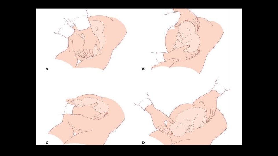
Vaginal delivery of breech Pre-requisites for vaginal breech delivery: • The presentation should be either extended (hips flexed, knees extended) or flexed • There should be no evidence of feto-pelvic disproportion and an estimated fetal weight of <3, 500 g (ultrasound or clinical measurement). • There should be no evidence of hyperextension of the fetal head, and fetal abnormalities

Vaginal delivery of breech Management of labour • Fetal wellbeing and progress of labour should be carefully monitored. • An epidural analgesia is not essential but may be advantageous? • Fetal blood sampling from the buttocks • Available operator experienced in delivery. Technique • It should be characterized by ‘masterly inactivity’ (handsoff). • Problems are more likely to arise when the obstetrician tries to speed up the process by pulling on the baby, and this should be avoided.

Vaginal delivery of breech • Delivery of the legs and lower body If the legs are flexed ? If extended > Pinard’s manoeuvre. ? • Delivery of the buttocks

• Delivery of the shoulders > Loveset’s manoeuvre • Delivery of the head > Mauriceau–Smellie–Veit manoeuvre

Head related abnormal presentations Face Brow

Head attitude Degree of extension-flexion of the fetal head with cephalic presentation. The most common attitude is vertex. • Vertex: head is maximally flexed • Military: head is partially flexed • Brow: head is partially extended • Face: head is maximally extended

Flexed Suboccipito bregmatic Deflexed Occipito-frontal Brow Mento-vertical Face Submento-bregmatic

Face presentation • Incidence and aetiology 1/500 • Associated with • Prematurity • Fetal goitre • Uterine anomalies • Polyhydramnios • Placenta praevia

Face: clinical finding • Diagnosis usually made in labour by vaginal examination • Landmarks: mandible, mouth, nose and orbital ridges • Avoid damage to the eyes on examination • Facial oedema: distinction between face and breech ? difficult • Ultrasound: if there is any doubt • Delay in the first or second stage of labour may occur

Face: management • Ultrasound: exclude fetal or pelvic abnormalities • Vaginal delivery: possible with the mento-anterior position • In the second stage : o Mento-anterior : head may deliver by flexion o Mento-posterior: may rotate during the second stage • Fetal risks: facial soft tissue trauma, causes feeding difficulties • Maternal risks: perineal injury, sphincter damage, CS • Augmentation: not advised • Lack of progress : prompt delivery by CS • Vacuum delivery: contra-indicated

Why mentoposterior does not deliver vaginally? • In vertex: delivery of fetal head occurs by extension • In face : head is already in maximum extension • In mentoposterior; the head, neck and shoulders enter the pelvis at the same time • The length of the sacrum is 10 cm • The length of neck is 5 cm • The shoulders get impacted • Labour is obstructed

Brow presentation Incidence and aetiology • 1/1000 deliveries • Due to a deflexed head Associated with o Prematurity o Fetal neck tumours

Brow: Clinical findings • In labour: failure to progress in first or second stage • Vaginal examination: forehead is the leading part • The anteroposterior diameter of the head is ‘mento-vertical’: about 13. 5 cm at term • The average anteroposterior and lateral diameters of the female mid-pelvis are 12 × 12 cm

Brow: management Diagnosis in the early first stage: • Expectant management for a short time (2– 3 hours) • May flex into a vertex or extend to face Diagnosis often made in late first or second stage: • Caesarean delivery is advised Augmentation with syntocinon: not advised “uterine Rupture” Mento-vertical dimensions may be smaller in a preterm fetus, allowing vaginal delivery

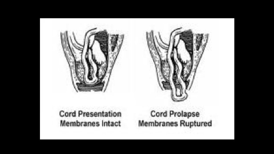
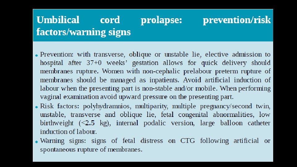
Cord presentation and prolapse Incidence and aetiology • Cord presentation/cord prolapse : 0. 1– 0. 6 % of all births • Cord presentation: Cord below presenting part, with the membranes intact • Cord prolapse: Cord descending through the cervix into the vagina with ruptured membranes • May follow fetal scalp electrode placement, stabilizing induction of labour, external cephalic version or internal podalic version

Clinical findings • Presence of a ‘high’ presenting part in early labour • Ultrasound: the presence of a cord presentation • In advanced labour, the findings are self-explanatory • The cord may be felt pulsating • Abnormal cardiotocography, should raise the possibility

Management Cord prolapse • An emergency delivery • Fetal hypoxia: o Pressure from the presenting part o Arterial spasm • The presenting part should be elevated (various methods) • The cord should be replaced in the vaginal with minimal handling • In the presence of viable fetus: Immediate delivery Cord presentation • May be seen by USS in preterm fetuses: No intervention • Usually diagnosed in labour by VE • If in labour: CS

Shoulder / compound presentation Compound presentation • More than one fetal part presenting Shoulder presentation • Shoulder is presenting Both • Associated with prematurity • Complicate unstable , ‘high’ head or breech Delivery: • Shoulder : CS if in labour • Compound: depends on the combination

Shoulder / compound presentation Compound presentation • More than one fetal part presenting Shoulder presentation • Shoulder is presenting Both • Associated with prematurity • Complicate unstable , ‘high’ head or breech Delivery: • Shoulder : CS if in labour • Compound: depends on the combination
")
Malpositions “Occipito-Posterior” (OP)

OP Prevalence • 15 to 32% at the onset of labour • 10 to 20% early in the second stage • 5 to 8% at delivery

OP: Consequences in Labour • Membranes rupture early • The forces push the head posteriorly: backache and urge to push before full dilatation • The occipito-frontal diameter reaches 10 cm; passage through the pelvis may be more difficult • The 1 st and 2 nd stages of labour may be prolonged • May rotate to OA or persists • If persistent OP in labour: o May deliver spontaneously: o If not: consider assisted delivery Rotation to OA by: Ventouse, Kielland Forceps or manual

Abnormal lie

Unstable/ transverse/ Oblique lies Incidence and aetiology 1/320 Association Multiparity Polyhydramnios Placenta praevia Pelvic tumour Uterine anomaly Contracted maternal pelvis Hydrocephalus and fetal neck tumours Fetal neuromuscular dysfunction “reduced FM”

Abnormal lies / Clinical findings The absence of a fetal pole in the pelvis on abdominal or vaginal examination

Abnormal lie: management Ultrasound scan • Confirm findings • Look for fetal-anomaly • Measure liquor volume • Check placental site • Pelvic tumours or uterine anomalies may be difficult to identify in late pregnancy

Abnormal lie: management • In the majority of cases: spontaneous version to longitudinal lie will occur prior to membrane rupture or labour onset • Inpatient management : from 37 weeks “ risk of cord prolapse” • Conservative Mx: Lie stabilised longitudinally for 48 H • Active Mx: ECV • ECV for unstable lie should only be done with immediate induction ‘stabilizing induction” • Stabilizing induction requires a favourable cervix • Should the patient present in early labour, ECV can be attempted

Abnormal lie: management Caesarean section: Should be planned at the appropriate gestational age ? 38 weeks Risk of cord prolapse in the event of contractions or rupture of membranes

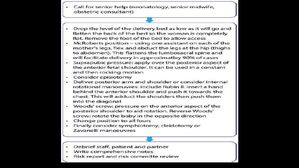
Shoulder dystocia • It is defined as a vaginal cephalic delivery that requires additional obstetric manoeuvres to deliver the fetus after the head has delivered and gentle traction has been unsuccessful in delivering the shoulders • It is associated with significant morbidity both for the mother and fetus.
 • postpartum")
Shoulder dystocia Maternal complications • increased perineal trauma (third- and fourthdegree tear) • postpartum haemorrhage • psychological trauma. Fetal complications • brachial plexus injury (2– 7% at birth reducing to 1– 3% at 12 months of age) • fractured clavicle or humerus (1– 2%) • hypoxic brain injury.

Shoulder dystocia Prevention: • Diagnosis and optimal control of gestational and insulin-dependent diabetics, reduction of maternal obesity. • Careful plan for mode of delivery in women with previous shoulder dystocia delivery (recurrence rate 10– 15%). Risk factors: • • • macrosomia poorly controlled gestational and insulin-dependent diabetes maternal obesity previous shoulder dystocia instrumental. Warning signs: • failure of restitution of head following delivery of the head • retraction of the fetal head against the perineum (analogous to a turtle withdrawing into its shell).

Thank you
- Slides: 51