Abnormal presentations Shoulder dystocia MARTYNA STANKIEWICZ Lie Presentation

Abnormal presentations Shoulder dystocia MARTYNA STANKIEWICZ

Lie, Presentation, • Fetal lie: The relation of the long axis of the fetus to that of the mother • Longitudinal lie is found in 99% of labours at term • Predisposing factors for transverse lie/oblique lie multiparity, placenta previa, hydramnious, & uterine anomalies • Fetal Presentation: The presenting part is the portion of the body of the fetus that is foremost in the birth canal • The presenting part can be felt through the cervix on vaginal examination • Longitudinal lie cephalic presentation breech presentation • Transvrse lie shoulder presentation

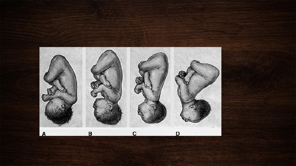
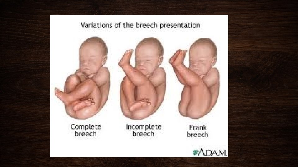
Lie, presentation, attitude, position CEPHALIC PRESENTATION • Head is flexed sharply vertex / occiput presentation • Head is extended sharply face presentation • Partially flexed bregma presenting (sinciput presentation) • Partially extended brow presentation BREECH PRESENTATION • Frank breech • Complete breech • Footling breech ATTITUDE • Posture of the fetus folded on itself to accommodate the shape of the uterus • Flexed head, thighs, knees &feet • The arms crossed over the chest • Face presentation extended concave contour of the vertebral column

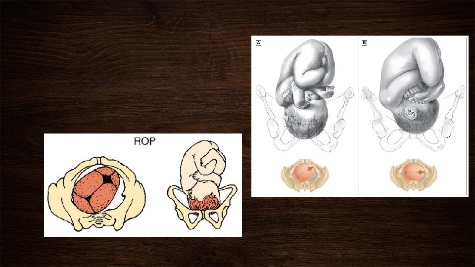
Position • The relations of an arbitrary chosen point of the fetal presenting part to the right or left side of the maternal birth canal • The chosen point: -Vertex presentation occiput -Face presentation ment Breech presentation sacrum Each presentation has two positions: right & left Each position has 3 varieties: anterior, transverse, posterior

Shoulder dystocia • occurs when, after delivery of the fetal head, the baby's anterior shoulder gets stuck behind the mother's pubic bone • the remainder of the baby does not follow the head easily out of the vagina as it usually does • generally reported to be between 0. 5 % and 1. 5% • Some studies have shown that only 25% to 50% of shoulder dystocias -- as noted by objective observers in a delivery room -are recorded by the delivering physician

Shoulder dystocia

"Is the rate of shoulder dystocia increasing? " • On average babies are significantly larger then in years past. The percentage of very large baby's (>4000 gms) in one study has gone up 300% between 1970 and 1988 • Over the last several decades there has been a marked increase in average maternal weight, average maternal weight gain during pregnancy, and the number of diabetic women having babies. All of these factors could be expected to increase the incidence of shoulder dystocia • The increased focus of attention among obstetricians about shoulder dystocia deliveries may have heightened awareness about it and increased reporting of it

Pelvic anatomy related to shoulder dystocia • The front-most bone is the symphysis pubis. It is on this structure that a baby's anterior shoulder gets caught during a delivery complicated by shoulder dystocia • The bone at the back of the maternal pelvis is the sacrum • The side walls of the maternal pelvis, although very important in determining the ease of the process of labor in general, usually do not contribute to shoulder dystocia

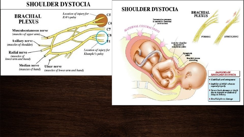
• THE BABY'S SHOULDERS, LIKEWISE BEING FLEXIBLE, USUALLY FOLLOW THE DELIVERY OF THE BABY'S HEAD QUICKLY AND EASILY • BUT FOR THIS TO HAPPEN, THE AXIS OF THE FETAL SHOULDERS MUST DESCEND INTO THE MATERNAL PELVIS AT AN ANGLE OBLIQUE TO THE PELVIS'S ANTERIOR-POSTERIOR DIMENSION • THIS POSITION AFFORDS THE SHOULDERS THE MOST ROOM FOR THEIR PASSAGE • IF INSTEAD THE SHOULDERS LINE UP IN A STRAIGHT FRONT-TOBACK ORIENTATION AS THEY ARE ABOUT TO EMERGE FROM THE MOTHER'S PELVIS • THERE WILL OFTEN BE INSUFFICIENT ROOM FOR THEM TO SQUEEZE THROUGH THE BACK OF THE MOTHER'S PUBIC BONE THEN FORMS A SHELF ON WHICH THE BABY'S ANTERIOR SHOULDER CAN GET CAUGHT If this happens, the shoulders cannot deliver and a shoulder dystocia results

Shoulder dystocia • Can also occur if the posterior shoulder of a baby gets caught on its mother's sacrum • Usually it is the fetal head that has the largest dimensions • Thus if it can pass through the maternal pelvis without difficulty, the rest of the baby usually follows easily • When the dimensions of the fetal shoulders or chest rival those of its head, the chances of a shoulder dystocia occurring are much increased • Such situations occur more frequently both in large babies and in babies of diabetic mothers • In large babes ( especially in diabetic mothers) the dimensions of the shoulders and chest tend to be disproportionately larger than those of the head • the risk of recurrent shoulder dystocia is substantial: 10 to 15%

Brachial plexus injury

• Preconceptual risk factors for shoulder dystocia-before pregnancy 1. Previous shoulder dystocia( 1112% risk) 2. 2. Maternal obesity 3. Maternal age Risk factors Shoulder dystocia 4. Abnormal pelvis-there is no evidence linking the "abnormal pelvis" to shoulder dystocia 5. Multiparity

Infant weight in Nondiabetic women Percent shoulder dystocia Less than 4000 g 1. 1% 4000 g - 4499 g 10. 0% Greater than 4500 22. 6% g • Antepartum factors risk factors for shoulder dystocia-during pregnancy 1. Macrosomia 2. Shoulder/chest/abdomen ratios 3. Diabetes 4. Maternal weight gain Risk factors Shoulder dystocia 5. Multiparity 6. Post- dates

• Intrapartum risk factors-during labor 1. Instrumental delivery 2. Experience of the deliverer 3. Labor abnormalities -extended/ rapid second stage Risk factors Shoulder dystocia

• The baby's body does not emerge with standard moderate traction and maternal pushing after delivery of the fetal head Recognition Shoulder dystocia • The "turtle sign". This is when the fetal head suddenly retracts back against the mother's perineum after it emerges from the vagina. The baby's cheeks bulge out, resembling a turtle pulling its head back into its shell. This retraction of the fetal head is caused by the baby's anterior shoulder being caught on the back of the maternal pubic bone, preventing delivery of the remainder of the baby

Recogniotion Shoulder dystocia

Management Shoulder Dystocia

• Involves sharply flexing the legs upon the maternal abdomen • By doing this, the symphysis pubis is rotated cephalad and the sacrum is straightened Mc. Roberts maneuver Shoulder dystocia • In a high percentage of cases this by itself suffices to free the impacted anterior shoulder

• The attempt to manually dislodge the anterior shoulder from behind the symphysis pubis during a shoulder dystocia • It is performed by an attendant making a fist, placing it just above the maternal pubic bone, and pushing the fetal shoulder in one direction or the other Suprapubic pressure Shoulder dystocia • Since shoulder dystocias are caused by an infant's shoulders entering the pelvis in a direct anterior-posterior orientation instead of the more physiologic oblique diameter, pushing the baby's anterior shoulder to one side or the other from above can often change its position to the oblique which will allow its delivery • Suprapubic pressure in conjunction with Mc. Roberts maneuver is often all that is needed to resolve 50 -60% of shoulder dystocias
")
• Pressure is applied on the posterior shoulder's anterior surface (Wood's Screw maneuver) • A variation of this -- the Rubin's maneuver -- involves pushing on the posterior surface of the posterior shoulder Rotate posterior arm Shoulder dystocia • In addition to the corkscrew effect, pressure on the posterior shoulder has the advantage of flexing the shoulders across the chest • This decreases the distance between the shoulders, thus decreasing the dimension that must fit out through the pelvis

• the obstetrician places his or her hand behind the posterior shoulder of the fetus and locates the arm • this arm is then swept across the fetal chest and delivered Delivery of the posterior shoulder Shoulder dystocia • with the posterior arm and shoulder now delivered, it is relatively easy to rotate the baby, dislodge the anterior shoulder, and allow delivery of the remainder of the baby • the major risk of this procedure is that of fracturing the humerus

• flexing the fetal head and pushing it back up into the vagina • get the fetal head back into the pelvis, perform an emergency cesarean section, and deliver a live baby • the head must first be rotated back to its pre-restitution position -- that is, occiput anterior -- and then flexed Zavanelli maneuver Shoulder dystocia • constant firm pressure is applied while pushing the head back into the vagina • tocolytic agents or uterine-relaxing general anesthesia may be administered to facilitate this process • cesarean section must be performed immediately after replacement of the head

Deliberate fracture of the clavicle Shoulder dystocia • although the fracture of the clavicle certainly would decrease the transverse diameter of the chest and shoulders, the potential of damaging the great vessels, fetal lungs, and other structures make this an extremely hazardous procedure even if it were possible to perform easily

• only in areas remote from the ability to perform Cesarean sections on a rapid basis • thing of a renaissance in the literature in recent years. The theory is that by transecting the firm ligaments joining the left and right symphyseal bones, an additional 2 -3 cm in pelvic circumference can be gained Symphysiotomy Shoulder dystocia • in most cases this will allow the anterior shoulder to be delivered beneath the symphysis • the benefit : it can be performed rapidly (usually takes 5 minutes or less and can be done under local analgesia)

• involves placing the gravid mother on her hands and knees • another means of changing the angle of the symphysis in relation to the stuck shoulder Gaskin Maneuver Shoulder dystocia

Physical injuries following shoulder dystocia deliveries Shoulder dystocia • Brachial plexus injuries ( Erb palsy: C 5 -C 7; Klumpke palsy: C 7, C 8, Th 1) • Fractured clavicle • Fractured humerus • Contusions and lacerations • Fetal asphyxia

• Significant blood loss • Vaginal and vulvar lacerations Maternal injuries Shoulder dystocia

The End
- Slides: 32