ABDOMINAL XRAY RADIOLOGICAL SIGNS Normal AXR Liver 11


ABDOMINAL X-RAY RADIOLOGICAL SIGNS

Normal AXR Liver 11 th rib T 12 Gas in stomach Splenic flexure Psoas margin Left kidney Hepatic flexure Transverse colon Iliac crest Gas in sigmoid Sacrum Gas in caecum Bladder SI joint Femoral head

Focus on film - what looking for? VIEW LOOK FOR SUPINE ABDOMEN Bowel gas pattern Calcifications Masses PRONE ABDOMEN Gas in rectosigmoid Gas in ascending and descending colon UPRIGHT ABDOMEN Free air, air-fluid levels UPRIGHT CHEST Free air, lung pathology secondary to intraabdominal process
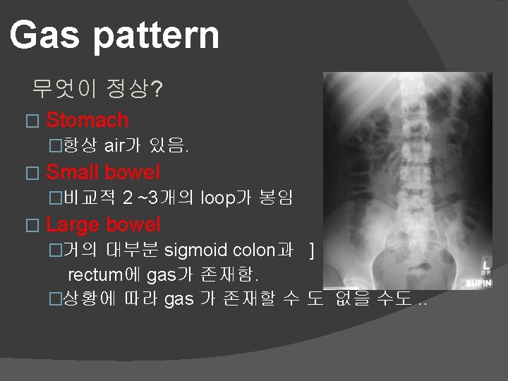

Normal fluid levels � Stomach �Upright position에서는 항상 � Small bowel �Upright position에서 2 ~3 개의 loop가 보임 � Large bowel �없음. (생리적으로 fluid를 재흡수 하는 곳)

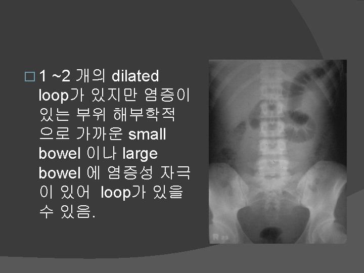
Abnormal Gas Patterns � Functional ileus �One or more bowel loops become aperistaltic usually due to local irritation or inflammation ○ Localised “sentinel loops” (one or two loops) ○ Generalised (all loops of large and small bowel) � Mechanical obstruction �Intraluminal or extraluminal ○ Small bowel obstruction ○ Large bowel obstruction

3, 6, 9 RULE Maximum Normal Diameter of bowel Small bowel 3 cm Large bowel 6 cm Caecum 9 cm

Causes of Localised Ileus by location SITE OF DILATED LOOPS Right upper quadrant Left upper quadrant Right lower quadrant CAUSE Cholecystitis Pancreatitis Appendicitis Left lower quadrant Mid-abdomen Diverticulitis Ulcer or kidney/ureteric calculi

Generalised ileus Key features 전체적인 장음이 낮아진 상태 aperistaltic/hypoperistaltic � 소대장이 전장에 걸쳐 Dilated 된 것을 볼 수 있음. (rectum/sigmoid 에 gas 가 없을 수 있음. ) � Long air-fluid levels � CAUSE REMARK *Postoperative Usually abdominal surgery Electrolyte imbalance Diabetic ketoacidosis * almost always

Generalised adynamic ileus

Mechanical Small Bowel Obstruction � Dilated small bowel � air-fluid levels at different levels � Little gas in colon, especially rectum

SBO Erect Air fluid levels SBO Supine

Causes of Mechanical SBO Adhesions Hernia* Malignancy Gallstone ileus* Intussesception Inflammatory bowel disease

Step ladder appearance

Coil spring sign
 and is")
String of pearls sign Considered diagnostic of obstruction (as opposed to ileus) and is caused by small bubbles of air trapped in the valvulae of the small bowel.

Mechanical Large Bowel Obstruction � Leading point 이전에 Colon dilation � Little/no air fluid levels (colon reabsorbs water) � Little or no air in rectum/sigmoid

Causes of Mechanical LBO TUMOUR VOLVULUS HERNIA DIVERTICULITIS INTUSSUSCEPTION
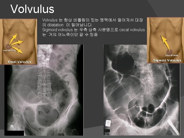

Coffee Bean Sigmoid volvulus Massively dilated sigmoid loop

Apple core sign contrast material enema 시행 시 보이 focal defect 가 보이는 것. � 환상(annular) 의 대장 암 이 m/c. �

Extraluminal air � TYPES �Pneumoperitoneum/free air/intraperitoneal air �Retroperintoneal air �Air in the bowel wall (pneumatosis intestinalis) �Air in the biliary system (pneumobilia)

Pneumoperitoenum 의 원인 Rupture of a hollow viscus �Perforated peptic ulcer �Trauma �Perforated diverticulitis (usually seals off) �Perforated carcinoma

Signs of free air � Crescent sign � Chilaiditis sign � Riglers (and False Rigler’s) � Football sign � Falciform ligament sign � Triangle sign � Cupola sign � Lesser sac sign
 에서 보이며 Rt.")
Crescent Sign Free air under the diaphragm erect view(erect abdomen, chest) 에서 보이며 Rt. Side 에서 잘 보임

Retroperitoneal Air � Recognised by: �후복벽의 장기 outline을 따라 보여지며 air가 얼룩 덜룩하게 보여짐. �상대적으로 고정되어 있음. � May outline: �Psoas muscles �Kidneys, ureters, bladder �Aorta or IVC �Subphrenic spaces
 – Duodeum 2 nd")
Causes of retroperitoneal air � Bowel perforation (appendix, ileum, colon) – Duodeum 2 nd portioin perforation � Trauma (blunt or penetrating) � Iatrogenic � Foreign body � Gas producing infection

Pneumatosis intestinalis � � Intramural air Old age 에무증상 으로 있는 경우 있 지만 드문 경우 수 술 후 혹은 ischemic bowel disease 가 의심되 는 환자에 있어서 acidosis 가 동반되 면 fetal 한 결과 오 며 특히 neonatal에 서 necrotizing intestinalis 의 소견 일 수 있다.

Air in the biliary tree � One or two tube-like branching lucencies in the RUQ, conform to location of major bile ducts

Causes � Sphincter of Oddi incompetence – old age � 이전 ERCP난 수술 에서 sphincterotomy를 했 거나 transplantation of CBD 을 한 경우 � Pathology (uncommon) �Gallstone ileus: GB 와 인접해 있는 기관( duodeum, ileum 등. . )에 염증과 유착으로 인해 gallstone 이 장관으로 들어가 기계적 장폐쇄증 (mechanical ileus)를 일으키는 것.

Abdominal Calcifications Location Pattern

Location � � � Vascular Liver Gallbladder Spleen Pancreas Lymph nodes Adrenals Kidneys Ureters Bladder Prostate

Rim-like � Calcification that has occurred in the wall of a hollow viscus �Cysts ○ renal, splenic, hepatic �Aneurysms ○ aortic, splenic, renal artery �Saccular organs ○ Gallbladder ○ Urinary bladder Calcified hydatid cysts

Linear/Track � Calcification in walls of tubular structures �Arteries �Fallopian tubes �Vas deferens �Ureter Aortoiliac calcification

Chinese Dragon Sign Calcified splenic artery

Calcified vas deferens

Bladder calculi Lamellar

Renal calculi Pelvicalyceal calcifications

Staghorn Calcification Tubular Renal stones are often small, but if large can fill the renal pelvis or a calyx, taking on its shape which is likened to a staghorn.

Renal calculi Parenchymal calcification Nephrocalcinosis Uncommonly the renal parenchyma can become calcified. This is known as nephrocalcinosis, a condition found in disease entities such as medullary sponge kidney or hyperparathyroidism. Flocculent
 � Autonephrectomy")
Putty Kidney � "Putty kidney" – sacs of casseous, necrotic material (TB) � Autonephrectomy – small, shrunken kidney with dystrophic calcification Flocculent

Calcified gallstones Lamellar

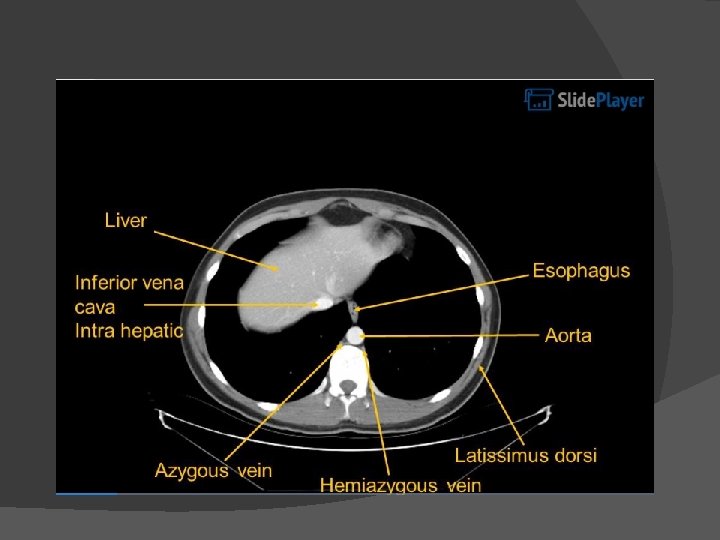
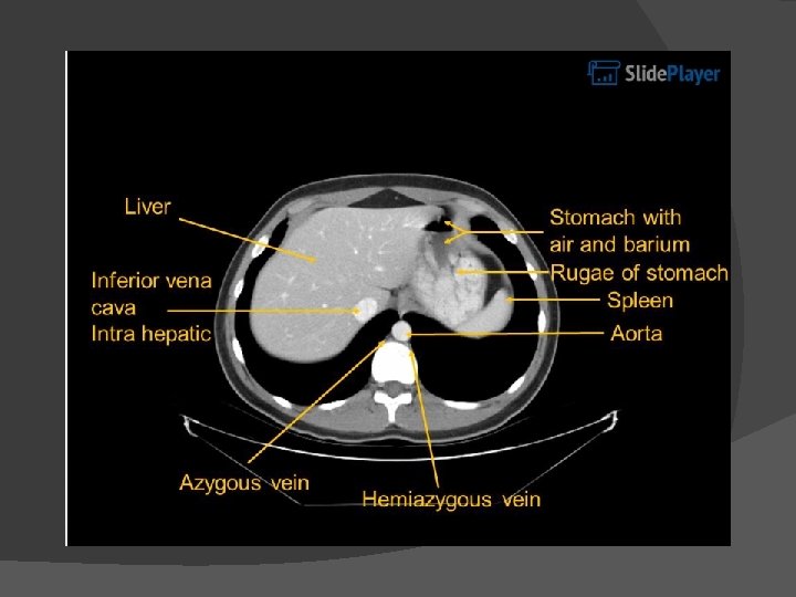
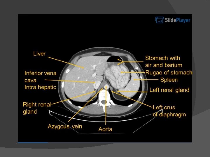
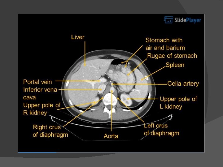
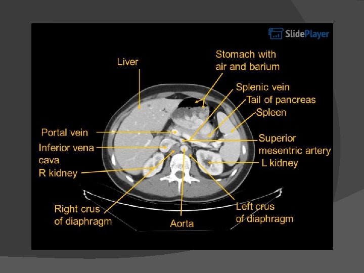
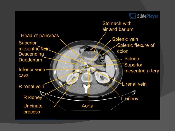
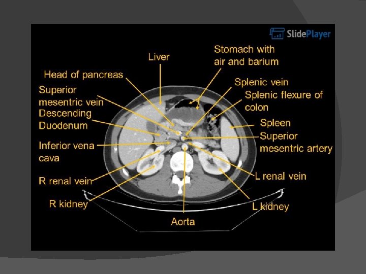
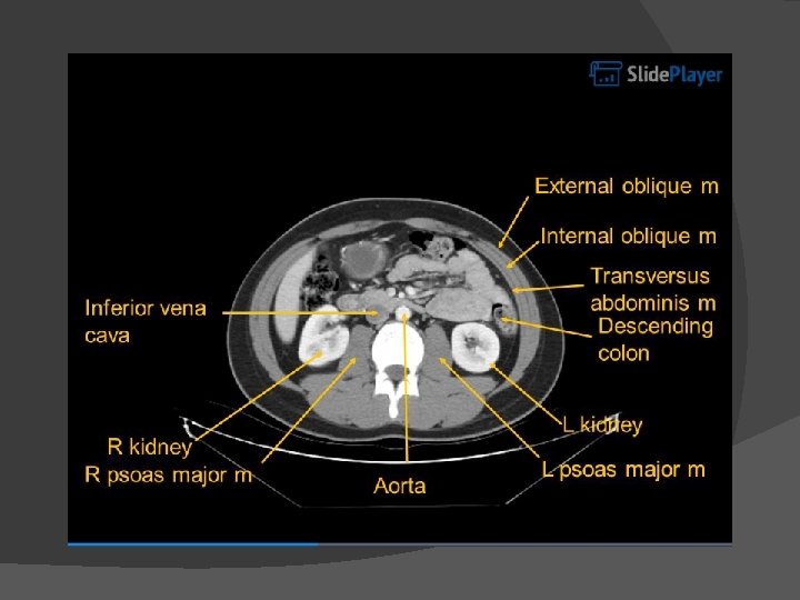
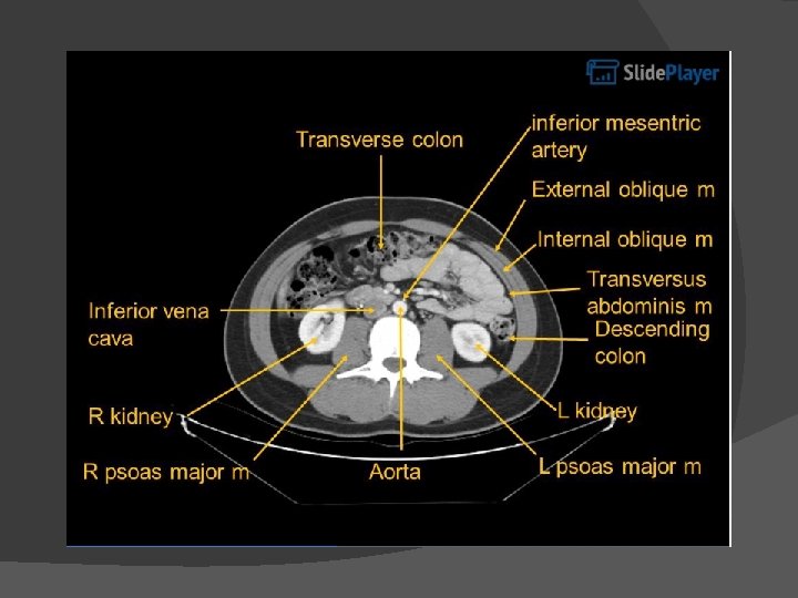
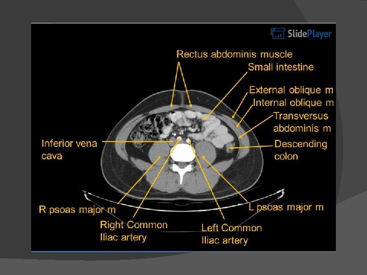
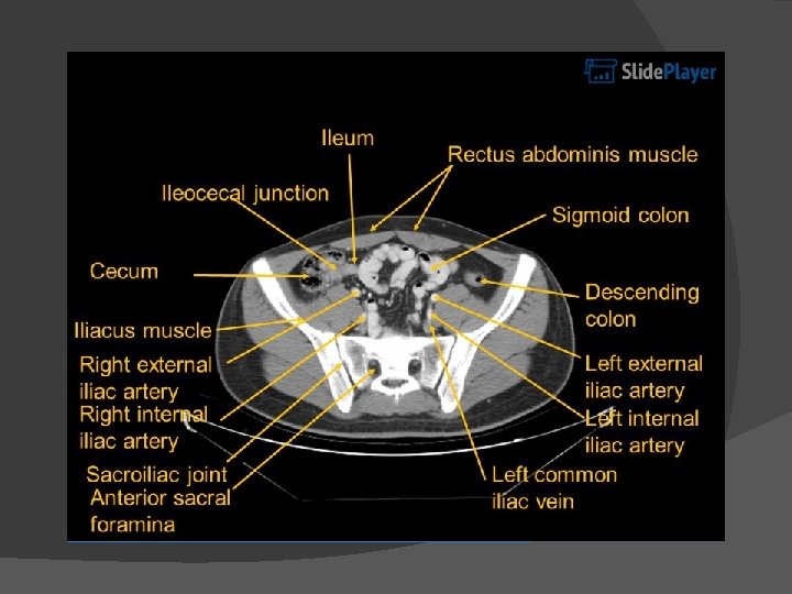
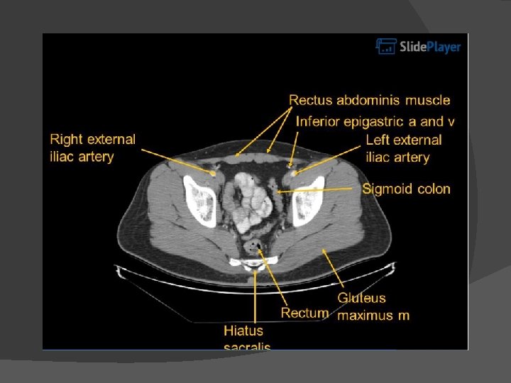
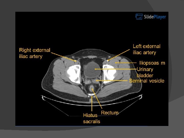
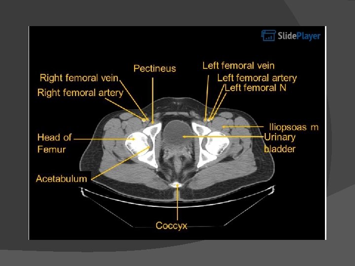
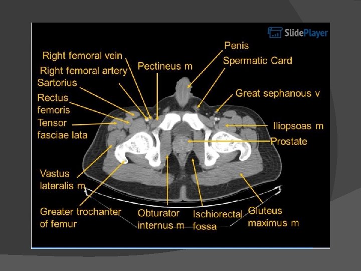
Abdominal CT

Gallbladder

GB wall thickening Cholecystitis - acute - chronic - acalculous � GB cancer � Adenomyomatosis � Liver cirrhosis � Hepatitis � Congestive right hear failure � Renal failure � Pancreatitis �

Acute cholecystitis

Chronic cholecystitis

GB stone, GB polyp USG

Adenomayomatosis

GB cancer

Choledocholithiasis 담도관내 담석증 Non-contrast image CBD dilatation 동반.

Liver

Liver cirrhosis

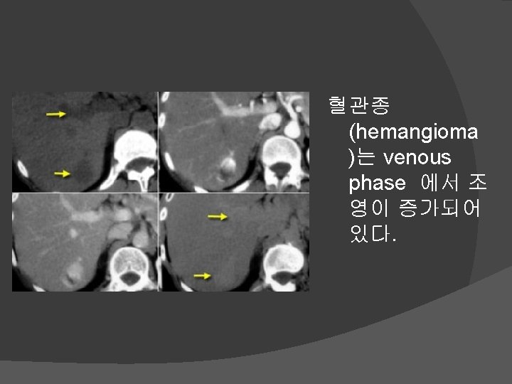
Liver mass HCC Vs hamangioma � Dynamic CT 상에서 Hepatoma 는 arterial phase 시 종영이 증 강이 되다가 venous phase 시 급속도록 조영제가 빠져 나가 mass가 보이지 않는다.

Acute pancreatitis

Chronic pancreatitis

Pancreatic cancer

Renal cyst
")
Stomach mass (r/o stomach ca. )

Ulcer perforation

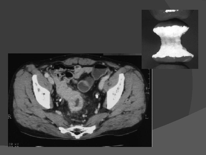
Appendicitis

Cecum colitis

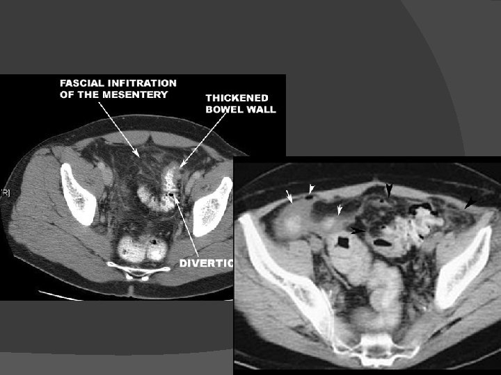
Diverticulitis Diverticulosis
")
Intestinal obstruction (mechanical ileus)

Colon cancer

Rectal cancer
- Slides: 90