Abdominal Wall Reconstruction 43 rd Annual Phoenix Surgical

Abdominal Wall Reconstruction 43 rd Annual Phoenix Surgical Symposium Scottsdale, AZ Elvis Presley Memorial Trauma Center UTHSC Department of Surgery Memphis, TN

I have no conflicts to disclose

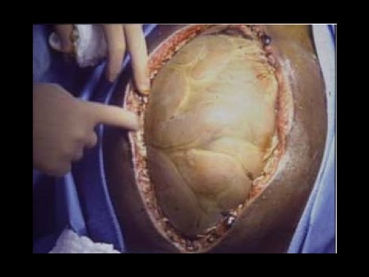
Etiology of Defect • Abdominal wall loss Necrotizing fascial infection § Close range shotgun blast § • Planned re-exploration § Diffuse suppurative peritonitis Pancreatic sepsis • Massive edema § § Resuscitation from shock § Resuscitation from sepsis

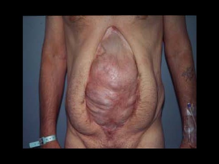
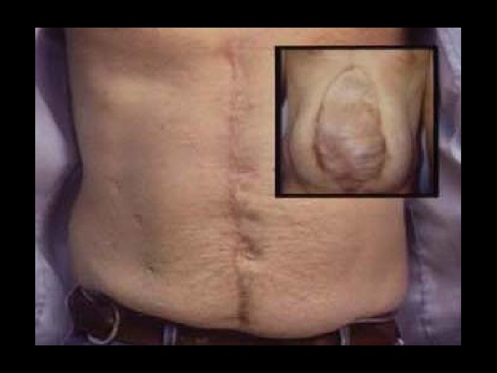
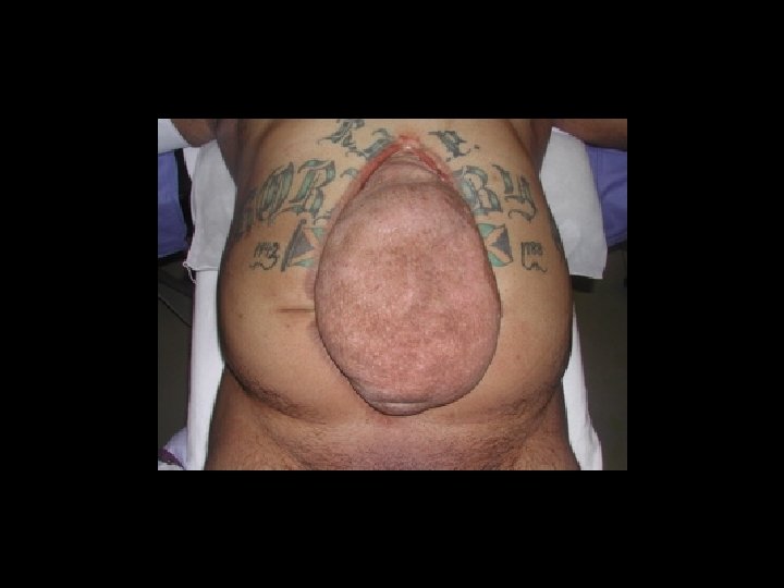
Abdominal Wall Reconstruction - Staged Management Technique - Stage I Prosthetic insertion 14 - 21 days Stage II Planned ventral hernia 8 - 12 months Stage III Definitive reconstruction

Abdominal Wall Reconstruction - Staged Management Technique - Stage I Prosthetic insertion Stage II Planned ventral hernia Stage III Definitive reconstruction

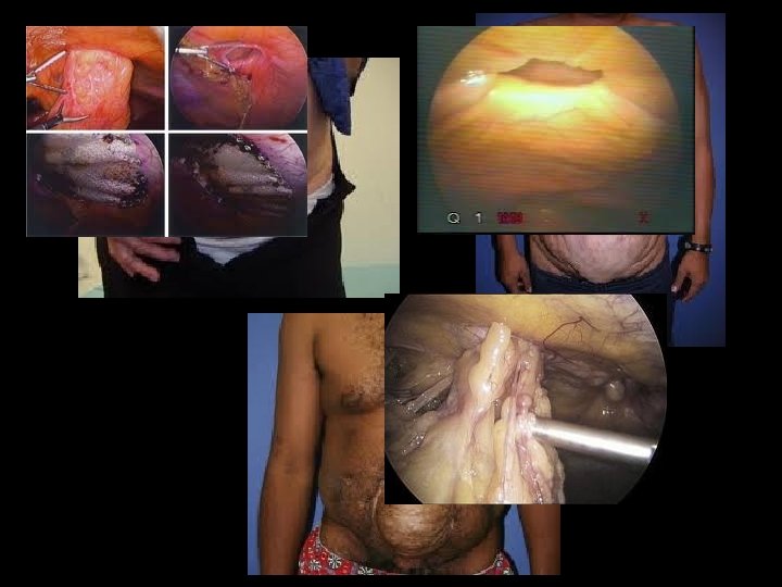
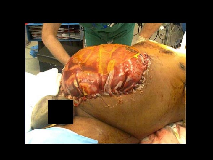
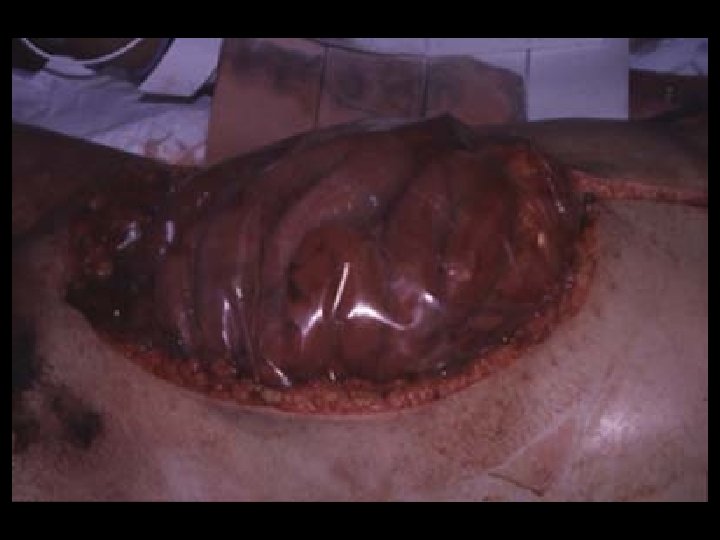
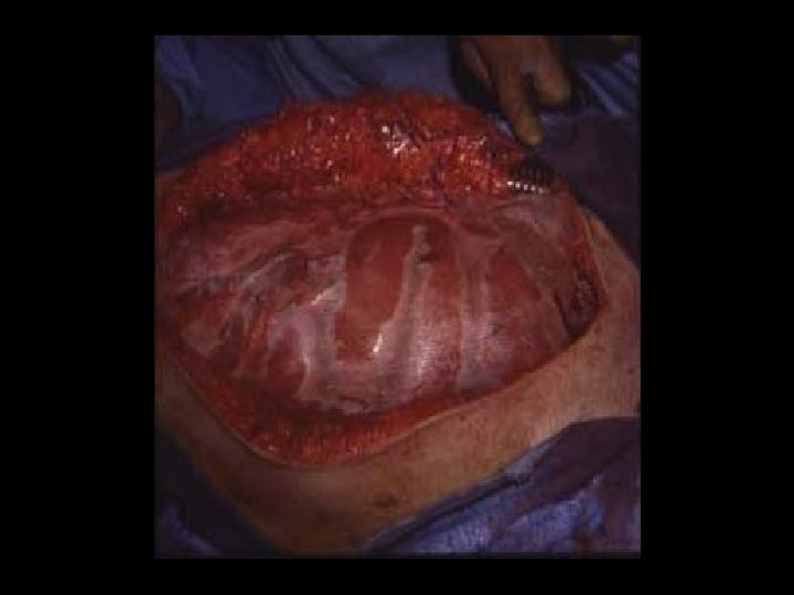
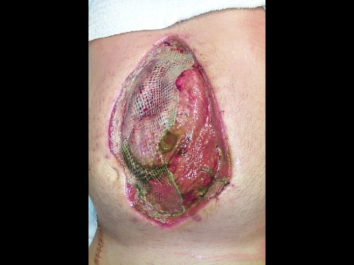
Multiple Methods for Coverage

Acute Coverage Recommendations - Plastic • Massive visceral edema • Gauze packing • ? Planned re-exploration

Acute Coverage Recommendations - Polypropylene -
 No 2 --")
Fate of Marlex Mesh Closure Retention Hernia Fistula Extrus Primary (6) No 2 -- -- Granulation (6) Yes 1 1 5 STSG (9) Yes 3 3 9 Skin & subcut (3) Yes 1 -- -- Voyles, Ann Surg 1981

PPM Use in Emergency Setting - Summary of 14 Reports - Patients 128 Fistula (%) 23 Hernia (%) 20 Extrusion (%) 12 Jones & Jurkovich, Am Surg 1989

Acute Coverage Recommendations - PTFE -

Acute Coverage Recommendations - Absorbable • Massive visceral edema • Abdominal wall loss • Planned re-exploration • Gauze packing

Vacuum Assisted

Delayed Fascial Closure - negative pressure - 53 with open abdomen 43 attempted closure 38 closed 88% Miller, Ann Surg 2004

Delayed Fascial Closure - negative pressure 74% sepsis 209 patients 12% ischemia 14% other 112 patients 35% incisional hernia Brandl, Hernia 2013


Patient Population - April 2003 to July 2007 - 17, 379 patients admitted 1343 emergent laparotomy 248 open abdominal management 51 met inclusion criteria 31 VAC 20 MESH

Outcomes VAC MESH % Abdominal abscess 12 9 % Delayed fascial closure 31 26 % Fistula 21 5

GI Tract Fistulae SB suture line MESH VAC Tube Enterostomy 0 1 2 3

Summary • Benefits of VAC - quick placement - sutureless - able to remove excess fluid - able to reexamine abdominal contents regularly • Benefits of MESH - transparent - pleated by bedside - permanent feeding tubes not a problem - place on all body habitus and loss of domain

Summary • Disadvantages of VAC - not practical with tube enterostomy - not transparent - commercial version expensive - not useful with loss of domain • Disadvantages of MESH - fascial sutures - difficult to quantitate fluid loss - more expensive than traditional VAC

Vac/Mesh Abdominal Closure - delayed fascial closure Prospective study, 4 sites 151 open abdomen 111 vac/mesh 99 fascial closure 89% Acosta , Br J Surg 2011

Vac/Mesh Abdominal Closure - 1 year follow up. Prospective study, 4 sites 111 patients with vac/mesh 64 survivors 23 clinical hernia 36% 23 no hernia 36% 18 CT hernia 36% Bjarnason, World J Surg 2013

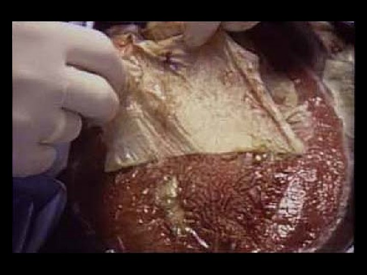
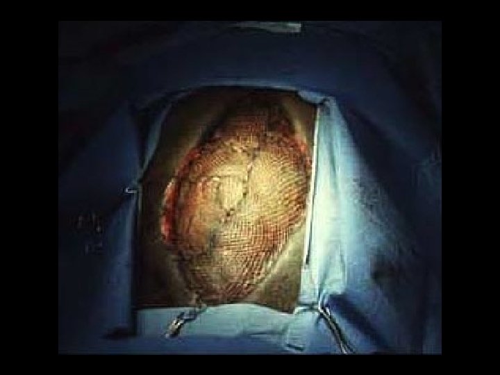
Abdominal Wall Reconstruction - Staged Management Technique - Stage I Absorbable mesh insertion 14 - 21 days Stage II Planned ventral hernia Stage III Definitive reconstruction


Timing of Wound Coverage and Fistulae Mesh days No fistula n = 101 Fistula n = 14 p - value 18. 1 < 0. 04 26. 5 Jernigan Ann Surg 2003

Timing of Fistula Development in the Open Abdomen Pre-Mesh With Mesh Post-Mesh Fischer, et al. J Trauma 2009

Wound VAC- “Floating Stoma”

Abdominal Wall Reconstruction - Staged Management Technique - Stage I Absorbable mesh insertion 14 - 21 days Stage II Planned ventral hernia 8 - 12 months Stage III Definitive reconstruction

Reconstructive Techniques Advantages Disadvantages Prosthetic materials simple hernia, fistulae, infection Biomaterials simple dissolution, long term integrity? Muscle flaps autogenous complex, little experience Components separation autogenous, simple inadequate for large defects Modified comp. separation autogenous, simple hernia, fistula rates?

Single Stage Components Repair - 26 patients • All with contaminated abdominal wall defects – 19 abdominal sepsis – 4 visceral edema – 3 wound issues • Mean defect size 267 cm 2 • 15% adjunctive mesh • 8% recurrence rate @ mean 27 months (13 -78 months) van Geffen, J Am Coll Surg, 2005

Components + Dermal Allograft - 16 patients • Incisional hernias • 56% with infection • Component separation repair with under& overlay with Allo. Derm • No recurrent hernias, mean 16 months (9 -23 months) Kolker, Ann Plas Surg, 2005

Reported Series n Months Follow-up Recurrence De Vries Reilingh 2003 43 15. 6 28% Lowe 2003 30 9. 5 10% Jernigan 2003 73 24 6% De Vries Reilingh 2007 37 36 57% Rodriguez 2007 23 7 9% De Moya 2008 6 12 100% Diaz 2009 165 10. 4 25% Ko 2009 200 10. 3 22% Total 577 13. 9 23%

Extreme Variability • Fascial approximation – tension • Mesh bridge – Fistula, infection • Mesh under/overlay – Infection • Biomaterial bridge – Laxity, wound issues • Biomaterial under/overlay – Wound issues

The gazelle doesn’t have to be as fast as the cheetah, it only has to be faster than the slowest gazelle.

Components Separation • Release of external oblique to gain extra length in midline (3 -5 cm on each side) Ramirez et al, 1990

Components Separation • Variable rate of recurrence, depending on follow-up interval • May require prosthetic for large defects Ramirez et al, 1990

Modified Components Separation • Components separation, plus: – Dissection of posterior sheath from the rectus – Division of anterior component of internal oblique down to arcuate line – Suture medial border of posterior rectus sheath to lateral border of anterior rectus sheath Fabian et al, 1994

Modified Components Separation • Gives additional length, 10 -15 cm • Often avoids the use of a prosthetic Fabian, Ann Surg, 1994

Timing of Reconstruction not considered until skin graft is easily separated from viscera

Release of External Oblique

Posterior sheath mobilized from rectus abdominus

Division of Posterior Components

Approximation of the separated components

Completed repair

152 Patients 38 Lost to follow-up 114 Long term follow-up 98 Successful Repair 75% 16 Recurrence 11 Salvage repair
 NS + prosthetic Components Separation (CS) CS + prosthetic")
Surgical Techniques No Separation (NS) NS + prosthetic Components Separation (CS) CS + prosthetic Modified Components Separation (MCS) MCS + prosthetic Di. Cocco J Am Coll Surg 2010

Study Population - 114 patients Nontrauma Female Blunt Penetrating Male Age 35 BMI 27 Defect size 620 cm 2

Methods of Repair NS CS MCS n=13 n=34 n=67 Age 32 33 37 BMI 29 27 27 % Female 15 15 18 % Prosthetic use 46 26 45 % With ostomy 23 21 15 % With fistula 23 18 16 % Recurrence 15 18 12

Methods of Repair No Separation Components Separation Modified Components

Methods of Repair 5% 20% 8% 0% 44% 33% No Separation Components Separation Modified Components

Timing of Reconstruction % with prosthetic < 12 months p - value 6 0. 04 > 12 months 25

n Months Follow-up Recurrence De Vries Reilingh 2003 43 15. 6 28% Lowe 2003 30 9. 5 10% Jernigan 2003 73 24 6% De Vries Reilingh 2007 37 36 57% Rodriguez 2007 23 7 9% De Moya 2008 6 12 100% Diaz 2009 165 10. 4 25% Ko 2009 200 10. 3 22% Total 577 13. 9 23% Memphis Study 114 63. 6 14% p=0. 03

Timing of Recurrences Previous studies limitations: • Inadequate follow-up • Missed recurrences • Higher recurrence rates

I approve!

Summary • Open abdomens save lives • Fistulas associated with prolonged time to coverage • Optimal time for reconstruction is 8 – 12 months to avoid loss of domain • Try to cover the mesh • Lowest long term recurrence rate seen with modified components separation

- Slides: 70