Abdominal Sonography Part II Lecture 17 Abdominal Organ

Abdominal Sonography Part II Lecture 17 Abdominal Organ Transplants �Liver �Kidneys �Pancreas Holdorf

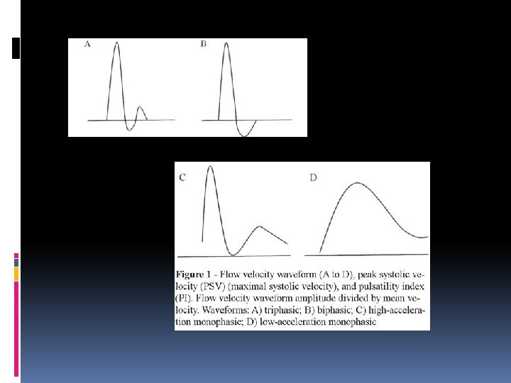
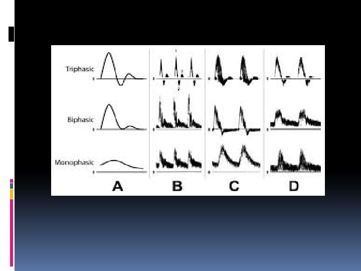
A brief Doppler Spectral analysis recap: Biphasic Triphasic Monophasic

Vascular Technology: Doppler has three sounds: monophasic, biphasic and triphasic. These sounds relate to how well the vascular system is functioning. How do the changes in the vascular system cause the different sounds that are produced? Regarding blood flow in peripheral arteries the typical pattern of flow in a resting state is to accelerate quickly then stop and even briefly flow backwards as a pressure wave traveling faster than the blood itself reflects back from the smaller arterioles near the end of the pathway blood follows.

Because the reflected pressure wave stops and even briefly reverses the flow of blood in the artery, two components are heard/seen. That type of pattern is called biphasic and is normal. Also normal is a pattern where the blood goes forward, reverses briefly then forward again (because the large artery leaving the heart - the aorta - has an elastic wall and stretches like a balloon when the heart contracts. A valve at the start of the aorta stops blood from flowing back into the heart. The elastic walls returning to their normal shape propel blood away from the heart throughout the entire heartbeat, not just when the heart is squeezing).

This elastic recoil pushes the blood forward again giving the Doppler noise we hear a third component (triphasic). If the artery being listened to have a severe blockage, the arterioles will open (vasodilation) to allow more flow in an attempt to deliver the amount of blood required by parts of the body downstream. That stops the reflection of the pressure wave giving a flow pattern with only one sound - in a forward direction - which is described as monophasic.

Biphasic and triphasic flow patterns are normal in a resting person. Monophasic flow generally indicates significant peripheral vascular disease. A person can have peripheral vascular disease even if their Doppler waveforms are bi or triphasic but some form of exercise testing may be required to reveal it. This type of testing is best done in an accredited vascular lab.

Triphasic Doppler Waveform

Ultrasound Examination of Solid-Organ Transplants Indications/Contraindications Indications for an ultrasound examination of a solid-organ transplant include but are not limited to the following:

A. Liver Transplant 1. Performance of a screening ultrasound examination to establish a baseline after transplantation as per the hospital surveillance protocol 2. Follow-up of abnormal findings on a prior transplant ultrasound examination; 3. Assessment of the transplant in the setting of abnormal liver function tests 4. Evaluation for pain, fever, sepsis, or laboratory abnormalities 5. Evaluation for a possible fluid collection or assessment of drainage catheter output; 6. Assessment of the biliary tree for dilatation, a stricture, Biloma, or an abscess; 7. Evaluation for vascular patency; and 8. Evaluation for malignancy, either recurrent or post-transplant lymphoproliferative disorder

B. Renal Transplant 1. Performance of a screening ultrasound examination to establish a baseline after transplantation as per the hospital surveillance protocol; 2. Follow-up of abnormal findings on a prior transplant ultrasound examination; 3. Evaluation for pain, fever, sepsis, or abnormal laboratory or clinical values (eg, elevated creatinine and low or decreased urine output); 4. Evaluation for vascular patency; 5. Assessment of hematuria or known or suspected hydronephrosis, hydroureter, or bladder abnormality; 6. Evaluation for a possible fluid collection or assessment of drainage catheter output; 7. Evaluation of the transplant in the setting of hypertension or bruit; 8. Evaluation for iatrogenic injury or complications after biopsy of a transplanted kidney; and 9. Evaluation for malignancy, either recurrent or post-transplant lymphoproliferative disorder.

C. Pancreas Transplant 1. Performance of a screening ultrasound examination to establish a baseline after transplantation as per the hospital surveillance protocol; 2. Follow-up of abnormal findings on a prior transplant ultrasound examination. 3. Assessment of graft dysfunction in patients with abnormal laboratory values or clinical parameters (eg, elevated blood glucose); 4. Evaluation for suspected stenosis or thrombosis of the vasculature; 5. Evaluation of pain at or near the surgical site; 6. Evaluation of the response to treatment (eg, immunosuppressive therapy in the setting of rejection); 7. Evaluation for iatrogenic injury or complications after biopsy of a transplanted pancreas; and 8. Assessment of the transplant in the setting of infection or pancreatitis.
, or pancreas should be performed when")
An ultrasound examination of the transplanted liver, kidney(s), or pancreas should be performed when there is a valid medical reason.

Living Donor

Orthotopic liver transplantation refers to a procedure in which a failed liver is removed from the patient's body and a healthy donor liver is transplanted into the same location. The procedure is the most common method used to transplant livers.

Liver Donor Requirements Any member of the family, parent, sibling, child, spouse or a volunteer can donate their liver. The criteria for a liver donation include: Being in good health Having a blood type that matches or is compatible with the recipient's, although some centers now perform blood group incompatible transplants with special immun suppression protocols Having a charitable desire of donation without financial motivation Being between 18 and 60 years old Being of similar or bigger size than the recipient Before one becomes a living donor, the donor must undergo testing to ensure that the individual is physically fit. Sometimes CT scans or MRIs are done to image the liver. In most cases, the work up is done in 2– 3 weeks.

Indications for Liver Transplant Primary or secondary biliary cirrhosis Chronic active hepatitis with cirrhosis Liver abscesses Fatty liver infiltrates Liver Ca 18

Liver Transplant Complications Graft rejection Manifestations of graft rejection are: Fever Tachycardia RUQ or flank pain Diminished bile flow through t-tube or change in bile color Increased bilirubin Increased jaundice

Treatment for Cirrhosis of Liver Injection sclerotherapy: varices are sclerosed TIPS: Transjugular intrahepatic portosystemic shunt – placement of a portal shunt for esophageal varices Surgical bypass shunting procedures: Last resort: Ascites is shunted from the abdominal cavity to the superior vena cava Liver Transplantation 20

Liver Cancer Risk Factors Cirrhosis Metastasis from another site Dx (Patient has a history of…) AFP: Alpha-fetoprotein tumor marker Liver enzymes ALP elevated Liver biopsy: definitive diagnosis 21

Liver Cancer HCC- hepatocellular carcinoma most common of liver cancer Most liver tumors are unrespectable 5 year survival rate is less than 9%. Clinical manifestations 22

Treatment For Liver Cancer Chemotherapy via an surgically implanted infusion pump Liver transplantation Portions of a liver are transplanted and will regenerate Transplantation surgery- 12 hours Immunosuppressant drugs Steroids Monitor for infection and organ rejection 23

Vascular Anatomy that matters during Liver transplants Main hepatic artery Right and left intrahepatic arteries Hepatic veins IVC Main Portal Vein Intrahepatic portal veins

Main Hepatic Artery

Right and Left Intrahepatic Arteries

Hepatic Veins

IVC

Main Portal Vein

Intrahepatic Portal Veins

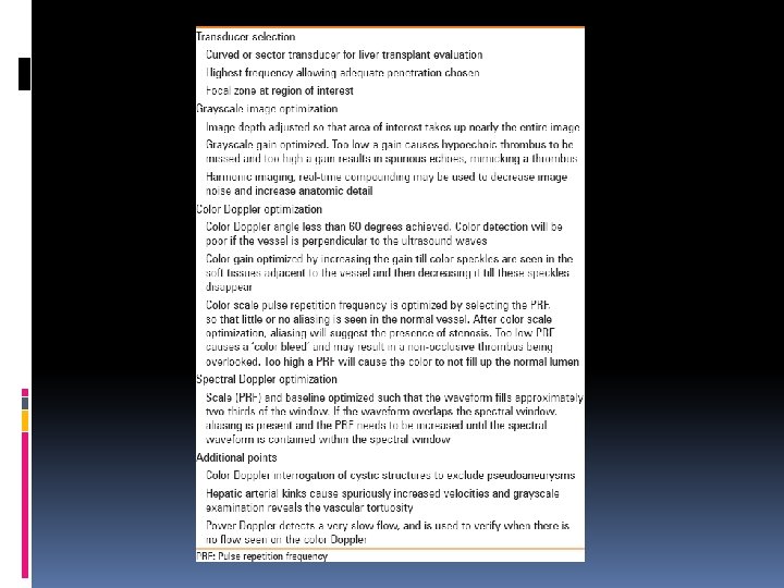
Liver Transplant Grayscale, color Doppler, and spectral Doppler examinations of the liver transplant vasculature should be performed. Before the ultrasound examination, the surgical anatomy and reconstructive techniques for that particular patient should be confirmed when this information is available. Comparisons with prior examinations should be made when possible.

Grayscale Evaluation of the Transplanted Liver A complete grayscale examination of the liver should be performed, including long-axis and transverse views. The liver parenchyma should be assessed for focal and/or diffuse abnormalities, and the echogenicity and echo texture of the liver should be noted. The biliary tree should be evaluated and the caliber of the common duct measured when possible. The subphrenic and subhepatic spaces should be investigated for possible fluid collections. Grayscale images of the hepatic vessels, including the portal vein, hepatic veins, and inferior vena cava (IVC), should be obtained.

Doppler Evaluation of the Transplanted Liver The vessels that should be examined include the main hepatic artery and right and left intrahepatic arteries, hepatic veins, IVC, main portal vein, and intrahepatic portal veins. The vascular anastomoses (hepatic arterial, portal venous, hepatic venous, and IVC) should be interrogated.

Hepatic Arteries The hepatic arteries should be interrogated to confirm normal flow and exclude complications such as hepatic artery thrombosis, stenosis, a pseudo -aneurysm, or an arteriovenous fistula. Both the main hepatic artery and the intrahepatic arteries should be evaluated when possible.

Main hepatic artery The main hepatic artery should be imaged along its length when possible. An attempt should be made to interrogate the native artery, region of the anastomosis, and donor artery. A Doppler evaluation should be obtained to demonstrate the presence of flow, configuration of the vessel, and any possible areas of color Doppler aliasing, which may suggest turbulent or high velocity flow. Spectral Doppler waveform morphology should be assessed. Velocity measurements may be obtained at the anastomosis and within the native and donor portions of the hepatic artery and at any areas of color flow aliasing. Doppler indices calculated from spectral Doppler waveforms obtained at these locations may include the peak systolic velocity (PSV), RI (systolic velocity – diastolic velocity/systolic velocity), and AT (time between end diastole and the first systolic peak)

Intrahepatic arteries The presence of flow should be confirmed in the intrahepatic (right and left hepatic) arteries. Resistive indices should be calculated from spectral Doppler waveforms obtained at these locations. Spectral Doppler waveform morphology should be assessed visually. Acceleration times can also be measured if the waveform appears abnormal, as in a tardus parvus waveform. Comparisons with prior examinations should be made when possible. Although the hepatic arterial waveform may change normally with time, some changes in the waveform configuration, RI, or PSV may require further evaluation.

Portal Vein The main portal vein and right and left branches should be scanned in their entirety, including the portal vein anastomosis. Images should document the presence of flow, the direction of flow, and any areas of possible color Doppler aliasing. Spectral Doppler evaluation should include an assessment of the waveform as well as angle-corrected peak velocity measurements proximal to, at, and distal to the main portal vein anastomosis. If there appears to be a discrepancy in velocities within the portal vein, an anastomotic-to-preanastomotic velocity ratio can be performed.
 should be")
Hepatic Veins and IVC The type of surgical anastomosis (piggyback versus interposition) should be determined before scanning when possible. Color and spectral Doppler tracings should be obtained from the right, middle, and left hepatic veins and the IVC in whole-liver transplants and from the existing hepatic veins and IVC in partial-liver transplants. In the case of a piggyback hepatic venous anastomosis, both the recipient IVC and the piggybacked hepatic vein confluence/donor IVC segment should be interrogated. Flow should be verified and the waveform assessed for the degree of phasicity. Comparisons with prior examinations should be made when possible. Follow-up examinations may be helpful if the initial ultrasound examination shows an abnormal waveform.
")
The type of surgical anastomosis (piggyback versus interposition)
 in adult patients has traditionally been performed using conventional caval")
Orthotopic liver transplantation (OLT) in adult patients has traditionally been performed using conventional caval reconstruction technique (CV) with veno-venous bypass. Recently, the piggyback technique (PB) without veno-venous bypass has begun to be widely used in order to preserve the Cava

Postoperative Doppler evaluation of liver transplants Doppler ultrasound plays an important role in the postoperative management of hepatic transplantation, by enabling early detection and treatment of various vascular complications. Doppler ultrasound is the modality of choice for evaluating post liver transplant patients for vascular complications. Awareness of the normal postoperative Doppler findings and timely identification of various vascular complications is essential for improving outcome of liver transplantation.

Liver transplantation is the only curative treatment for end-stage liver disease. The common indications for liver transplantation include cirrhosis secondary to alcoholic liver disease, hepatitis, hepatocellular carcinoma, nonalcoholic and metabolic diseases, and hepatic failure. Over the last few decades, advancement in surgical techniques and perioperative management has greatly improved the outcomes of liver transplantation. Early detection of vascular complications by postoperative Doppler imaging has played a vital role in decreasing the incidence of graft failure.

The allograft for liver transplantation can be obtained either from a deceased donor or from a living donor after performing a partial hepatectomy. Deceased donor liver transplantation is by far the most prevalent method of transplantation in much of the western world. For example, approximately 300 living donor liver transplants (LDLT) are performed every year in the US compared to 6000 cadaveric donor transplants. In countries where cadaveric organ procurement is difficult, LDLT is more common than cadaveric donor transplants. Although the preoperative workup and technique of cadaveric donor and LDLT are different, the principles of postoperative Doppler imaging, performed to evaluate the vascular patency, are similar.
 anastomosis is followed by portal venous,")
During liver transplantation surgery, inferior vena cava (IVC) anastomosis is followed by portal venous, arterial, and biliary anastomoses. For deceased donors, IVC anastomosis can be performed using the 'standard' or 'piggyback' techniques. In the standard technique, the recipient liver is removed along with the IVC. End-to-end anastomoses, between the donor and recipient IVCs, are performed above and below the allograft. In the piggyback technique, the donor hepatic venous confluence is anastomosed with the recipient IVC. Piggyback anastomosis helps maintain hemodynamic stability during surgery. The piggyback technique is used for all LDLTs.

Vascular complications are a common cause for allograft failure after hepatic transplantation. Early detection and treatment of vascular complications help reduce the incidence of graft failure. Doppler ultrasound is a noninvasive test that allows real time dynamic evaluation of the allograft vasculature and is extensively used in the postoperative evaluation of allografts.


Gray-scale ultrasound of the right upper quadrant precedes Doppler interrogation of the allograft vasculature. Along with evaluation of the hepatic parenchyma, attention is paid to the presence any peri-hepatic fluid collection. Perihepatic fluid collections may represent postoperative hematomas, seromas or bilomas. Superinfection of any fluid collection cause abscess formation. Pneumobilia can be present in patients with an anastomosis. Presence of biliary dilation may indicate development of biliary stenosis. Doppler examination of the liver transplant involves interrogation of the main hepatic artery and its intrahepatic branches, main portal vein and its branches, hepatic veins, and the IVC.

The first post-transplant Doppler examination is often performed within the first 24 hours of surgery, with follow-up exams, as clinically indicated. The main, right, and left hepatic arteries are evaluated and should have a similar appearance. When examining the hepatic arterial waveform, attention is directed to the systolic upstroke, peak systolic velocity, and resistive index (RI)
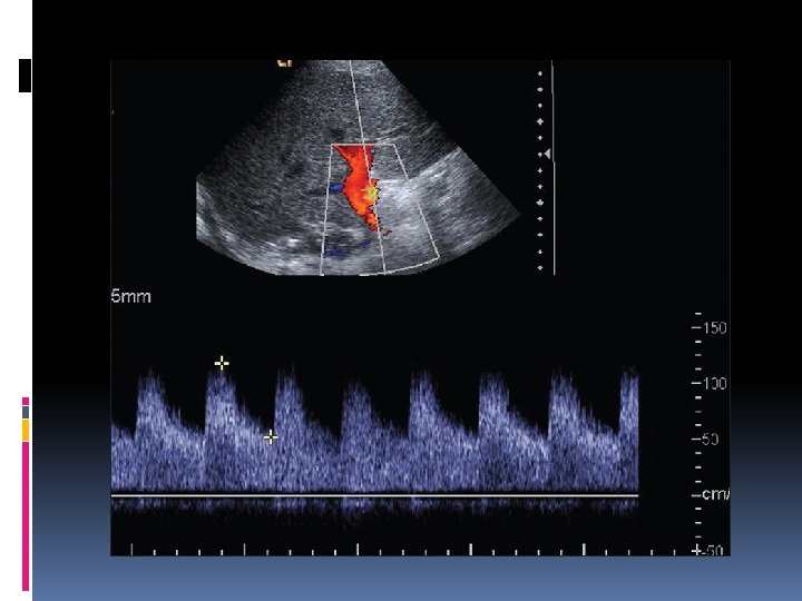

Figure 1: previous slide Normal main hepatic arterial waveform. Doppler image of the main hepatic artery post liver transplant shows continuous antegrade flow with rapid systolic upstroke, RI of 0. 60 and PSV of 110 cm/s

The normal hepatic arterial waveform has a rapid systolic upstroke. The systolic acceleration time (SAT) - the interval from end diastole to the first systolic peak - is a measure of the rapidity of the upstroke. A SAT greater than 80 m/s is abnormal in a post -transplant patient. However, there is a significant variability in the measurement of SAT, a fact that needs to be kept in mind when interpreting small changes in its value.
 is an important tool to assess the hepatic")
The hepatic arterial resistive index (RI) is an important tool to assess the hepatic arterial waveform. RI is determined by the following formula: (Peak systolic velocity-end diastolic velocity)/Peak systolic velocity. The RI increases as the diastolic flow decreases in the hepatic artery and absent or reversed diastolic flow corresponds to an RI of 1. 0. In a post-transplant patient, the normal hepatic arterial RI ranges from 0. 55 to 0. 80.
 is 103 cm/s. However, a wide")
The mean hepatic arterial peak systolic velocity (PSV) is 103 cm/s. However, a wide variability in PSV may be noted in the immediate postoperative period in the absence of any hepatic arterial complication.

On account of the hemodynamic changes associated with transplantation, the normal hepatic arterial waveform described earlier, is often not present in the immediate postoperative Doppler ultrasound. The hepatic arterial abnormalities seen in the immediate postoperative Doppler are often transient and resolve on follow-up scans. The first postoperative Doppler serves as baseline for future Doppler examinations.

The most common transient hepatic arterial waveform abnormality seen in the immediate postoperative period is increased hepatic arterial RI, due to decreased diastolic flow. Increased RI (RI > 0. 8, absent or even reversed diastolic flow) is seen in almost half the patients immediately after liver transplantation. This can involve the main hepatic artery as well as its intrahepatic branches. The RI usually normalizes within 7 -15 days and is not associated with poor graft function. This transient elevation of RI is likely secondary to allograft edema, increased cold ischemia time, increased portal flow or vessel spasm
: (A) Transient increased hepatic arterial RI. Doppler ultrasound performed")
Figure 2 (A and B): (A) Transient increased hepatic arterial RI. Doppler ultrasound performed on the first postoperative day after liver transplantation shows absent diastolic flow (RI = 1) in the main hepatic artery (B) Transient increased hepatic arterial RI. Follow-up

Doppler ultrasound performed after three days shows improvement of the waveform with increase in diastolic flow (RI 0. 85). Increased RI is a common finding in the immediate postoperative period, which usually resolves on follow-up imaging
 due to increased diastolic")
A decrease in hepatic arterial RI (RI < 0. 55) due to increased diastolic flow is a more ominous finding than increased RI, and is usually of concern for arterial complications. Decreased RI may occasionally be a transient finding on an immediate postoperative scan, which resolves in a few days. This is probably due to anastomotic edema in the postoperative period.

Vascular kinks due to vessel redundancy can lead to spuriously elevated values of peak systolic velocity. Correction of the angle of interrogation can help differentiate this from true stenosis. Anastomotic edema may also occasionally cause temporarily increased hepatic arterial velocity in the immediate postoperative scans.

The diagnosis of a hepatic arterial complication is seldom established on the basis of a single immediate postoperative Doppler ultrasound. Arterial waveform abnormalities on the immediate postoperative scans should be followed and correlated with the patient's clinical findings including liver function tests. Transient waveform changes usually resolve in 7 -15 days. Arterial complications are detected by persistence of abnormalities or further deterioration of the waveform, on follow-up Doppler studies.

An important point to note while interpreting post-transplant Doppler studies is that deterioration of a waveform on a follow-up study is highly suggestive of an arterial complication. The first postoperative Doppler serves as a baseline and any worsening of the hepatic arterial waveform on follow-up is always due to an arterial complication.

The reduction in portal venous resistance in the immediate post-transplant period can cause increased portal venous flow. This manifests as a high portal venous velocity, which gradually normalizes in the postoperative period, as the body adapts to the new hemodynamics. Compression of the portal vein by transient postoperative collections can also cause temporarily increased portal venous velocity, which normalizes as the postoperative fluid resolves.

A transient increase in portal venous velocity should not be misdiagnosed as portal venous stenosis, which is a rather rare complication. Hepatic veins normally have a triphasic waveform, but monophasic or biphasic waveforms are commonly seen in the postoperative period, secondary to graft edema or compression by the adjacent fluid collection. This usually normalizes on follow-up studies.
: (A) Transient increase in portal venous velocity. Doppler ultrasound")
Figure 3 (A and B): (A) Transient increase in portal venous velocity. Doppler ultrasound performed on the first postoperative day after liver transplantation shows increased main portal venous velocity (123 cm/s) (B) Transient increase in portal venous velocity. Follow-up Doppler ultrasound performed five days later shows decrease in the main portal venous velocity (59 cm/s). Increased portal venous velocity is a common transient finding after liver transplantation, which normalizes on follow-up imaging
, hepatic arterial stenosis (HAS), and pseudo-aneurysm")
Hepatic arterial complications include hepatic arterial thrombosis (HAT), hepatic arterial stenosis (HAS), and pseudo-aneurysm formation. Hepatic arterial complications are particularly significant in post liver transplant patients. The biliary tree derives its blood supply solely from the hepatic arteries after transplantation. Any hepatic arterial complication cause biliary ischemia, resulting in biliary strictures, necrosis, abscesses, and allograft failure. Biliary changes usually take time to develop and are usually not evident on ultrasound in the early stages.
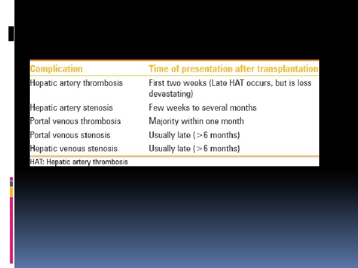

HAT is the most common and devastating complication after liver transplantation, which results in high mortality. Early HAT occurs in the first few weeks and presents with rapid clinical deterioration. Thrombosis occurring after the first month has a more insidious clinical course. HAT requires re-transplantation in majority of the cases. Surgical revascularization may be attempted when the diagnosis is established early in the postoperative course.

Ultrasound is diagnostic in 92% of cases of HAT. On Doppler ultrasound, HAT is diagnosed by the absence of flow in the hepatic arteries. As the allograft edema in the postoperative period can make visualization of the hepatic arteries difficult, it is important to ensure that the gain settings are optimized and a thorough search for hepatic arteries is performed to prevent a false positive diagnosis. A high resistance waveform (RI = 1) may be obtained in the hepatic artery if it is sampled proximal to the site of the thrombosis. On account of the catastrophic nature of this complication, computed tomography (CT) or digital subtraction angiography (DSA) angiography is usually performed to confirm the diagnosis.


Hepatic arterial collaterals can develop after HAT, particularly in the late cases. These collaterals can cause tardus parvus waveforms in the right and left hepatic arteries. Demonstration of such waveforms in the downstream hepatic arteries must not be considered evidence of patency of the more proximal hepatic artery. The tardus parvus waveform does not exclude HAT, but rather indicates the presence of either upstream stenosis or thrombosis.
: (A) Hepatic artery thrombosis. CT angiogram performed six months")
Figure 4 (A and B): (A) Hepatic artery thrombosis. CT angiogram performed six months after liver transplantation shows thrombosis of the main hepatic artery (arrow) (B) Hepatic artery thrombosis. Doppler ultrasound of the same patient shows tardus parvus waveform (delayed upstroke with increased diastolic flow) in the left hepatic artery, due to formation of collaterals following hepatic arterial thrombosis. Detection of flow in the left hepatic artery does not exclude HAT
 can cause complications similar to Hepatic Artery Thrombosis (HAT), but")
Hepatic Artery Stenosis (HAS) can cause complications similar to Hepatic Artery Thrombosis (HAT), but it has a more insidious course with symptoms developing over days to weeks after transplantation. HAS usually occurs at the anastomosis, but can occur anywhere in the graft artery. Patients usually present with abnormal liver enzymes or non-anastomotic biliary strictures. Timely detection of HAS on a postoperative Doppler is extremely important. Unlike HAT, this condition is treatable by percutaneous angioplasty and stenting. HAS itself causes graft ischemia and untreated stenosis can progress to the even more devastating HAT.

On Doppler ultrasound, HAS can be diagnosed by direct visualization of the stenotic segment or by identification of the characteristic downstream changes in the distal arteries. The stenotic segment is identified on color Doppler as an area of turbulence and aliasing. The PSV at the stenotic segment is usually elevated, greater than 200 cm/s. Secondary changes seen downstream to the stenosis are also very useful in establishing the diagnosis of HAS. There is decreased resistance of the arterial tree downstream to the stenosis. This results in an increased diastolic flow and a corresponding decrease in RI, to less than 0. 55. The usually rapid hepatic arterial systolic upstroke gets delayed (systolic acceleration time greater than 80 m/s). A tardus parvus waveform may be seen distal to the stenotic segment.
: (A) Hepatic artery stenosis. Doppler ultrasound of the right hepatic artery")
Figure 5 (A-D): (A) Hepatic artery stenosis. Doppler ultrasound of the right hepatic artery shows increased diastolic flow with an abnormally low RI of 0. 48 (B) Hepatic artery stenosis. Doppler ultrasound of the proximal main hepatic artery near the anastomosis shows focal area of aliasing and increased peak systolic velocity (>350 cm/s) consistent with focal stenosis (C) Hepatic artery stenosis. CT angiogram shows a focal severely stenotic segment in the main hepatic artery (arrow) (D) Hepatic artery stenosis. Percutaneous stenting of the stenotic segment was performed. DSA angiogram image shows a patent stent (arrowheads) across the previously stenotic segment

Hepatic arterial pseudoaneurysm formation is a very rare complication. Pseudoaneurysm formation can be due to infection or can be iatrogenic, secondary to a biopsy or angioplasty. Doppler interrogation of any cystic structure encountered near the hepatic hilum is performed to exclude a pseudoaneurysm. On Doppler ultrasound, a pseudoaneurysm is identified as a cystic structure along the hepatic artery, with a disorganized flow within it.

Splenic Artery Steal Syndrome In some patients with pre-existing portal hypertension and splenomegaly, following liver transplantation, the hypertrophied splenic artery may shift the blood flow away from the liver toward the spleen, resulting in hepatic hypoperfusion. This is known as the splenic artery steal syndrome. On Doppler, this manifests as a high-resistance waveform in the main, left, and right hepatic arteries. Increased portal vein velocity and hyperdynamic splenic arterial flow are present.

The imaging findings are not specific, as these may be present in normal patients in the immediate postoperative period and usually normalize on follow-up imaging. However, if these findings persist on follow-up examinations and patients have graft dysfunction, the presence of a splenic artery steal needs to be considered. Angiography is performed to confirm the diagnosis and the condition is treated by splenic arterial embolization

After transplantation, the normal portal vein has antegrade flow. The portal venous velocity is variable and tends to decrease on serial examinations after transplantation. Portal vein thrombosis is the most common portal venous complication after surgery. The thrombus is identified as either anechoic or echogenic material within the portal vein, on gray-scale ultrasound, and no flow is identified on Doppler interrogation. The portal vein should be interrogated with power Doppler so that the slow flow is not mistaken as thrombosis.

In a majority of the patients, portal venous thrombosis is diagnosed within one month of transplantation. It may present with signs of portal hypertension or nonspecific abnormalities of liver function tests. The treatment options in acute cases include catheter-guided thrombolysis and thrombectomy, as well as, anastomotic revision and re-transplantation.

Figure 6: Portal vein thrombosis. Doppler ultrasound of the main portal vein following liver transplant shows echogenic thrombus within the main portal vein with lack of colorfilling, consistent with portal vein thrombosis (arrows)

Portal vein stenosis is another complication occasionally seen after transplantation. Clinically significant portal vein stenosis after transplantation is rare and usually occurs in the pediatric and living donor population, due to the small graft vein size. Portal vein stenosis presenting within six months is likely due to technical reasons. The presentation is similar to portal vein thrombosis with portal hypertension or hepatic failure. Aliasing and a threeto-four fold increase in velocity is noted at the stenotic segment of the portal vein. A venogram, with measurement of the pressure gradient across the stenotic segment can be performed to assess the functional significance of the narrowing.
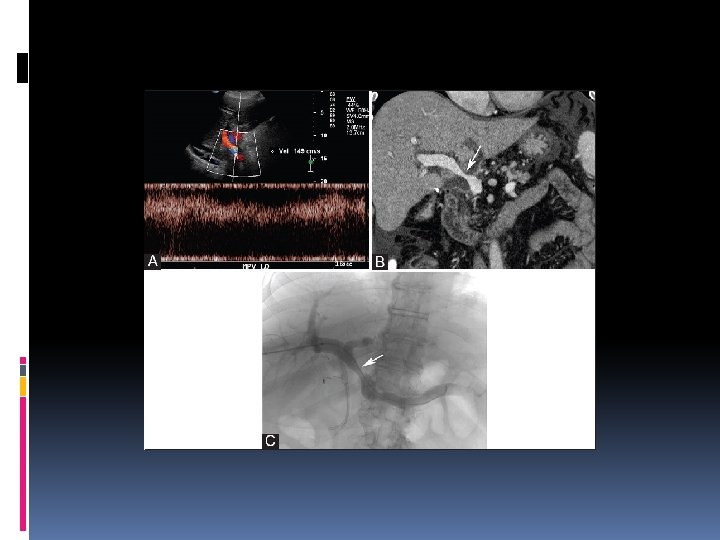
: (A) Portal vein stenosis. Doppler ultrasound of the main")
Previous slide Figure 7 (A-C): (A) Portal vein stenosis. Doppler ultrasound of the main portal vein following liver transplant shows a focal area of aliasing and increased velocity (149 cm/s). The adjacent segment of the portal vein has a velocity of 40 cm/s. This was considered suspicious for main portal vein stenosis (B) Portal vein stenosis. CT scan confirmed moderately severe narrowing of the main portal vein (arrow) (C) Portal vein stenosis. Patient subsequently underwent a portal venogram with pressure gradient measurement. Although the portal vein narrowing was re-demonstrated (arrow), there was a negligible pressure gradient across the stenotic segment. There was no evidence of portal hypertension. The stenosis was deemed not to be functionally significant and no therapeutic intervention was performed

Hepatic Venous and Inferior Vena Cava Complications Thrombosis and stenosis are two rare complications of the IVC and hepatic veins, after hepatic transplantation. The venous complications tend to occur at the anastomosis and knowledge of the surgical anatomy is useful. The incidence of hepatic venous outflow stenosis is higher in living donors and split graft liver pediatric grafts than in whole liver grafts, likely due to the size mismatch and small anastomosis. Most hepatic venous stenosis present late, many months to years after transplantation.

Normal hepatic veins have a triphasic waveform due to transmission of cardiac pulsations from the heart. Hepatic venous stenosis prevents transmission of the cardiac pulsations, resulting in loss of triphasicity. However, loss of triphasicity is a very nonspecific finding and is often seen in normal postoperative patients. Therefore, while the presence of hepatic venous triphasicity can be used to exclude hepatic venous stenosis, loss of triphasicity does not imply the presence of a hepatic venous complication. Clinically significant hepatic venous outflow stenosis can be treated with venous angioplasty and stenosis.

Overall, portal venous and hepatic venous outflow stenosis are rare. Ultrasound findings suggestive of venous stenosis should be correlated with the patients' clinical presentation.

Figure 8: IVC stenosis. Doppler ultrasound performed seven months after a cadaveric donor liver transplantation demonstrates aliasing and increased velocity (up to 263 cm/s) at the caudal IVC anastomosis, consistent with stenosis. IVC anastomoses had been performed using the standard technique, in which two end-to-end anastomoses were performed. Although the IVC was stenotic on imaging at the caudal anastomosis, the patient remained asymptomatic and no intervention was carried out

A word about T-Tubes A narrow flexible tube in the form of a T that is used for drainage of the common bile duct. The cross head is placed in the Common Bile duct while the stem is connected to a small pouch (bile Bag). It is used as a temporary post-operative drainage of the CBD (post Cholecystectomy)

A word about T-Tubes

Kidney/Renal Transplant
 should")
Renal Transplant Grayscale, color Doppler, and spectral Doppler examinations of the renal transplant(s) should be performed. Before the ultrasound examination, the surgical anatomy should be confirmed when this information is available. Comparison with prior examinations should be made when possible.

Grayscale Evaluation of the Transplanted Kidney Longitudinal and transverse views of the transplanted kidney and bladder should be obtained, and the longest renal length should be measured. The renal collecting system should be assessed for evidence of hydronephrosis, and, if present, the level of obstruction should be determined. The perinephric space should be assessed for evidence of fluid collections. Transverse and longitudinal images of the urinary bladder should be included. If a ureteral stent is in place, an attempt should be made to determine the proximal and distal extent of the stent. For patients in whom more than 1 transplanted kidney is present and evaluation of more than 1 transplant is required, each component of the examination should be performed for each renal transplant.

Images for each graft should be clearly labeled in such situations as appropriate (eg, “medial kidney” and “lateral kidney”).

Doppler Evaluation of the Transplanted Kidney Doppler evaluation of the transplanted kidney or kidneys should be performed for assessment of transplant vascularity. The vessels that should be examined include the main renal artery and vein, including anastomoses whenever possible, the adjacent external iliac artery and vein, and the intrarenal arteries of the transplanted kidney.

Main Renal Artery or Arteries The number of main renal arteries should be recorded. If more than 1 artery is present with separate anastomoses, each anastomosis should be similarly evaluated. Color Doppler images of the main renal artery or arteries from the transplanted kidney to the anastomosis should be obtained wherever possible. Velocity measurements should be obtained at the anastomosis and distal to the anastomosis whenever possible and at any areas of color flow aliasing suggestive of high-velocity flow. Doppler indices should include the PSV and may include the AT, RI, and/or pulsatility index.

Main Renal Vein Color Doppler images should be obtained from the transplant renal vein throughout its course from the kidney to anastomosis. Spectral Doppler images should be obtained from the transplant renal vein at the anastomosis and distal to the anastomosis.

External Iliac Artery and Vein Color and spectral Doppler images of the external iliac artery and vein should be obtained cephalad to the main renal artery and main renal vein anastomoses. Calculation of the renal artery-to-iliac artery PSV ratio may be helpful in evaluating for renal artery stenosis.

Intrarenal Arteries Color or power Doppler images of the entire kidney should be obtained to provide a global assessment of transplant renal perfusion and to assess for vascular abnormalities. Doppler indices calculated from spectral Doppler waveforms obtained in the interlobar or segmental arteries in the upper pole, interpolar region, and lower pole of the transplanted kidney should include the RI and may include the AT if a tardus parvus waveform is present.

Intrarenal Veins Color Doppler images and/or spectral Doppler waveforms may be obtained to assess venous flow within the transplant.

Pancreas Transplant

Pancreas Transplant Grayscale, color Doppler, and spectral Doppler examinations of a pancreas transplant should be performed. Before the ultrasound examination, the surgical anatomy should be confirmed when this information is available. Comparisons with prior examinations should be made when possible. The sonographic evaluation of the transplanted pancreas may be limited by reduced acoustic windows, which may impact the feasibility of obtaining the suggested images.

Grayscale Evaluation of the Transplanted Pancreas Imaging of the entire pancreas transplant should be performed in transverse and longitudinal planes. The echogenicity and echo texture of the pancreatic parenchyma should be assessed. The orientation of the graft should be ascertained, and grayscale images of the arterial Y-graft, arterial vasculature, and donor portal vein should be obtained to assess for evidence of intraluminal abnormalities

Diagram of Y-graft used for arterial anastomosis between pancreatic vessels and the recipient's CIA (CIA: Common iliac artery, IIA: Internal iliac Artery.

The pancreatic duct should be assessed. The peritransplant space should be assessed for fluid collections. For patients with enteric drainage of the transplanted pancreas, evaluation of the adjacent bowel may be helpful to assess for areas of dilatation, which may suggest obstruction. For patients with urinary bladder drainage of the transplanted pancreas, images of the urinary bladder should be obtained in transverse and longitudinal planes. If a pancreatic stent is in place, attempts should be made to determine the location of the proximal and distal portions of the stent.

Doppler Evaluation of the Transplanted Pancreas The structures that should be examined include the transplant arterial Y-graft, the transplant superior mesenteric artery and splenic artery, the recipient artery (typically the common or external iliac artery), the transplant superior mesenteric vein, splenic vein, and portal vein, and the recipient vein (typically an iliac vein or superior mesenteric vein).

Pancreatic Cancer Vague symptoms Usually diagnosed in late stages after liver or gallbladder problems Cause is unknown Occurs 60 -80 years of age Risk factors 107

Surgical Procedures Whipple procedure: Removal of the head of the pancreas, duodenum, parts of the jejunum and stomach, gallbladder, and possibly the spleen The pancreatic duct is connected to the common bile duct and the stomach is connected to the jejunum Post-op care 108
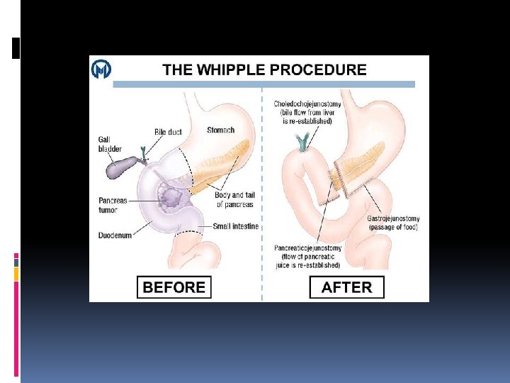

Postoperative Care Whipple Surgery Vital signs Respiratory status: Incentive spirometer, O 2 Pain Management Monitor NG -tube Surgical drainage: Protect the surgical sites Semi-fowlers: facilitate lung expansion and to decrease stress on the suture line Monitor glucose levels and administer insulin prn Provide nutritional support Standard post-op care for GI surgery 110

Complications Whipple Procedure 1. Fistulas Due to breakdown of a site of anastomosis 2. Peritonitis Internal leakage of corrosive pancreatic fluid Elevated WBCs, fever, abdominal pain, rebound tenderness, alteration in bowel sounds, shoulder pain Administer antibiotics 3. Venous thromboembolism: most common complication of pancreatic cancer 111

Color Doppler images of the Y-graft from the recipient arterial anastomosis, across both limbs of the Y-graft to both the superior mesenteric artery and splenic arterial anastomoses, should be obtained. Images should be assessed for any areas of color flow aliasing. Spectral Doppler images should be obtained within the recipient artery proximal to the Y-graft anastomosis and within the Y-graft itself, and the waveforms should be assessed for morphology. Spectral Doppler images with angle correction should be obtained within the splenic artery and superior mesenteric artery of the transplanted pancreas and at any areas of color flow aliasing. Doppler indices obtained at these locations should include the PSV and may include RIs.

Color or power Doppler images of the entire pancreas transplant should be obtained to assess the global vascularity of the graft. A spectral Doppler evaluation of Intraparenchymal pancreatic arteries should be performed in the pancreatic head, body, and tail, and RIs may be calculated.

Transplant Veins: Pancreas Color and spectral Doppler images of the graft splenic vein, superior mesenteric vein, and portal vein to the recipient venous anastomosis should be obtained. Spectral Doppler assessment with angle correction and measurement of the peak velocity may be performed within the graft portal vein, at the graft portal vein–venous anastomosis, and distal to the anastomosis within the recipient vein. Additional measurements at areas of color flow aliasing may be helpful. Intraparenchymal venous flow should also be documented in the head and tail of the transplanted pancreas.

Transplants?
- Slides: 115