Abdominal Pain During Pregnancy LAdel Shafik Pain Definition

Abdominal Pain During Pregnancy L/Adel Shafik

Pain Definition : It is a varying degree of unpleasant sensation. It may be acute with rapid onset and ends in days or chronic that recurs or persists for more than six months. There are three basic types of pain – Visceral , Somatic and referred.

• Visceral pain comes from outer covering of abdominal viscera which are innervated by autonomic nervous fibers , pain is due to distension or muscular contraction of a viscera It is typically vague , dull and nauseating , perceived in areas corresponding to embryonic dermatomal origin. • Somatic pain comes from parietal peritoneum which are innervated by somatic nervous fibers. Pain is due to irritation from inflammation , infection or chemical irritation. It is typically sharp and well localized. • Referred pain is pain perceived distant from its source and result from convergence of nerve fibers at spinal cord

Abdomen • The abdomen is roofed by the diaphragm, which separates it from the thorax (chest). • At the front and sides is the abdominal wall: skin, fat and muscles. • At the back, the spine (vertebral column). • It is completely filled with the abdominal organs: These are all covered by a thin membrane, continuous with that which also lines the inside of the abdominal wall (peritoneum). • The abdominal cavity and its peritoneal lining are continuous below with those of the pelvis.
 Pregnancy Related Pain: • Early pregnancy")
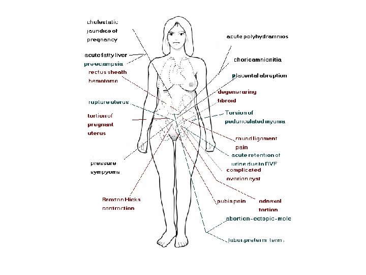
CAUSES OF ABDOMINAL PAIN DURING PREGNANCY • (A) Pregnancy Related Pain: • Early pregnancy o Abortion: Inevitable, incomplete or septic abortions o Vesicular mole: when expulsion starts. o Ectopic pregnancy: pain precedes bleeding. Later pregnancy o Braxton-Hicks Contraction o Round Ligament Pain o Pressure symptoms o Cholestasis of pregnancy o Placental abruption o Placenta percreta o Acute Fatty Liver o Pre-eclampsia , HELLP o Spontaneous rupture of the liver o Uterine rupture o Chorioamnionitis o Acute Polyhydramnios o Labor ( Term , Preterm ) (B)Conditions associated with pregnancy • © Non-Pregnancy Related Pain • Gastrointestinal o Acute appendicitis o Peptic ulcer o Gastroenteritis o Hepatitis o Inflammatory Bowel Complication (Crohn’s &Ulcerative Colitis ) o Bowel obstruction o Bowel perforation o Herniation o Meckel diverticulitis o Toxic megacolon o Pancreatic pseudocyst Hepato. Bliary o Biliary Stones o Acute Hepatitis o Acute Cholecystitis o Acute pancreatitis Rupture of rectus abdominus muscle Torsion of the pregnant uterus Acute urinary retention due to retroverted gravid uterus Musculo. Skeletal ( Pubic Symphysis pain-sacroiliac – back pain ). Red degeneration of myoma. Torsion of pedunculated myoma Ovarian cyst rupture. Adnexal torsion Genitourinary o Ureteral calculus o Acute pyelonephritis o Acute cystitis o Rupture of renal pelvis o Ureteral obstruction Vascular o Superior mesenteric artery syndrome o Thrombosis/infarction - Specifically mesenteric venous thrombosis o Ruptured visceral artery aneurysm Respiratory o Pneumonia Other o Intraperitoneal hemorrhage o Splenic rupture o Abdominal trauma o Acute intermittent porphyria o Diabetic ketoacidosis o Sickle cell disease

Physical examination Findings may be less prominent compared to those of non-pregnant patients with the same disorder. • Peritoneal signs are often absent in pregnancy because of the lifting and stretching of the anterior abdominal wall. The underlying inflammation has no direct contact with the parietal peritoneum, which precludes any muscular response or guarding that would otherwise be expected • The uterus can also obstruct and inhibit the movement of the omentum to an area of inflammation, distorting the clinical picture. • To help distinguish extrauterine tenderness from uterine tenderness, performing the examination with the patient in the right or left decubitus position, thus displacing the gravid uterus to one side, may prove helpful. • When performing a physical examination of the gravid abdomen, it is essential to recall the changing positions of the intra-abdominal contents at different gestational ages. For example, the appendix is located at the Mc. Burney point in patients in early pregnancy and in nonpregnant patients. After the first trimester, the appendix is progressively displaced upward and laterally, until it is closer to the gallbladder in late pregnancy. • Such alterations in physical assessment can delay diagnosis, and many authorities attribute the increased morbidity and mortality of acute abdomen in gravid patients to this delay. • When evaluating the gravid patient, the clinician must evaluate 2 patients at the same time, the mother and the fetus.

Investigations of abdominal pain in pregnancy • When evaluating the gravid patient with acute abdominal pain, remember that some very commonly used laboratory tests have altered reference ranges in pregnancy. These changes can make the initial evaluation process somewhat more difficult. • Fetal monitoring • Urinanalysis, MSU: infection, proteinuria in pre-eclampsia • Full blood count: raised white cell count suggestive of infection, although the white cell count is normally slighty raised in pregnancy • Liver function tests • Ultrasound: may demonstrate ectopic pregnancy, abruption, miscarriage • Magnetic resonance imaging : The intrinsic safety of MRI and its ability to accurately show abdominal and pelvic disease in pregnant patients make it highly useful in the evaluation of these patients. • Laparoscopy to confirm ectopic pregnancy - Laparoscopy has become increasingly popular in the treatment and evaluation of acute abdomen. In the past, pregnancy was considered a contraindication for laparoscopy , Care must be taken to minimize manipulation of the uterus. Adjust the location of trocar placement based on uterine size. Monitor fetal heart tones during the surgical procedure

Management of abdominal pain in pregnancy • A thorough assessment of the wellbeing of the mother and fetus, as well as the possible underlying cause is required. • Treatment of cause: urgent hospital referral if uncertain cause, and/or maternal or fetal distress. • If surgery is required but is considered elective, waiting until after the pregnancy is completed is prudent. • A pregnancy in a woman with an intra-abdominal inflammatory disease will not be harmed by proper surgical treatment. The fetus is more likely to be damaged if the properation is delayed. • Laparotomy (or perhaps laparoscopy but not in late pregnancy) is indicated if the diagnosis is in doubt or if there is shock.

Delivery • Base delivery decisions on obstetric indications • The mode of delivery used should also be decided based on obstetric indications. • If continuation of the pregnancy is expected to lead to maternal morbidity or mortality, delivery is indicated. • If improvement of the maternal condition cannot be expected with delivery, treat the patient with the fetus in utero • The prophylactic effect of tocolytics remains unproven in these patients. If used, tocolytics should be administered with care • If preterm delivery is likely, glucocorticoids can be administered to the mother to decrease the risk of neonatal complications. • Avoid glucocorticoids if the mother is at serious risk for significant infection

Braxton Hicks contractions • • • They are sporadic uterine contractions that actually start at about 6 weeks, although women won't be able to feel them that early. if they notice them at all (some women don't). They get their name from John Braxton Hicks, an English doctor who first described them in 1872. As pregnancy progresses, Braxton Hicks contractions tend to come somewhat more often, but they remain infrequent, irregular, and essentially painless. Sometimes, though, Braxton Hicks are hard to distinguish from early signs of preterm labor
 • Irregular, • Not progressively")
False labour pain ( Late Braxton Hicks contractions ) • Irregular, • Not progressively increasing • Not associated with bulging of forebag of water or dilatation of the cervix. • Respond to anlgesics • Cause women confusion as to whether or not they were going into actual labor. • They are thought to be part of the process of effacement, the thinning and dilation of the cervix
 • The evaluation of all pregnant women with abdominal discomfort")
Labour (term or preterm) • The evaluation of all pregnant women with abdominal discomfort must always include uterine contractions as an etiology. • Pain from labor is generally intermittent, occurring at decreasing intervals. However, tetanic uterine contractions, often evidence of uterine irritability, may produce sustained pain. An accurate estimation of gestational age is crucial to distinguish the normal labor anticipated in a term pregnancy from preterm labor. • • Preterm contractions are contractions that are painful and occur by definition before 37 weeks of gestational age; preterm labor is defined as preterm contractions with associated cervical change. • When contractions are suspected as a cause of abdominal pain, a cervical examination should be performed to evaluate the cervix for dilation, effacement, and if possible the station of the presenting part.

Round Ligament Pain with advancing gestational age as the uterine size increases. The round ligaments, found on the right and left sides of the uterus, attach to the pubic bone and help support the placement of the uterus in the abdominal cavity. these ligaments endure continual stretching and are a common source of pain in the latter part of pregnancy. Pain, either a sharp spasm or dull ache continuous, and may be described as a stretching or pulling sensation, is felt on one, or sometimes both, sides of the lower abdomen , often described as "round ligament pain"; however the exact origin of this pain many vary from patient to patient. This pain may be relieved by heat or acetaminophen, It is a benign and usually self-limiting occurrence that commonly causes discomfort in the second trimester
 • The most common")
Red Degeneration Of A Uterine Myoma (syndrome of painful myoma) • The most common complication is the syndrome of ‘painful myoma’; this is due to red or carneous degeneration and occurs in 5– 8% of myomas during pregnancy • This complication is associated with localized pain of rapid onset, nausea, vomiting and fever, tenderness, and an elevated white blood cell count • It usually occurs during the second trimester of pregnancy • ultrasound findings reveal cystic spaces in the myomas • The management of leiomyoma during pregnancy is medical, (symptomatic control of pain with acetaminophen and narcotics if required )

Uterine Torsion • The uterus rotates axially 30 °- 40 °to the right in 80% of normal pregnancies. • Rarely, it rotates > 90 °causing acute uterine torsion in mid or late pregnancy with abdominal pain, shock, a tense uterus, and urinary retention (catheterization may reveal a displaced urethra in twisted vagina). • Fibroids, adnexal masses, or congenital asymmetrical uterine anomalies are present in 90%. • Diagnosis is usually at laparotomy. • Delivery is by caesarean section

Pressure symptoms • Upper abdominal pressure --- pain due to flaring of the ribs particularly in breech presentation - The ribcage expands enormously during pregnancy to help make room for the expanding uterus and to maintain adequate lung capacity. Many pregnant women experience rib discomfort from this expansion, as well as the occasional little foot or knee of fetus that might habitually press against the ribs. • Mid abdominal pressure --- distension of the abdominal wall ( Twins , polyhydramnios ) • Lower abdominal pressure --engagement of the head

Hepatic rupture • Hepatic rupture is a rare but catastrophic occurrence in pregnancy it may be spontaneous but most are associated with preeclampsia and eclampsia associated disseminated intravascular coagulation. • Rupture usually occurs close to term or immediately postpartum. • Right upper quadrant pain and tendernes, hemorrhagic shock , distended abdomen • Rupture of the liver capsule is thought to result from subcapsular bleeding and can be confirmed by sonography

PROM/Chorioamnionitis • Choioamniotitis usually precipitated by pre-term premature rupture of membranes

Ectopic Pregnancy • The most common obstetric cause of an acute abdomen in the first trimester is ectopic pregnancy. • History, pelivc examination, serum ß-h. CG, culdocentesis, vaginal sonography, and laparoscopy can all be valuable in establishing the diagnosis. • Treatment is surgical, with laparoscopy or laparotomy. Linear salpingotomy, linear salpingostomy, or salpingectomy can be performed

A ruptured corpus luteum In pregnancy • A ruptured corpus luteum in pregnancy may produce intraabdominal bleeding and even an acute abdomen. • In early pregnancy before an intrauterine pregnancy can be confirmed by US, a corpus luteum may be very difficult to distinguish from an ectopic pregnancy or adnexal torsion, and frequently patients in the first trimester with pain and an adnexal mass on ultrasonography require a laparoscopy for definitive diagnosis

Placental Abruption • Placental abruption is premature separation of the placenta from the uterus, resulting in bleeding from the site of placental attachment (concealed , revealed. Combined ) • Abruption The triad of abdominal pain, uterine rigidity, and vaginal bleeding suggests this. • It occurs in between 1 in 80 and 1 in 200 pregnancies. • fetal monitoring may indicate fetal tachycardia and Fetal loss is high (up to 60%). • A tender uterus is highly suggestive and may be identified on tocography • Ultrasound may be diagnostic (but not necessarily so). • Minor abruptions with no maternal or fetal compromise may be managed conservatively, while others are true obstetric emergencies

Severe Preeclampsia • Right upper quadrant pain may be a manifestation of severe pregnancy -induced hypertension or preeclampsia, and is related to a combination of hepatic edema and ischemia -- subcapsular hepatic hematoma even hepatic rupture • Women with severe pre-eclampsia must be delivered rapidly

Acute polyhydramnios • • • Polyhydramnios is generally due to maternal diabetes (around 20% of cases) and results from hyperglycemia of the fetus which results in fetal polyuria (fetal urine is a major source of amniotic fluid). About 20% of the cases of polyhydramnios are associated with fetal anomalies that impairs the ability of the fetus to swallow (because the fetus normally swallows amniotic fluid and absorbs it through its intestinal villi). This includes: gastrointestinal abnormalities such as esophageal atresia and duodenal atresia , chromosomal abnormalities such as Down's syndrome (which is itself often associated with GI abnormalities) , neurological abnormalities such as anencephaly, which impair the swallowing reflex In a multiple gestation pregnancy, twin-twin transfusion syndrome is usually the cause. Fetuses with polyhydramnios are at risk for a number of other problems including cord prolapse, placental abruption and perinatal death. At delivery the baby should be checked for congenital abnormalities. Treatment -- amnioreduction has been used in response to polyhydramnios by frequent transabdominal removal of relatively small amniotic fluid volumes was associated with prolongation of pregnancy

Peptic ulcer disease • Complications of peptic ulcer disease such as perforation, hemorrhage, and obstruction are extremely rare during pregnancy • however, when they occur, they do so late either in the third trimester during pregnancy or early in the postpartum period

Bowel Obstruction e. g. adhesions, volvulus • Intestinal obstruction in the pregnant patients occurs in 1 out of 2500 to 3000 pregnancies. • Most cases occur in patients who have had a prior operative procedure, most commonly appendectomy or gynecologic surgery, and presumably result from pressure on preexisting adhesions by the enlarging uterus. • Obstruction is most common in the third trimester and least common in the first. • Symptoms of obstruction are similar to those of the nonpregnant patient.

Acute Appendicitis • It affects 1 in 1500 pregnancies, less common than in non-pregnant women , mortality is higher (esp. from 20 weeks), Perforation is commoner (15â-20%), Fetal mortality is ~1. 5% for simple appendicitis , ~30% if perforation. • Diagnosis is complicated by change in position of appendix as it migrates upwards, outwards and posteriorly as pregnancy progresses, so pain is less well localized (often paraumbilical or subcostal but right lower quadrant still commonest) and tenderness, rebound, and guarding less obvious. Peritonitis can make the uterus tense and woody-hard. • Leucocytosis is suggestive. • Operative delay is dangerous. • Laparotomy over site of maximal tenderness with patient tilted 30° to the left should be performed by an experienced obstetric surgeon (or by laparoscopy). • Appendicitis is not diagnosed in 1 in 5 cases in pregnant women until the appendix has ruptured causing peritonitis, which can cause premature labour or abortion.

Urinary Stones approximately 1 in 1500 pregnancies The classic presentation is flank pain, but patients may also present with nonspecific abdominal pain. Uterine contractions may also be present, further complicating the diagnosis, as the onset of preterm labor in patients with urinary calculi has been described. Fever may also be present and should raise the index of suspicion for a coexisting urinary tact infection. Urolithiasis should also be suspected in a patient with suspected pyelonephritis who fails to respond to antibiotic management. Every pregnant patient with flank or abdominal pain should have urine microscopic analysis performed. The presence of blood in the absence of recent urinary tract instrumentation raises the possibility of urinary calculi and appropriate imaging studies should be used to aid in the diagnosis. Other indications for imaging the renal system include a negative urine culture with the clinical diagnosis of pyelonephritis, suspected pyelonephritis with persistent fever after 48 hours of appropriate antibiotics, increasing blood urea nitrogen (BUN) and creatinine levels, and protracted pain. Initial imaging may include a renal ultrasound. However, the finding of hydroureteronephrosis in pregnancy is often a nonspecific physiologic finding related to bolus fluid administration, mechanical obstruction from the uterus, or the smooth muscle relaxing effect of progesterone. Identification of ureteral jets with Doppler sonography may help in excluding ureteral obstruction. If the diagnosis is still uncertain after sonographic evaluation, a noncontrast abdominal (renal protocol) CT or intravenous pyelogram (IVP) should be done. A limited IVP, a scout film and an image at 20 minutes, will result in a dose of less than 200 mrad, significantly less than the teratogenic level Patients with urolithiasis should be hydrated and be given appropriate pain control. Antibiotics are indicated for a concomitant upper or lower urinary tract infection. The urine may also be strained to identify when the stone has passed. This conservative approach will be successful in the majority of patients Surgical intervention may be required when conservative management fails or complications of ureteral obstruction develop such as worsening renal function or a persistent infection proximal to the stone.

Acute Pyelonephritis • • Acute pyelonephritis must always be considered in the differential diagnosis of abdominal or flank pain in pregnancy, and must also be considered in any patient with obstructive urinary calculi. Classic symptoms include back or flank pain in association with fever, chills, nausea, vomiting, and malaise. Patients may also complain of uterine contractions. On examination the patient is often febrile, looks ill, and will have costovertebral angle tenderness on the affected side. Clinical presentation may range from mild, nonspecific discomfort to urosepsis. Initial evaluation of the patient with suspected acute pyelonephritis should include a microscopic analysis of the unspun urine. Pyuria is almost always present and a Gram stain will often reveal bacteria, indicating >105 colony forming units per milliliter. Early identification of either gram-negative or gram-positive bacteria in this manner can also help guide initial antibiotic selection. 79, 80 Leukocytosis will generally be present, often with an increase in immature forms, or bands, and urine and blood cultures should also be obtained prior to initiation of antibiotics. A serum BUN and creatinine should also be obtained for assessment of baseline renal function in any patient with suspected pyelonephritis. While urine cultures of >105 colony forming units per milliliter are generally seen with pyelonephritis, 20 percent of patients will have urine cultures with lower colony counts. 79, 80 Pregnant women with pyelonephritis generally warrant inpatient therapy. Many will require intravenous hydration, and may be too ill to tolerate oral antibiotics, and those who have reached the stage of fetal viability will require close observation for the first 24 to 48 hours for preterm labor. Initial antibiotic therapy may be guided by the Gram stain before the urine culture results are available. Second- or third-generation cephalosporins are generally effective against gramnegative bacteria, although local resistance patterns should always be considered. Demonstration of gram-positive bacteria on Gram stain is concerning for enterococci and so empiric therapy with ampicillin and gentamicin should be instituted.

Adnexal And Ovarian complications • • • Adnexal disorders requiring surgical intervention occur in approximately one in 1000 pregnancies. Ovarian masses may be problematic during pregnancy because of their risk for torsion, rupture, or hemorrhage. The size of the mass is an important consideration as complications increase with increasing size; large ovarian lesions may also become impacted in the pelvis and even obstruct labor. While most adnexal masses in pregnancy are functional cysts that resolve by 18 weeks' gestation, The most common diagnosis in an ovarian mass large enough to warrant surgical intervention after 18 weeks' gestation is a benign cystic teratoma As most ovarian masses in pregnancy are smaller, functional cysts that are asymptomatic, they are usually identified as incidental findings on pelvic examinations in early pregnancy or on obstetric or pelvic ultrasounds ordered for other reasons. As the uterus enlarges, identification of adnexal pathology on bimanual examination becomes increasingly more difficult. Complications of adnexal lesions, such as adnexal torsion, rupture, or hemorrhage may present with pain and also with signs of peritoneal irritation such as nausea and vomiting. The best method for evaluating the adnexa in pregnancy is ultrasound. Simple cysts smaller than 6 cm are more likely to be functional, but extremely large functional cysts may sometimes be seen. , also be used when adnexal torsion is suspected. Adnexal torsion is more likely in the Masses greater than 6 cm that persist should generally be removed in the early second trimester to reduce the risk of complications such as rupture, torsion, or hemorrhage. Large masses that are symptomatic may sometimes require earlier intervention

Back Pain • Factors that influence back pain during pregnancy • The spine is vulnerable due to the following factors during pregnancy: -Hormone production during pregnancy makes joints less stable (to allow the pelvis to spread as the baby grows) -Typical weight gain of 25 -35 pounds during pregnancy, with the majority or extra weight distributed around the abdomen -Increase in postural strain as the body compensates for changes in the pregnant woman’s center of gravity

To help prevent or ease back pain • • • Try to be aware of how she stands, sits, and moves. Wear low-heeled (but not flat) shoes with good arch support. Ask for help when lifting heavy objects. When standing for long periods, place one foot on a stool or box. If her bed is too soft, have someone help her place a board between the mattress and box spring. Don't bend over from the waist to pick things up—squat down, bend your knees, and keep herr back straight. Sit in chairs with good back support, or use a small pillow behind the low part of her back. Try to sleep on her side with one or two pillows between her legs for support. Try to wear Maternity Belt comfortably supports the lumbar and abdominal regions Apply heat or cold to the painful area or massage it.

Rectus sheath haematoma • Very rarely, bleeding within the rectus sheath and haematoma formation can occur • with coughing (or spontaneously) in late pregnancy causing produces a painful, tender swelling that can mimic an intraperitoneal mass with features of an acute abdomen. • Ultrasound is helpful. • Conservative treatment

• • • Key Points Diagnostic delays are more common with surgical disorders in pregnancy, increasing both maternal and fetal morbidity and mortality. Physical finding of a surgical abdomen may be more difficult to elicit in pregnancy. Obstetric causes must always be considered in the pregnant patient with abdominal or pelvic pain, regardless of gestational age. With any pain in pregnancy think, could this be the onset of labour? Abdominal pain may be from ligament stretching or from symphysis pubis strain. In early pregnancy remember miscarriage and ectopics Suspected appendicitis is the most common nonobstetric indication for surgery in pregnancy. Adnexal masses that persist beyond 18 weeks' gestation are rarely functional. Ultrasound (US) is the most useful imaging tool in the evaluation of abdominal pain in pregnancy. Do not hesitate to involve a surgeon, obstetrician/gynecologist, and a specialist in maternal-fetal medicine when dealing with this challenging situation.
- Slides: 34