
Abdominal pain A site specific approach to radiologic
Abdominal pain A site specific approach to radiologic diagnosis Dr. Ahmed Refaey MBBCh, MS, FRCR Consultant Radiologist Department of Radiology Riyadh Militry Hospital
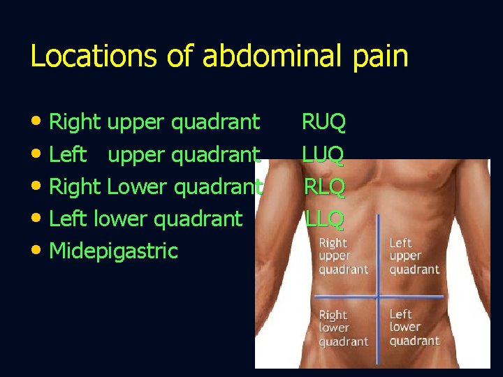
Locations of abdominal pain • Right upper quadrant • Left upper quadrant • Right Lower quadrant • Left lower quadrant • Midepigastric RUQ LUQ RLQ LLQ
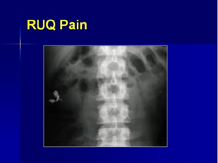
Acute RUQ pain • Acute cholecystitis – most common • DD - PUD - acute hepatitis - liver abscess - RLL pneumonia
RUQ pain imaging evaluation • Ultrasound • Acute cholecystitis – most common • DD - PUD - acute hepatitis - liver abscess - RLL pneumonia
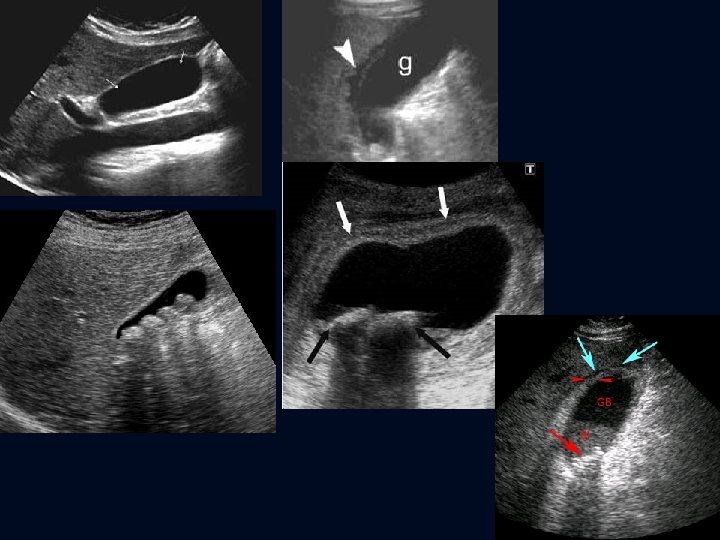
US in acute cholecystitis • Gall stones with …. ( sonographic Murphy’s sign ) • distended gall bladder • Pericholecystic fluid collection • Obstructing stone in GB neck or in cystic duct • gall bladder wall thickening (> 3 mm )
LUQ pain
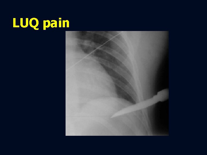
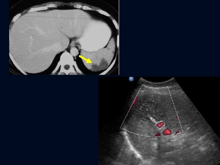
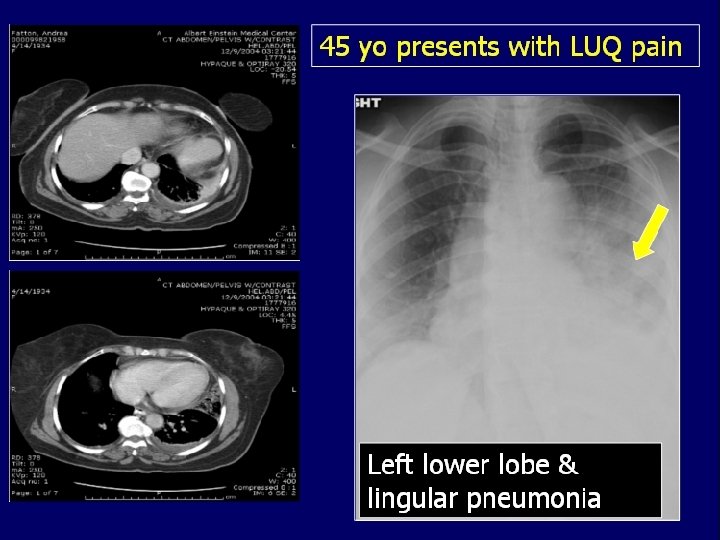
LUQ pain • Peptic ulcer disease • Splenic infarct • LLL pneumonia
Diagnostic tests • Plain X-ray chest -- pneumonia • US - splenic infarct • UGI - peptic ulcer disease
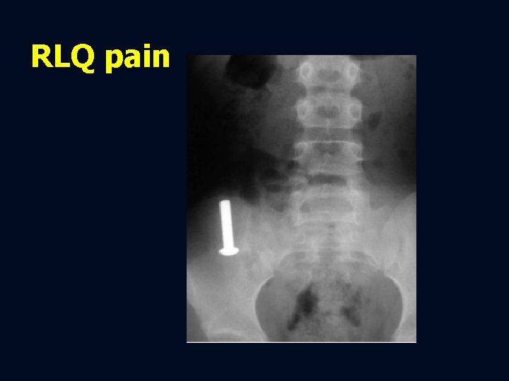
RLQ pain
RLQ pain • Acute appendicitis • Mesenteric adenitis • Diverticulitis • Urinary tract calculi • Gynacologic disease
Diagnostic imaging • US • CT • Exceptions for CT * children * pregnant women • Acute appendicitis • • Mesenteric adenitis Diverticulitis Urinary tract calculi Gynacologic disease
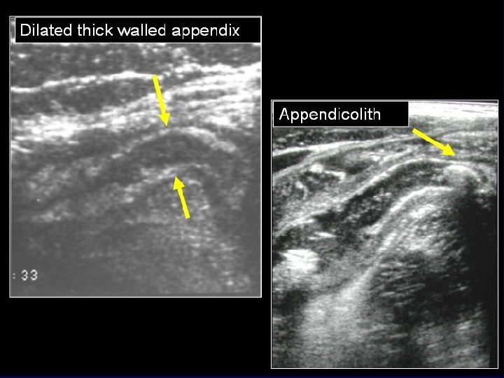
CT findings in acute appendicitis • Dilated appendix ( > 6 mm ) • Non filling of the appendix with contrast • Peri-appendiceal inflammatory changes • Increased enhancement of the wall • Appendicolith • Accuracy of CT is 94 -98 %
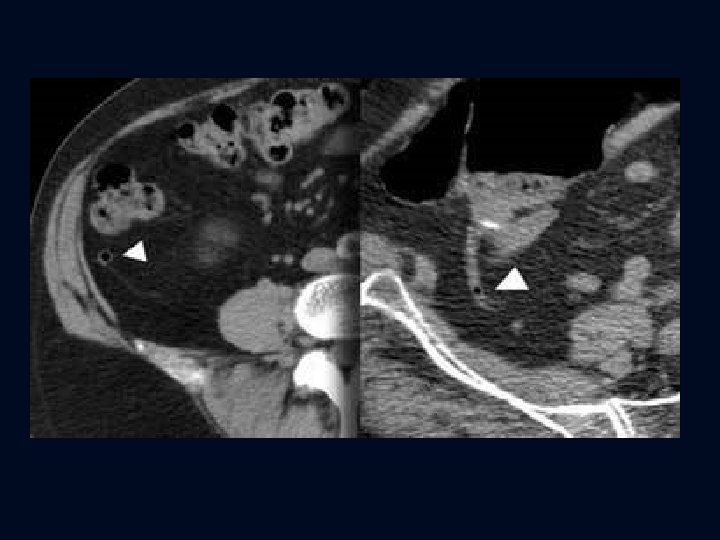
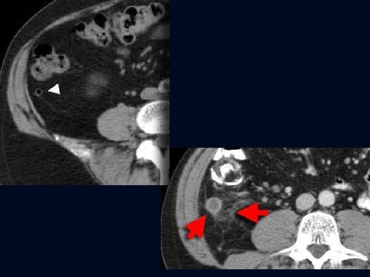
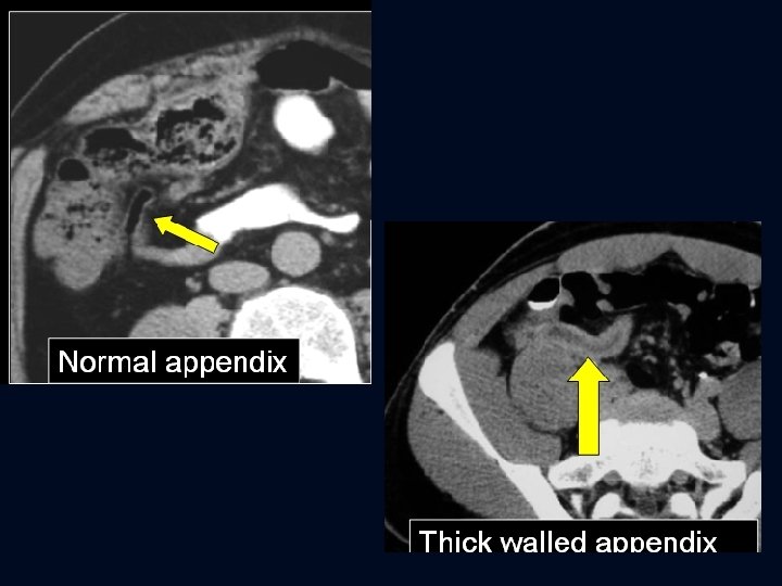
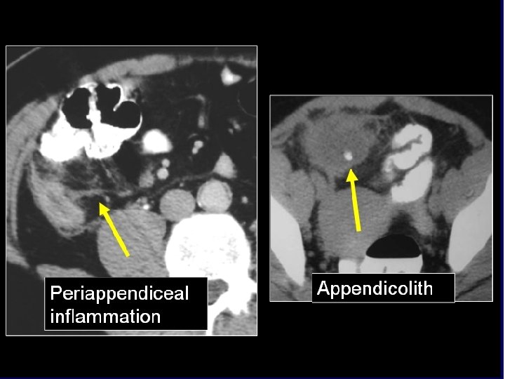
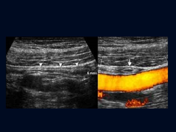
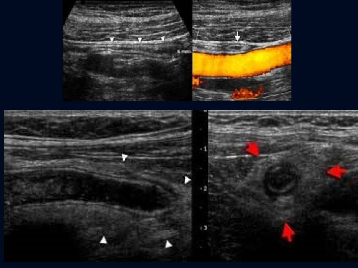
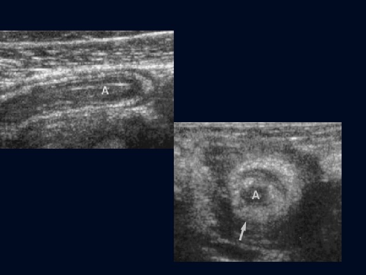
US findings in acute appendicitis • Non-compressible • Dilated appendix ( > 6 mm ) • Hyperemic thick wall • Appendicolith • Surrounded by omentum • Peri-appendiceal fluid collection
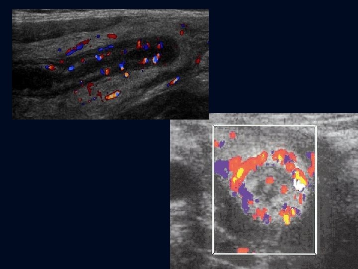
Mesenteric adenitis • Clinical presentation similar to appendicitis • inflammatory / infectious process • Children > adults • Most common alternative condition identified at surgery with removal of normal appendix
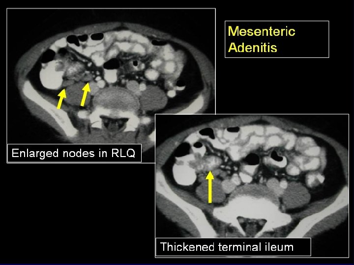
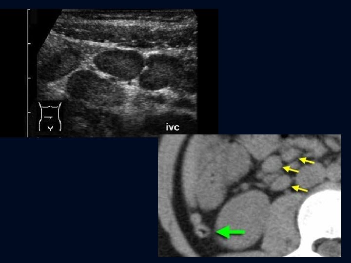
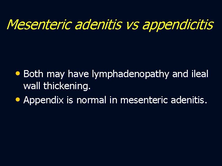
Mesenteric adenitis vs appendicitis • Both may have lymphadenopathy and ileal wall thickening. • Appendix is normal in mesenteric adenitis.
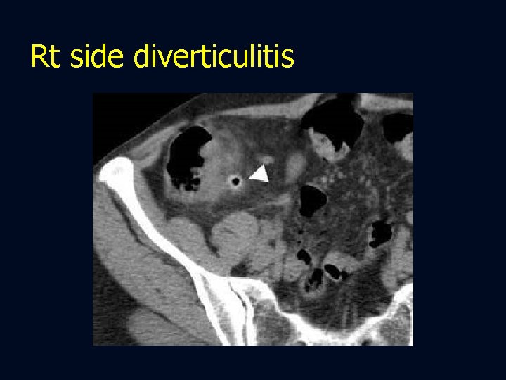
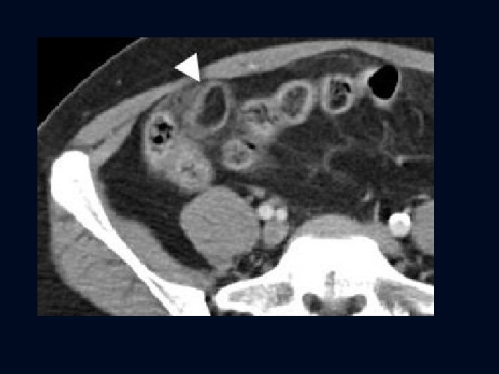
Rt side diverticulitis
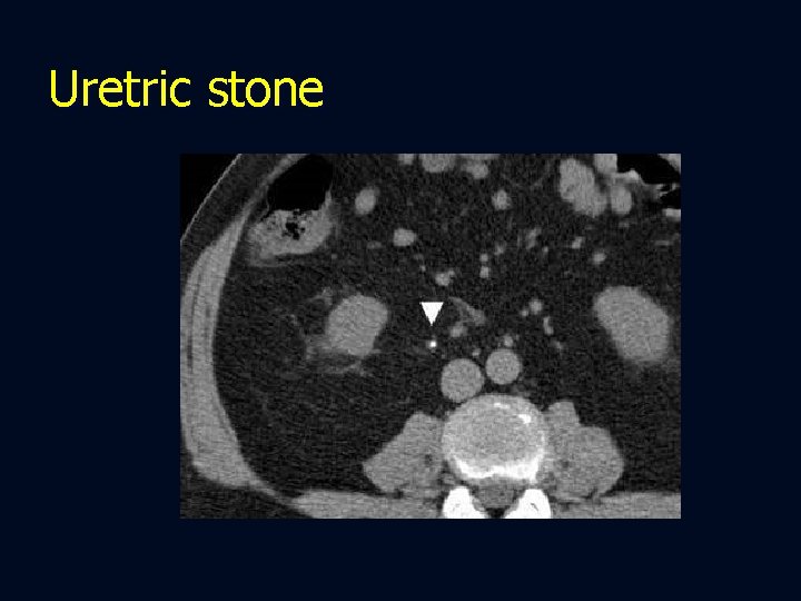
Uretric stone
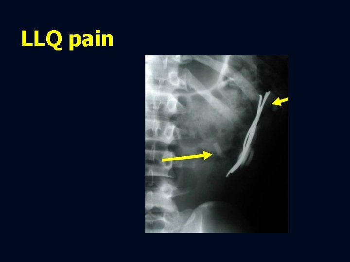
LLQ pain
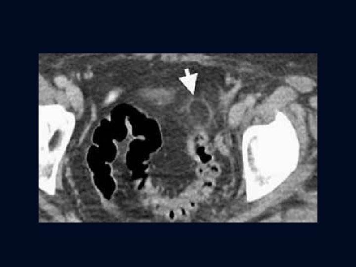
LLQ pain • Divericulitis • Epiploic appendagitis • Perforated colon cancer • Urinary tract calculi • Gynacologic disease
Diagnostic imaging • CT: best overall examination • US : better for females
CT in diverticulitis • Diverticulosis • Wall thickening • Inflammatory changes in pericolonic fat • Extraluminal air or abscess • Soft tissue inflammatory mass ( phlegmon) • Evidence of colovesical fistula
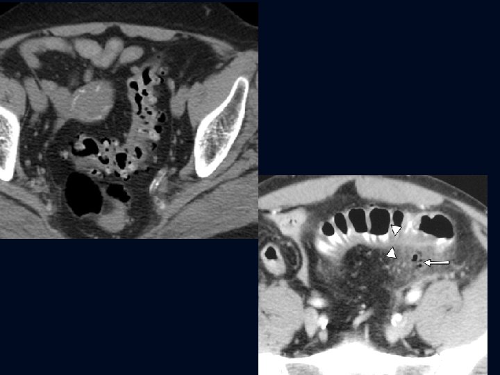
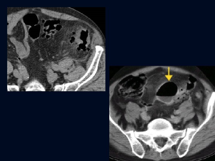
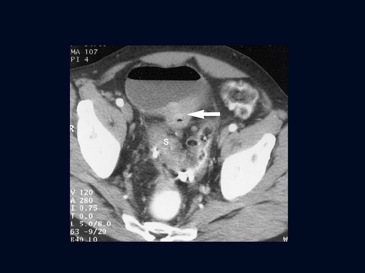
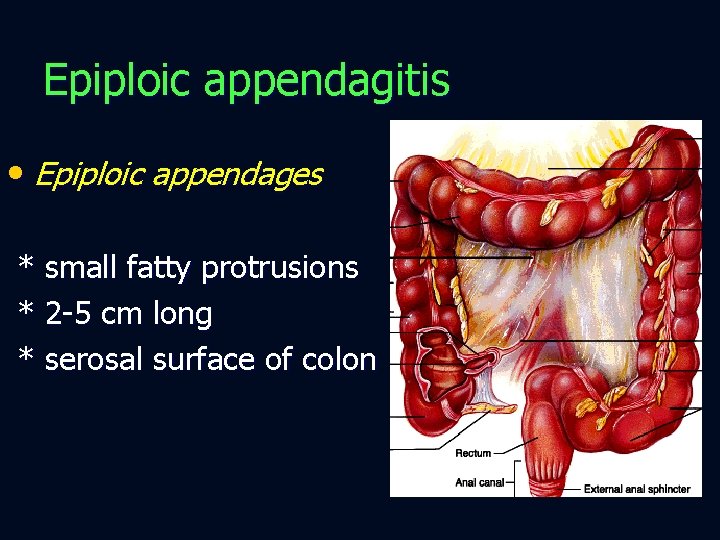
Epiploic appendagitis • Epiploic appendages * small fatty protrusions * 2 -5 cm long * serosal surface of colon
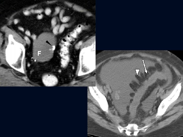
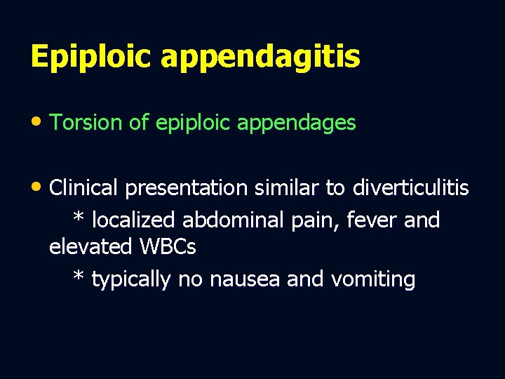
Epiploic appendagitis • Torsion of epiploic appendages • Clinical presentation similar to diverticulitis * localized abdominal pain, fever and elevated WBCs * typically no nausea and vomiting
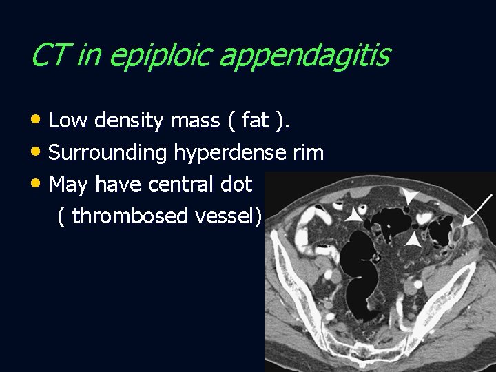
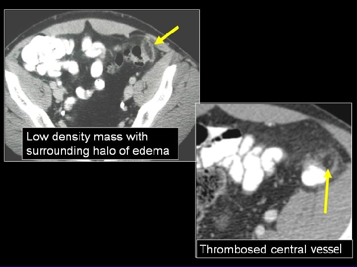
CT in epiploic appendagitis • Low density mass ( fat ). • Surrounding hyperdense rim • May have central dot ( thrombosed vessel)
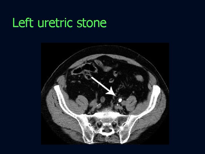
Left uretric stone
Qynacologic causes of LQ/pelvic pain • PID • Ruptured ovarian cyst • Hemorrhagic ovarian cyst • Adenexal torsion • Ectopic pregnancy
Diagnostic imaging • US is the primary imaging modality
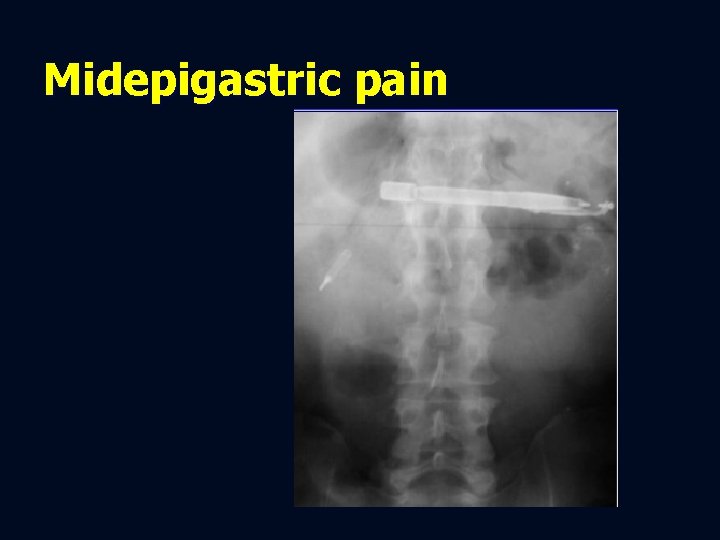
Midepigastric pain
Midepigastric pain • PUD • Acute pancreatitis • Aortic dissection • Aortic aneurysm rupture
Diagnostic imaging • CT the best for all except PUD Acute pancreatitis Aortic dissection Aortic aneurysm rupture
Acute pancreatitis diagnostic imaging • CT is the modality of choice • US may be useful screening tool
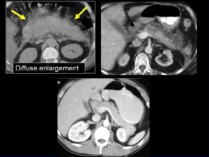
CT in acute pancreatitis • Focal or diffuse enlargement • Peripancreatic inflammation • Areas of necrosis • Peripancreatic collection
Abdominal aortic aneurysm • Without rupture, may present with pulsatile abdominal mass without pain • With rupture, presents with midabdominal and back pain
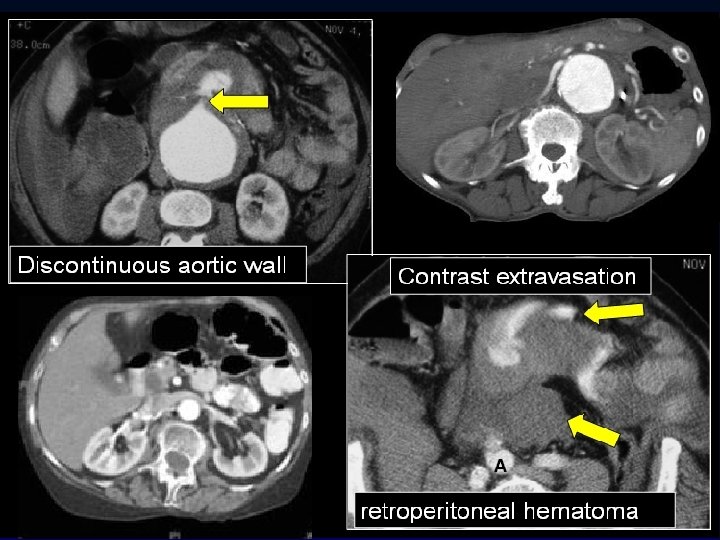
CT diagnosis of AAA rupture • Retroperitoneal hematoma • Contrast extravasation
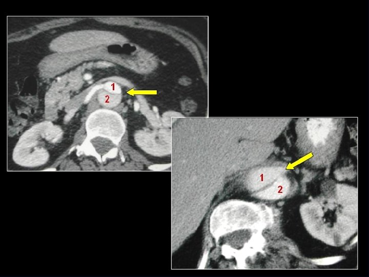
Abdominal aortic dissection • CT is the modality of choice
Abdominal aortic dissection CT diagnosis • Contrast in 2 channels • Intervening intimal flap
Summary • RUQ pain acute cholecystitis US • LUQ pain splenic/gastric UGI/US • RLQ pain A. appendicitis CT/US • LLQ pain diverticulitis CT • Midepigastric pancreas/aorta CT • Young female pelvic pain US • Children use US whenever possible

- Slides: 63