ABDOMINAL MUSCLES ABDOMINAL WALL Kaan Ycel M D

ABDOMINAL MUSCLES ABDOMINAL WALL Kaan Yücel M. D. , Ph. D. 24. April. 2014 Thursday

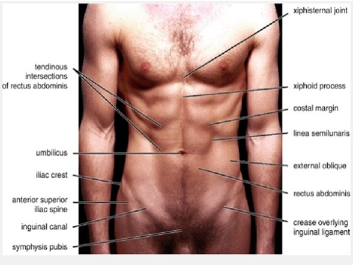
Anterior Lateral Posterior Anterolateral ABDOMINAL WALL enclose and protect abdominal contents while providing the flexibility required by respiration, posture, and locomotion.

Rectus abdominis

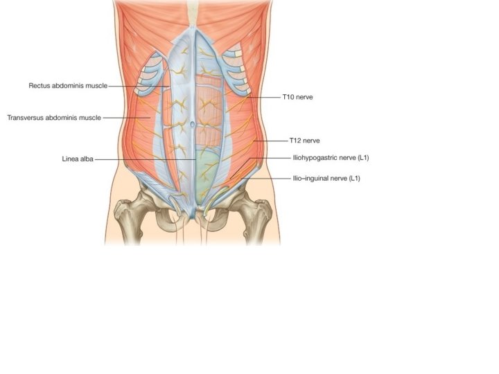
Pyramidalis muscle From pubic bone to linea alba Anterior rami of T 12
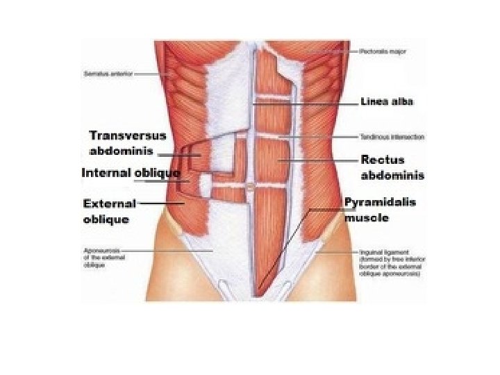
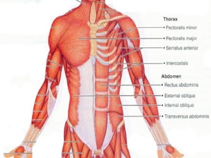

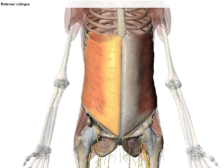
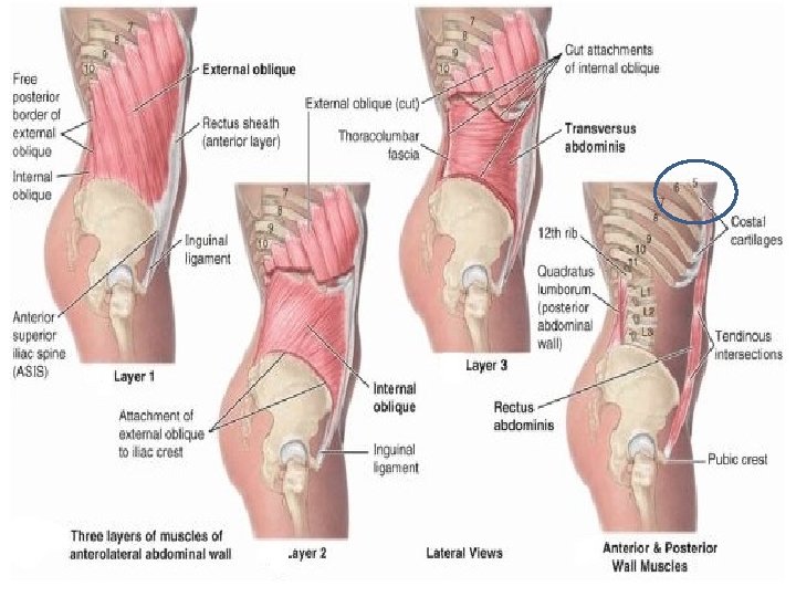
EXTERNAL OBLIQUE O External surfaces of 5 th-12 th ribs I Linea alba Pubic tubercle Anterior half of iliac crest T 7 -T 11 spinal nerves and subcostal nerve
")
The lower border of the external oblique aponeurosis forms the inguinal ligament (Poupart’s ligament) thickened reinforced free edge of the external oblique aponeurosis between anterior superior iliac spine laterally and pubic tubercle medially. It folds under itself forming a trough, which plays an important role in the formation of the inguinal canal.

from extensions of the fibers at the medial end of the inguinal ligament: lacunar ligament crescent-shaped extension of fibers at the medial end of the inguinal ligament pass backward attach to pecten pubis on the superior ramus of the pubic bone pectineal (Cooper's) ligament from the lacunar ligament along the pecten pubis of the pelvic brim
 and first lumbar nerves INTERNAL")
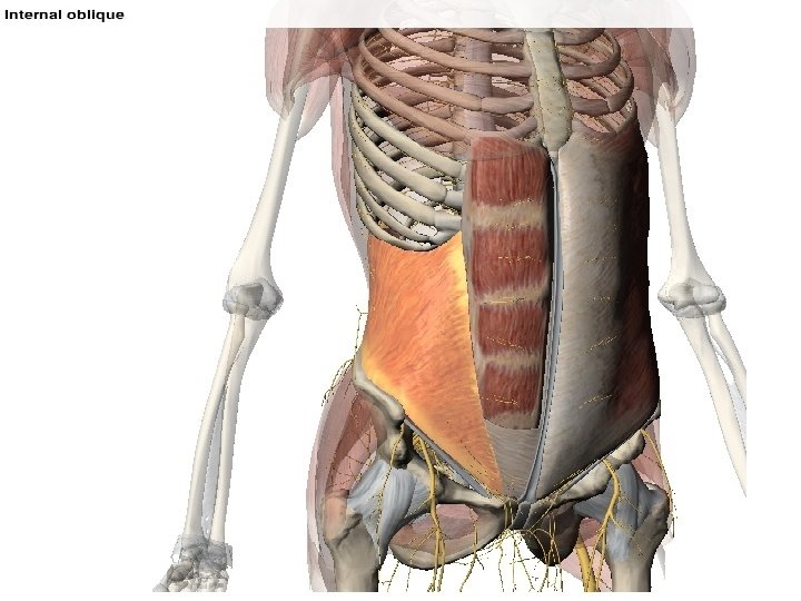
Anterior rami of T 6 -T 12 spinal nerves) and first lumbar nerves INTERNAL OBLIQUE I Inferior borders of 10 th-12 th ribs Linea alba Pecten pubis via conjoint tendon O Thoracolumbar fascia Anterior 2/3 of iliac crest Connective tissue deep to lateral 1/3 of inguinal ligament
 and first lumbar")
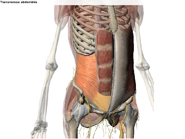
TRANSVERSUS ABDOMINIS Anterior rami of T 6 -T 12 spinal nerves) and first lumbar nerves I Linea alba with aponeurosis of internal oblique Pubic crest Pecten pubis via conjoint tendon O Internal surfaces of 7 th-12 th costal cartilages Thoracolumbar fascia Iliac crest Connective tissue deep to lateral 1/3 of inguinal ligament

RECTUS ABDOMINIS O: Pubic symphysis Pubic crest I: Xp 5 -7 costal cartilages Anterior rami of T 6 -T 12 spinal nerves) and first lumbar nerves

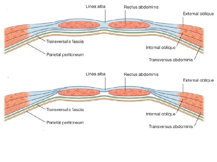
Rectus sheath unique layering of the aponeuroses of the external and internal oblique, and transversus abdominis muscles Ant. + Post. of ¾ rectus abdominis closed. Post. of ¼ rectus abdominis closed.

no sheath covers the posterior surface of the lower quarter of the rectus abdominis at this point is in direct contact with the transversalis fascia Marking this point of transition is an arch of fibers (arcuate line).

Rectus sheath & Transversalis fascia Rectus sheath anterior wall aponeurosis of external oblique & half of aponeurosis of internal oblique, splits at lateral margin of rectus abdominis posterior wall other half of aponeurosis of internal oblique & aponeurosis of transversus abdominis

FUNCTIONS AND ACTIONS OF ANTEROLATERAL ABDOMINAL MUSCLES üForm a strong expandable support for the anterolateral abdominal wall. üSupport the abdominal viscera and protect them from most injuries. üCompress the abdominal contents to maintain or increase the intra-abdominal pressure and, in so doing, oppose the diaphragm (increased intra-abdominal pressure facilitates expulsion). üMove the trunk and help to maintain posture.

Rectus abdominis is a powerful flexor of the thoracic and especially lumbar regions of the vertebral column. SPINE FLEXION VIDEO HERE

SPINE LATERAL FLEXION The oblique abdominal muscles also assist in movements of the trunk, especially lateral flexion and rotation of the lumbar and lower thoracic vertebral column. VIDEO HERE

SPINE ROTATION VIDEO HERE

ABDOMINAL CAVITY quiet and forced expiration by pushing the viscera upward (helps push the relaxed diaphragm further into the thoracic cavity) coughing vomiting sneezing eructation screaming parturition micturition defecation flatus
 Quadratus lumborum Iliacus")
Psoas major Psoas minor (80%) Quadratus lumborum Iliacus

Psoas major fills the space between vertebral bodies &transverse processes Anterior rami of L 1, L 2, L 3 Flexion of thigh Lateral flexion of spine Psoas minor (80%) Anterior rami of L 1 Weak flexion of lumbar vertebral column

Iliacus Femoral nerve fills the iliac fossa on each side. Psoas major + Iliacus = Iliopsoas chief flexor of the thigh Lesser troachanter of femur

Quadratus lumborum fill the space between ribs XII and the iliac crest on both sides of the vertebral column. I Transverse process of first four lumbar vertebrae and the inferior border of rib XII O Transverse process of L 5, Iliolumbar ligament, iliac crest Depress and stabilize 12 th ribs Lateral bending of the trunk. Acting together, extend lumbar part of the spine. anterior rami of T 12 and L 1 to L 4 spinal nerves

VIDEO HERE http: //www. getbodysmart. com
 continuous over the inguinal ligament")
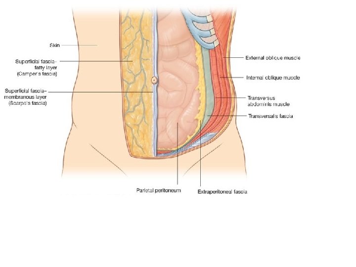
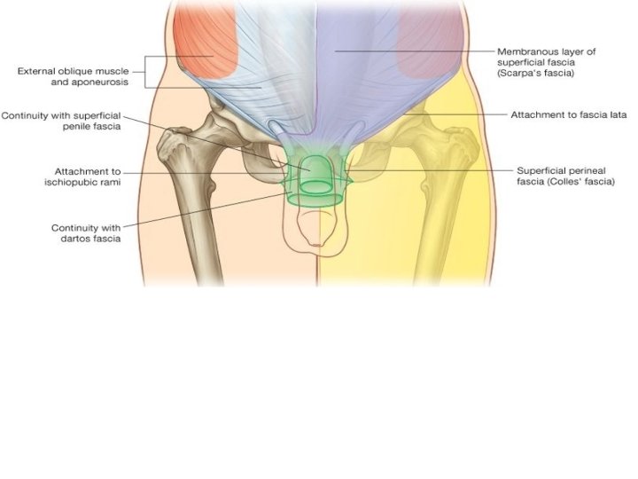
The superficial fatty layer of superficial fascia (Camper's fascia) continuous over the inguinal ligament with the superficial fascia of the thigh and with a similar layer in the perineum. In men, continues over the penis and, fusing with the deeper layer of superficial fascia, continues into the scrotum specialized fascial layer containing smooth muscle fibers (the dartos fascia). In women, retains some fat and is a component of the labia majora.
 thin and membranous contains little")
The deeper membranous layer of superficial fascia (Scarpa's fascia) thin and membranous contains little or no fat. Inferiorly, continues into the thigh, just below the inguinal ligament, fuses with deep fascia of the thigh (the fascia lata). In the midline, firmly attached to the linea alba & symphysis pubis. continues into the anterior part of the perineum where it is firmly attached to the ischiopubic rami and to the posterior margin of the perineal membrane. Here, referred to as superficial perineal fascia (Colles' fascia).
- Slides: 36